

, blocked with antigen-specific peptides.
Lane 2: HepG2 cells(serum starvation treatment).
Lane 3: Hela cells(lps 4h treatment).")
.
Bands result from membrane strip incubation.")

 by IF/ICC. The samples were fixed with PFA and permeabilized in 0.1% Triton X-100,then blocked in 10% serum for 45 minutes at 25°C. Samples were then incubated with primary Ab(AF5335) and mouse anti-beta tubulin Ab(T0023) for 1 hour at 37°C. An AlexaFluor594 conjugated goat anti-rabbit IgG(H+L) Ab(Red) and an AlexaFluor488 conjugated goat anti-mouse IgG(H+L) Ab(Green) were used as the secondary antibody.
The nuclear counter stain is DAPI(blue).")
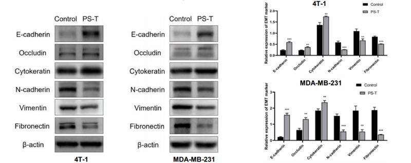
 inhibited JAM-A-transfected cell invasion (B) and EMT (C).")

. (A) Immunohistochemical validation of the essential extracellular
matrix proteins of the native testis (laminin, collagen type I, collagen type IV, and fibronectin) in the DTM. Scale bar, 200 µm. (B–E) Relative quantity of collagen I,
collagen IV, fibronectin, and laminin. ImageJ software was used to determine the average optical density (AOD). Data were shown as mean ± SD, n = 8. Differences
were considered statistically significant at *p < 0.05, **p < 0.01.")
 from day 7–13 after administrating BLM (A) Immunohistochemistry was used to analyze the expression levels of α-SMA, Col1 and Fn (n = 3). Quantitative analysis was shown beside. Scale bars: 50 μm (B–C) The expression levels of α-SMA and Col1 were detected by immunofluorescence in lung sections. The analyses of mean gray value were shown beside. Scale bars: 50 μm (D) Lung homogenization was used to analysis the α-SMA, LC3-II/I and p-mTOR (S2248), mTOR expression levels by Western blot (n = 6). Densitometric analyses were shown beside. Data in (A,D) are means ± Standard Error of Mean, *p < 0.05, **p < 0.01, ***p < 0.001, and NS: nonsignificant (one-way ANOVA). β-tubulin was used as a loading control.")
 Expression of proliferation- and migration-associated genes (PCNA, MMP9 and TIMP-1) were evaluated using western blotting in HEY cells. (C and D) Western blotting of proteins involved in integrin-β1-FAK signaling pathway in the KRT7-overexpressing HEY cells. (E) Expression of MMPs after knockdown of KRT7 in OVCAR433 cells. (F and G) Expression of the TGF-β signaling pathway-related proteins was evaluated by western blotting in KRT7-overexpressing HEY cells and KRT7-knockdown OVCAR433 cells. All experiments were performed at least three times. Results are presented as the mean ± standard deviation. **P<0.01. FAK, focal adhesion kinase; PCNA, proliferating cell nuclear antigen; FN, fibronectin; TIMP-1, TIMP metallopeptidase inhibitor 1; p-, phosphorylated; MMP, matrix metalloproteinase; KRT7, keratin 7; sh, short hairpin RNA; NC, negative control.")


, whereas proteins were detected using Cy3 (red). Scale bar, 100 µm. α‑SMA, α‑smooth muscle actin; ASIV, astragaloside IV; COL1A1, α1 type I collagen; FN, fibronectin.")
 COL1A1, (B) α‑SMA and (C) FN mRNA. (D) Protein expression level of α‑SMA and FN following ASIV treatment. *P<0.05, **P<0.01 and ***P<0.001. α‑SMA, α‑smooth muscle actin; ASIV, astragaloside IV; COL1A1, α1 type I collagen; FN, fibronectin.")
 and the presence or absence of TGF-b1 (5 ng/ml) for 24 h. (a and b) Real-time qPCR on related genes, including a-SMA and fibronectin. (c) Cell proliferation and apoptosis were measured by Western blot. Quantification of a-SMA and fibronectin expression is achieved using densitometric values as shown in(d).")
 Western blot analysis of the protein levels of Fn, Col1 and α-SMA in lung tissues.")
 Immunohistochemical staining of α-SMA, Col1 and Fn in lung tissues. Scale bar = 50 µm.")
 The total collagen content of the ECM-coated
titanium-modified surface after UV irradiation was detected by using a conventional hydroxyproline assay. *p < 0.05; **p < 0.01. (B)
Alizarin red staining was used to analyze the mineralization of the ECM-coated titanium-modified surface after UV irradiation. The
calcifying nodules were shown by green arrows. (C) The fibronectin on the ECM-coated titanium-modified surface after UV treatment
was detected by immunofluorescence. The fibronectin was indicated in green. (D) The topography of the biomimetic titanium implant
before screwing into bone (a–c) and after implant removal from bone (d–f) (50,000× magnification). (a) and (d) represent the ECM-Ctrl
group, (b) and (e) represent the ECM-SLA group, and (c) and (f) represent the ECM-TNT group.
Control: Polishing titanium implant; ECM-Ctrl: Polishing titanium implant coated with extracellular matrix; ECM-SLA: Sand blasting and
etching treated titanium implant coated with extracellular matrix; ECM-TNT: TiO2 nanotube topography implant coated with
extracellular matrix; SLA: Sand blasting and etching treated titanium implant; TNT: TiO2 nanotube topography implant; UV: Ultraviolet.")
 Effects of ketamine on parameters of oxidative stress in WT and KO mice at 4, 8 and 12 weeks as detected by ELISA. Negative control mice were treated with NS. *P<0.05 and **P<0.01. KO, knock-out; NS, normal saline; KHK, knock-out high-dose ketamine group; SOD, superoxide dismutase; GSH, glutathione-sulfhydryl; MDA, malondialdehyde; WHK, wild-type high-dose ketamine group; COX-2, cyclooxygenase 2; iNOS, inducible nitric oxide synthase; WT, wild-type; KNS, knock-out normal saline control group; WNS, wild-type normal saline control group; KLK, knock-out low-dose ketamine group; WLK, wild-type low-dose ketamine group; HK, high-dose ketamine; W, week; -, knock-out; +, wild-type. Ketamine increases the level of oxidative stress in aldh2 KO mice and aggravates pathological damage. (B) Representative hematoxylin and eosin staining images of bladder tissues from KO and WT mice in week 12. Magnification, x100 for the upper images; x400 for the lower images. (C) Representative immunohistochemical staining images of COX-2 and iNOS proteins in the bladder tissues of KO and WT mice in weeks 4 and 12. The cytoplasm and cell membranes exhibiting brown-yellow colors were considered as positive expression of the target protein, which were mainly confined to the bladder epithelium Magnification, x200. (D) Representative Masson trichrome staining images of bladder tissues of KO and WT mice in week 12. Collagen fibers stained green, muscle fibers stained red, and nucleus stained blue-brown. Magnification, x200. Data are presented as the mean ± standard error of the mean from ≥3 experimental repeats. *P<0.05 and **P<0.01. KO, knock-out; NS, normal saline; KHK, knock-out high-dose ketamine group; SOD, superoxide dismutase; GSH, glutathione-sulfhydryl; MDA, malondialdehyde; WHK, wild-type high-dose ketamine group; COX-2, cyclooxygenase 2; iNOS, inducible nitric oxide synthase; WT, wild-type; KNS, knock-out normal saline control group; WNS, wild-type normal saline control group; KLK, knock-out low-dose ketamine group; WLK, wild-type low-dose ketamine group; HK, high-dose ketamine; W, week; -, knock-out; +, wild-type.")


Immunohistochemistry staining of E-cadherin
and vimentin in lung tissues of mice. Scale bar
=50 μm. (b) The protein levels of E-cadherin,
fibronectin, α-SMA, and vimentin. Data were
expressed as mean ± SD. n = 6 in each group.
**p < 0.01 vs. control; ##p < 0.01 vs. BLM.")
 NIH-3T3 cells were co-treated with TGF-β1 (5 ng ml−1) and Remdesivir (12.5, 25, 50 μM) for 24 h. mRNA levels of α-SMA, Fibronectin and Collagen I were tested by RT-PCR in NIH-3T3 cells (B) NIH-3T3 cells were co-treated with TGF-β1 (5 ng ml−1) and Remdesivir (12.5, 25, 50 μM) for 24 h. α-SMA and Fibronectin were assessed using western blot, GAPDH was used as the internal control (C) PPF cells were co-treated with TGF-β1 (5 ng ml−1) and Remdesivir (12.5, 25, 50 μM) for 24 h. α-SMA and Fibronectin were assessed using western blot, β-tubulin was used as the internal control (D) Immunofluorescence staining of α-SMA were performed on NIH-3T3 cells treated with/without TGF-β1 (5 ng ml−1) and/or Remdesivir (12.5, 25, 50 μM) for 24 h. Data was noted as the means ± SD, n = 3. *p < 0.05, **p < 0.01, ***p < 0.001, **** p < 0.0001.")
 qRT-PCR was performed to compare the CSMD1 mRNA expression in fibroblasts transfected with Lenti-shRNA-CSMD1 (shCSMD1) and Lenti-GFP (shNC). All experiments were performed in triplicate and the data were shown as mean ± SD, *p < 0.05. (B) Immunofluorescence was performed to confirm the knockdown of CSMD1 protein expression in the CSMD1-silenced fibroblasts. Scale bar: 200 μm. (C) Histogram showing fluorescence intensity of CSMD1 in the shNC and shCSMD1 fibroblasts from the IF photos taken and analyzed using the NIS-Elements D software. (D-E) Transwell assays were performed to detect the migration of CSMD1-silenced fibroblasts. Quantification of numbers of migrated cells per field was presented as mean ± SD from three independent experiments in the right panel. Scale bar: 200 μm, **p < 0.01. (F-G) Wound healing assays were performed to detect the migration of CSMD1-silenced fibroblasts. The wound area at 0 h was set as 100%. Quantification of the healing rate was presented as mean ± SD from three independent experiments in the right panel. Scale bar: 200 μm, **p < 0.01. (H-I) qRT-PCR and western blot were performed to measure the ACTA2, COL1 and FN1 mRNA and protein levels respectively in the shNC and shCSMD1 fibroblasts. The results showed significantly upregulated mRNA levels of ACTA2, COL1 and FN1 upon CSMD1 knockdown, while only increased expression of FN1 at the protein level. *p < 0.05")
 Western blot of the effect of recombinant LAP on α-SMA, collagen I and FN expression in H9C2 cells induced by TGF-β1; (B–D) Semi-quantitative analysis of α-SMA, collagen I and FN expression presented as the relative ratio to β-actin. The expression levels of α-SMA, collagen I and FN in H9C2 cells treated with TGF-β1 were significantly higher than the control group (α-SMA, NC vs TGF-β1, P = 0.0013; collagen I, NC vs TGF-β1, P = 0.0004; FN, NC vs TGF-β1, P = 0.0005). Compared with the TGF-β1 group, the expressions of α-SMA, collagen I and FN in the LAP group were significantly reduced (α-SMA, TGF-β1 vs TGF-β1+LAP, P = 0.0311; collagen I, TGF-β1 vs TGF-β1+LAP, P = 0.0096; FN, TGF-β1 vs TGF-β1+LAP, P = 0.0276). NC: control group; TGF-β1: 10 ng/mL TGF-β1 group; TGF-β1+LAP: 10 ng/mL TGF-β1+60 μg/mL LAP group. n = 3, *P < 0.05 vs NC group; **P < 0.01 vs NC group; ***P < 0.001 vs NC group; #P < 0.05 vs TGF-β1 group; ##P < 0.01 vs TGF-β1 group.")
 collagen-1, (b) fibronectin (Fn), and (c) α-SMA. (d) Statistical expressions of fibrosis biomarkers in the immunohistochemical assay (∗∗p < 0.01 vs. the Sham group; ##p < 0.01 vs. the DKD group, n = 6). (e) Detection of fibrosis biomarker protein expressions by western blot analysis. (f) Statistical expressions of fibrosis biomarkers in the western blot assay (∗p < 0.05, ∗∗p < 0.01 vs. the Sham group; ##p < 0.01 vs. the DKD group, n = 3). The statistical results were compared with the Sham operation group and other groups.")
 collagen-1, (b) fibronectin (Fn), and (c) α-SMA. (d) Statistical expressions of fibrosis biomarkers in the immunohistochemical assay (∗∗p < 0.01 vs. the Sham group; ##p < 0.01 vs. the DKD group, n = 6). (e) Detection of fibrosis biomarker protein expressions by western blot analysis. (f) Statistical expressions of fibrosis biomarkers in the western blot assay (∗p < 0.05, ∗∗p < 0.01 vs. the Sham group; ##p < 0.01 vs. the DKD group, n = 3). The statistical results were compared with the Sham operation group and other groups.")

 and antibodies to α‑SMA, Collagen I and Fibronectin (red), immunofluorescence staining (magnification × 200), Scale bars = 40 μm. **P < 0.01 vs control group; ***P < 0.001 vs control group; #P < 0.05 vs TGF-β1 group; ##P < 0.01 vs TGF-β1 group")
 The representative band of Western blot. (b)–(h) The quantitative result of Western blot. Data were expressed as mean ± SD. #P < 0.05/##p < 0.01 vs. control; ∗p < 0.05/∗∗p < 0.01 vs. the BLM group. n = 3.")
 Representative images of Masson's trichrome stained kidney tissues of mice in the sham and ALD groups. Scale bar, 50 µm. (B) RT-qPCR analysis of miR-26a expression levels in the kidney tissues of mice in the sham and ALD groups; U6 was used for normalization. (C) RT-qPCR analysis of collagen I, α-SMA and LCN2 mRNA expression levels in the kidney tissues of mice from the sham and ALD groups; β-actin was used for normalization. (D) Representative western blotting images and semi-quantitative analysis of E-cadherin, collagen I, α-SMA, CTGF and LCN2 protein expression levels in the kidney tissue of mice in the sham and ALD groups. (E) Immunohistochemical analysis of E-cadherin (green), α-SMA (red) and fibronectin (green) in the kidney tissue of mice in the sham and ALD groups; DAPI (blue) was used to stain the nuclei. Scale bar, 50 µm. Data are presented as the mean ± SD; n=5 mice/group; *P<0.05 vs. sham. α-SMA, α-smooth muscle actin2; CTGF, connective tissue growth factor; LCN2, lipocalin; RT-qPCR, reverse transcription-quantitative PCR.")
 Schematic illustration presenting the preparation of 5% BSAMA–collagen I (B5-C), 5% BSAMA–fibronectin (B5-F), 5% BSAMA–collagen I–fibronectin (B5-CF). (B) FTIR spectra of unmodified and ECM protein-modified BSAMA cryogels. The broad peaks at 3118–3490 cm−1 resulting from a N-H stretching frequency of amine groups (-NH2). Peaks at 1648 cm−1, 1529 cm−1, and 1229 cm−1 correspond to the stretching/bending of amide-I, amide-II, and amide-III groups, respectively. (C) Immunostaining of collagen I and fibronectin in the ECM-coated BSAMA cryogels. Collagen I was stained with DyLight 488 (Green) and fibronectin was stained with DyLight 594 (red). Scale bar: 200 μm. (D) SEM images of ECM-modified BSAMA cryogels for ensuring that the overall porous structure of the cryogels appeared intact even after the coating. Scale bar: 100 μm (top); 40 μm (bottom).")
 The expression levels of collagen Ⅰ, fibronectin and α-SMA in the lung tissues of mice were detected by Western blotting. (E,F) Immunohistochemical staining analysis of collagen I, fibronectin, α-SMA and Ki-67, 20x, Scale bar = 50 μm. Data are shown as the mean ± SD. # represents the difference between the NaCl and model groups, # p < 0.05, ## p < 0.01, ### p < 0.001. * represents the difference between the model and treatment groups,")
 The expression levels of collagen Ⅰ, fibronectin and α-SMA in the lung tissues of mice were detected by Western blotting. (E,F) Immunohistochemical staining analysis of collagen I, fibronectin, α-SMA and Ki-67, 20x, Scale bar = 50 μm. Data are shown as the mean ± SD. # represents the difference between the NaCl and model groups, # p < 0.05, ## p < 0.01, ### p < 0.001. * represents the difference between the model and treatment groups,")
 α-SMA (b) COL6A2 (c) Fibrillin (d) Fibronectin (e) PAI-1 (f) UPR genes XBP1 and CCT4 (g) Protein expression of pro-fibrotic molecules after TGF-β1 treatment with densitometry analysis using ImageJ (h). (i) Vimentin expression after 10 ng/mL TGF-β1 treatment and densitometry analysis (j). The error bars represent standard deviation")
 The protein levels of ATG-5, LC3-I, LC3-II, SMA, vimentin, fibronectin, and fibrillin in whole-cell lysates of siRNA-mediated knockdown HTM cells with (10 ng/mL) and without TGF-β1. (B–G), relative quantification of the proteins obtained using densitometry analysis of the bands. The error bars represent the standard deviation")
 Representative images of α-SMA (top) and COL1A1 (bottom) immunostaining in HLFs co-treated with TGF-β1 (5 ng/ml) for 24 h in the presence of PBS (positive control), IL-11 W147C or IL-11 W147C dimer (50 ng/ml). HLFs treated without TGF-β1 were used as a negative control. Scale bars, 100 μm. (B-C) Total α-SMA and COL1A1 area were quantified by Image J, n = 3, the data were analyzed by t-test and are represented by means ± SEM (* p < 0.05, ** p < 0.01). (D) Western blotting analysis of the protein expression. (E-G) Expression levels (α-SMA, COL1A1 and fibronectin) were quantified by Image J, the data were analyzed by t-test (* p < 0.05, ** p < 0.01). (H) TF-1 cell proliferation experiment of the IL-11 W147C and IL-11 W147C dimer. (I-K) Competitive inhibition of wild type IL-11 (100ng/ml) mediated TF-1 proliferation by IL-11 W147C and IL-11 W147C dimer.")
 for 12 h to induce cell polarization. In some experiments, Ruxolitinib was applied to the cells at 5 and 10 μM. (A) Schematic diagram of the co-culture system. (B) Scratch assay of cells co-cultured with IL-4/13 (20 ng/mL) and Ruxolitinib (5 and 10 μM). Scratch closure was imaged at 0, 12, 24, and 48 h. (C) qPCR analysis of mRNA expression levels of Acta-2, Col-1, and Fn. (D) Western blot analysis of α-SMA, COL-1, and FN protein expression levels. Data are presented as mean ± SEM (n = 3). *P < 0.05, **P < 0.01, ***P < 0.001.")
 and three key targets (THBS2, MAFI2, and FGF9) were detected using western blotting. Each experiment was performed at least three times; *P < 0.05, **P < 0.01, ***P < 0.001.")
 The status of rat renal tissue was assessed using hematoxylin and eosin staining (bar = 20 μm). (b) BUN and Crea levels were tested by an automated biochemical analyzer (n = 5). (c) α-SMA and fibronectin levels were detected by immunohistochemistry (n = 3). op")
 Effect of Biejiaxiaozheng pills on the viability of LX-2 cells. (B) Effect of Biejiaxiaozheng pills on the Viability of TGF-β-induced activation model of LX-2 cells. (C) Effect of Biejiaxiaozheng pills on the fibrosis-related proteins Expression of LX-2 cells.")
 for 24 hours. The protein expression levels of α-SMA, CTGF, collagen I, collagen III and FN in fibroblasts were analyzed by Western blot (A–F). Data are expressed with mean ± SD; (B–F), *p < 0.05: Hypoxia group vs Control group, &p < 0.05: si-SPP1 group vs Hypoxia group, ns, Negative control group vs Hypoxia group. All experiments were repeated three times independently.")
 in long-term PD patients with peritoneal fibrosis. (a) Heatmap showing differential gene expression in PMCs from 1W-PD and 1Y-PD patients. (b) Representative immunofluorescence images of staining for POSTN, NF-κB P65, p-P65, CXCL8, α-smooth muscle actin (α-SMA), collagen I (Col-1), transforming growth factor-β (TGF-β), and FN in PMCs from 1W-PD and 1Y-PD patients. Scale bar = 50 µm. (c–i) mRNA expression levels in the two groups. (j–o) Representative Western blot images and quantitative analysis of POSTN, p-P65/P65, CXCL8, α-SMA, and Col-1 expression in the two groups.")
 The knockdown effectiveness of circNRIP1 was determined by qRT–PCR. (B and C) Cell migration was determined by wound healing assay in U87 and LN229 cells. The scale bars are 600 μm. (D and E) Cell invasion was determined by transwell assay in U87 and LN229 cells. The scale bars are 400 μm. (F) Cell proliferation was detected by CCK-8 assay in U87 and LN229 cells. (G and H) Protein levels of E-cadherin, N-cadherin, Fibronectin, and Vimentin were detected by western blotting. Data are presented as the mean ± SD of three independent experiments.")
 Image of subcutaneous xenograft tumors (n = 6 for each group). (B) Tumor volume was measured every 2 days. (C) Tumor weight was examined 28 days after injection. (D) Ki67, Fibronectin, N-cadherin, Vimentin, GPR133 IHC staining of xenograft tumors. The scale bars are 100 μm.")
| Product: | Fibronectin Antibody |
| Catalog: | AF5335 |
| Description: | Rabbit polyclonal antibody to Fibronectin |
| Application: | WB IHC IF/ICC |
| Cited expt.: | WB, IHC, IF/ICC |
| Reactivity: | Human, Mouse, Rat |
| Prediction: | Pig, Bovine, Sheep, Rabbit |
| Mol.Wt.: | 263 kDa, 200 kDa(Observed); 272kD(Calculated). |
| Uniprot: | P02751 |
| RRID: | AB_2837820 |
Control Products

Related Downloads
Protocols
Product Info
*The optimal dilutions should be determined by the end user. For optimal experimental results, antibody reuse is not recommended.
*Tips:
WB: For western blot detection of denatured protein samples. IHC: For immunohistochemical detection of paraffin sections (IHC-p) or frozen sections (IHC-f) of tissue samples. IF/ICC: For immunofluorescence detection of cell samples. ELISA(peptide): For ELISA detection of antigenic peptide.
Cite Format: Affinity Biosciences Cat# AF5335, RRID:AB_2837820.
Fold/Unfold
CIG; Cold insoluble globulin; Cold-insoluble globulin; DKFZp686F10164; DKFZp686H0342; DKFZp686I1370; DKFZp686O13149; ED B; Fibronectin 1; FINC; FINC_HUMAN; FN; FN1; FNZ; GFND; GFND2; LETS; Migration stimulating factor; MSF; Ugl-Y3;
Immunogens
A synthesized peptide derived from human Fibronectin, corresponding to a region within C-terminal amino acids.
Expressed in the inner limiting membrane and around blood vessels in the retina (at protein level) (PubMed:29777959). Plasma FN (soluble dimeric form) is secreted by hepatocytes. Cellular FN (dimeric or cross-linked multimeric forms), made by fibroblasts, epithelial and other cell types, is deposited as fibrils in the extracellular matrix. Ugl-Y1, Ugl-Y2 and Ugl-Y3 are found in urine (PubMed:17614963).
- P02751 FINC_HUMAN:
- Protein BLAST With
- NCBI/
- ExPASy/
- Uniprot
MLRGPGPGLLLLAVQCLGTAVPSTGASKSKRQAQQMVQPQSPVAVSQSKPGCYDNGKHYQINQQWERTYLGNALVCTCYGGSRGFNCESKPEAEETCFDKYTGNTYRVGDTYERPKDSMIWDCTCIGAGRGRISCTIANRCHEGGQSYKIGDTWRRPHETGGYMLECVCLGNGKGEWTCKPIAEKCFDHAAGTSYVVGETWEKPYQGWMMVDCTCLGEGSGRITCTSRNRCNDQDTRTSYRIGDTWSKKDNRGNLLQCICTGNGRGEWKCERHTSVQTTSSGSGPFTDVRAAVYQPQPHPQPPPYGHCVTDSGVVYSVGMQWLKTQGNKQMLCTCLGNGVSCQETAVTQTYGGNSNGEPCVLPFTYNGRTFYSCTTEGRQDGHLWCSTTSNYEQDQKYSFCTDHTVLVQTRGGNSNGALCHFPFLYNNHNYTDCTSEGRRDNMKWCGTTQNYDADQKFGFCPMAAHEEICTTNEGVMYRIGDQWDKQHDMGHMMRCTCVGNGRGEWTCIAYSQLRDQCIVDDITYNVNDTFHKRHEEGHMLNCTCFGQGRGRWKCDPVDQCQDSETGTFYQIGDSWEKYVHGVRYQCYCYGRGIGEWHCQPLQTYPSSSGPVEVFITETPSQPNSHPIQWNAPQPSHISKYILRWRPKNSVGRWKEATIPGHLNSYTIKGLKPGVVYEGQLISIQQYGHQEVTRFDFTTTSTSTPVTSNTVTGETTPFSPLVATSESVTEITASSFVVSWVSASDTVSGFRVEYELSEEGDEPQYLDLPSTATSVNIPDLLPGRKYIVNVYQISEDGEQSLILSTSQTTAPDAPPDTTVDQVDDTSIVVRWSRPQAPITGYRIVYSPSVEGSSTELNLPETANSVTLSDLQPGVQYNITIYAVEENQESTPVVIQQETTGTPRSDTVPSPRDLQFVEVTDVKVTIMWTPPESAVTGYRVDVIPVNLPGEHGQRLPISRNTFAEVTGLSPGVTYYFKVFAVSHGRESKPLTAQQTTKLDAPTNLQFVNETDSTVLVRWTPPRAQITGYRLTVGLTRRGQPRQYNVGPSVSKYPLRNLQPASEYTVSLVAIKGNQESPKATGVFTTLQPGSSIPPYNTEVTETTIVITWTPAPRIGFKLGVRPSQGGEAPREVTSDSGSIVVSGLTPGVEYVYTIQVLRDGQERDAPIVNKVVTPLSPPTNLHLEANPDTGVLTVSWERSTTPDITGYRITTTPTNGQQGNSLEEVVHADQSSCTFDNLSPGLEYNVSVYTVKDDKESVPISDTIIPEVPQLTDLSFVDITDSSIGLRWTPLNSSTIIGYRITVVAAGEGIPIFEDFVDSSVGYYTVTGLEPGIDYDISVITLINGGESAPTTLTQQTAVPPPTDLRFTNIGPDTMRVTWAPPPSIDLTNFLVRYSPVKNEEDVAELSISPSDNAVVLTNLLPGTEYVVSVSSVYEQHESTPLRGRQKTGLDSPTGIDFSDITANSFTVHWIAPRATITGYRIRHHPEHFSGRPREDRVPHSRNSITLTNLTPGTEYVVSIVALNGREESPLLIGQQSTVSDVPRDLEVVAATPTSLLISWDAPAVTVRYYRITYGETGGNSPVQEFTVPGSKSTATISGLKPGVDYTITVYAVTGRGDSPASSKPISINYRTEIDKPSQMQVTDVQDNSISVKWLPSSSPVTGYRVTTTPKNGPGPTKTKTAGPDQTEMTIEGLQPTVEYVVSVYAQNPSGESQPLVQTAVTNIDRPKGLAFTDVDVDSIKIAWESPQGQVSRYRVTYSSPEDGIHELFPAPDGEEDTAELQGLRPGSEYTVSVVALHDDMESQPLIGTQSTAIPAPTDLKFTQVTPTSLSAQWTPPNVQLTGYRVRVTPKEKTGPMKEINLAPDSSSVVVSGLMVATKYEVSVYALKDTLTSRPAQGVVTTLENVSPPRRARVTDATETTITISWRTKTETITGFQVDAVPANGQTPIQRTIKPDVRSYTITGLQPGTDYKIYLYTLNDNARSSPVVIDASTAIDAPSNLRFLATTPNSLLVSWQPPRARITGYIIKYEKPGSPPREVVPRPRPGVTEATITGLEPGTEYTIYVIALKNNQKSEPLIGRKKTDELPQLVTLPHPNLHGPEILDVPSTVQKTPFVTHPGYDTGNGIQLPGTSGQQPSVGQQMIFEEHGFRRTTPPTTATPIRHRPRPYPPNVGEEIQIGHIPREDVDYHLYPHGPGLNPNASTGQEALSQTTISWAPFQDTSEYIISCHPVGTDEEPLQFRVPGTSTSATLTGLTRGATYNVIVEALKDQQRHKVREEVVTVGNSVNEGLNQPTDDSCFDPYTVSHYAVGDEWERMSESGFKLLCQCLGFGSGHFRCDSSRWCHDNGVNYKIGEKWDRQGENGQMMSCTCLGNGKGEFKCDPHEATCYDDGKTYHVGEQWQKEYLGAICSCTCFGGQRGWRCDNCRRPGGEPSPEGTTGQSYNQYSQRYHQRTNTNVNCPIECFMPLDVQADREDSRE
Predictions
Score>80(red) has high confidence and is suggested to be used for WB detection. *The prediction model is mainly based on the alignment of immunogen sequences, the results are for reference only, not as the basis of quality assurance.
High(score>80) Medium(80>score>50) Low(score<50) No confidence
Research Backgrounds
Fibronectins bind cell surfaces and various compounds including collagen, fibrin, heparin, DNA, and actin. Fibronectins are involved in cell adhesion, cell motility, opsonization, wound healing, and maintenance of cell shape. Involved in osteoblast compaction through the fibronectin fibrillogenesis cell-mediated matrix assembly process, essential for osteoblast mineralization. Participates in the regulation of type I collagen deposition by osteoblasts.
Anastellin binds fibronectin and induces fibril formation. This fibronectin polymer, named superfibronectin, exhibits enhanced adhesive properties. Both anastellin and superfibronectin inhibit tumor growth, angiogenesis and metastasis. Anastellin activates p38 MAPK and inhibits lysophospholipid signaling.
Sulfated.
It is not known whether both or only one of Thr-2155 and Thr-2156 are/is glycosylated.
Forms covalent cross-links mediated by a transglutaminase, such as F13A or TGM2, between a glutamine and the epsilon-amino group of a lysine residue, forming homopolymers and heteropolymers (e.g. fibrinogen-fibronectin, collagen-fibronectin heteropolymers).
Phosphorylated by FAM20C in the extracellular medium.
Proteolytic processing produces the C-terminal NC1 peptide, anastellin.
Some lysine residues are oxidized to allysine by LOXL3, promoting fibronectin activation and matrix formation.
Secreted>Extracellular space>Extracellular matrix.
Expressed in the inner limiting membrane and around blood vessels in the retina (at protein level). Plasma FN (soluble dimeric form) is secreted by hepatocytes. Cellular FN (dimeric or cross-linked multimeric forms), made by fibroblasts, epithelial and other cell types, is deposited as fibrils in the extracellular matrix. Ugl-Y1, Ugl-Y2 and Ugl-Y3 are found in urine.
Research Fields
· Cellular Processes > Cellular community - eukaryotes > Focal adhesion. (View pathway)
· Cellular Processes > Cell motility > Regulation of actin cytoskeleton. (View pathway)
· Environmental Information Processing > Signal transduction > PI3K-Akt signaling pathway. (View pathway)
· Environmental Information Processing > Signaling molecules and interaction > ECM-receptor interaction. (View pathway)
· Human Diseases > Infectious diseases: Bacterial > Bacterial invasion of epithelial cells.
· Human Diseases > Infectious diseases: Parasitic > Amoebiasis.
· Human Diseases > Infectious diseases: Viral > Human papillomavirus infection.
· Human Diseases > Cancers: Overview > Pathways in cancer. (View pathway)
· Human Diseases > Cancers: Overview > Proteoglycans in cancer.
· Human Diseases > Cancers: Specific types > Small cell lung cancer. (View pathway)
References

Application: IF/ICC Species: human Sample: NK-92 cells

Application: WB Species: Mouse Sample:
Application: WB Species: human Sample: breast cancer cells

Application: WB Species: Human Sample: breast cancer cells

Application: WB Species: Mouse Sample:

Application: IHC Species: Rat Sample:
Restrictive clause
Affinity Biosciences tests all products strictly. Citations are provided as a resource for additional applications that have not been validated by Affinity Biosciences. Please choose the appropriate format for each application and consult Materials and Methods sections for additional details about the use of any product in these publications.
For Research Use Only.
Not for use in diagnostic or therapeutic procedures. Not for resale. Not for distribution without written consent. Affinity Biosciences will not be held responsible for patent infringement or other violations that may occur with the use of our products. Affinity Biosciences, Affinity Biosciences Logo and all other trademarks are the property of Affinity Biosciences LTD.