

, using PERK Antibody at 1/1000 dilution.")


, diluted 1/600 was used as secondary antibody.")
 and mouse anti-beta tubulin Ab(T0023 1:200) for 1 hour at 37°C. An AlexaFluor594 conjugated goat anti-rabbit IgG(H+L) Ab(Red) and an AlexaFluor488 conjugated goat anti-mouse IgG(H+L) Ab(Green) were used as the secondary antibody.
The nuclear counter stain is DAPI(blue).")
 and mouse anti-beta tubulin Ab(#T0023) for 1 hour at 37°C. An AlexaFluor594 conjugated goat anti-rabbit IgG Ab(Red) and an AlexaFluor488 conjugated goat anti-mouse IgG Ab(Green) were used as the secondary antibody.
The nuclear counter stain is DAPI (blue).")
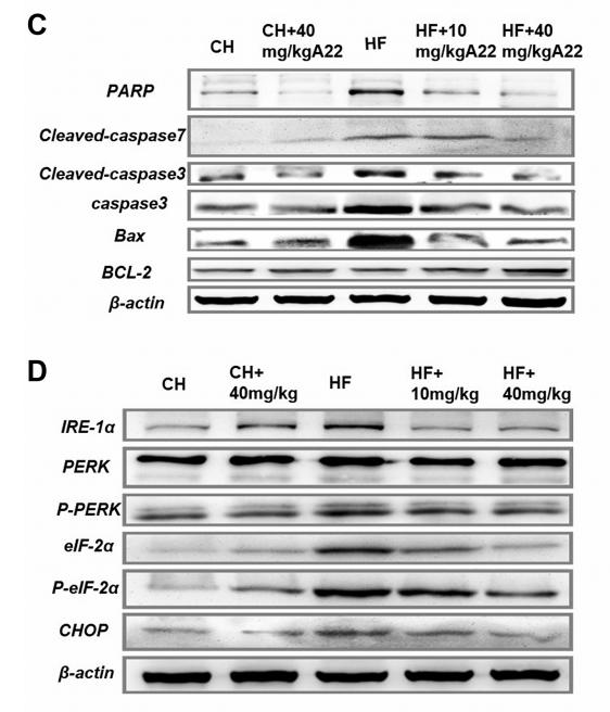
 the ER stress markers GRP 78, caspase 12, and CHOP protein expressions; (b–d) the ratios of p-PERK, p-eIF2α/eIF2α, and p-IRE 1/IRE 1 in the left ventricle")

via suppression of activated ER stress. NRCMs were pretreated with 5 mM 4-PBA (an inhibitor of ERS) or 10 ng/mL tunicamycin (Tm, an ERS
inducer) for 2 h and then exposed to glucose (33 mM) in the presence or absence of ZNS (3 μM) for 24 h. a–b Representative and quantitative
images showing the protein expression of ERS markers, including GRP78, XBP-1s, ATF6, p-PERK, PERK, ATF4, CHOP, and Hrd1.
c Immunofluorescence staining of cardiomyocytes with phalloidin (red) and cell nuclei with DAPI (blue), Scale bar = 50 μm. d Quantitative
analysis of cell surface area by ImageJ software. e–f Representative Western blotting and analysis of Bax and Bcl-2 expression. g–h
Representative and quantitative images of GRP78, ATF6, p-PERK, PERK, ATF4, and CHOP expression. All values are the fold changes normalized
to their control group. The results are presented as the means ± SEM (n = 6). *P < 0.05, **P < 0.01 vs. Con; #P < 0.05, ##P < 0.01 vs. HG; $P < 0.05,
$$P < 0.01 vs. HG + ZNS.")
 Western blot analysis of
p‐IRE1α, IRE1α, XBP1s, p‐PERK, PERK, p‐eIF2α, eIF2α, and CHOP proteins in the kidney tissues of mice. (b) Apoptosis in renal cortex was
visualized by TUNEL immunohistochemical staining. Data are presented as mean ± SD (n = 6). CHOP, C/EBP homologous protein; eIF2α,
eukaryotic initiation factor 2 α; IRE1α, inositol‐requiring transmembrane kinase/endoribonuclease 1α; TUNEL, terminal deoxynucleotidyl
transferase dUTP nick end labeling; XBP1, X‐box binding protein 1")
 Representative immunoblots of AdipoR1, p-AMPK, and T-AMPK. β-Actin was used as a loading control. (e)
Relative band intensity.")
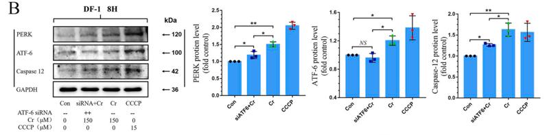
 RT‑qPCR and (D) western blotting. A549 cells were transfected with GRP78‑siRNA or negative control, hyperoxia was established subsequently for 24, 48 and 72 h after transfection. Sham siRNA: A549 cells treated with negative control siRNA; siRNA+N: A549 cells were treated with GRP78‑siRNA and nomaxia for 24 h; siRNA+H 24 h, siRNA+H 48 h, siRNA+H 72 h: A549 cells were treated with GRP78‑siRNA and hyperoxia for 24, 48 and 72 h. CHOP protein expression was slightly increased after 24 h under hypoxia after (E and F) shamRNA treatment.")
.")
 Tumor tissues from each group were processed for the proteins p‑PERK, PERK, p‑eIF2α, eIF2α, ATF4 and Lcn2 detection.")
 Representative immunoreactive bands of GRP78, PERK, p-PERK, IRE1α, p-IRE1α, and ATF6")
 Western blot analysis of p-PERK, PERK, p-eIF2α, eIF2α, and ATF4 proteins.Data are presented as the mean ± SD (n = 3) calculated from one-way ANOVA with Tukey’s test.")
 Western blot analysis was performed with antibodies against Sec61γ, BiP, p-PERK/PERK, p-eIF2α/eIF2α, and ATF4. The quantitative densitometric analysis of Sec61γ (B), BiP (C), p-PERK/PERK (D), p-eIF2α/eIF2α (E), and ATF4 (F) was carried out.")


B. Effect of G-1 treatment on CHOP expression in colitis (The figure above was the
original one, and the figure below was the statistical one, n=4, ***P<0.001,
**P<0.01).
C. Effect of G-1 treatment on PERK activity in colitis (The figure above was the
original one, and the figure below was the statistical one, n=4, *P <0.05).
D. Effect of G-1 treatment on IRE1αactivity in colitis (The figure above was the
original one, and the figure below was the statistical one, n=4, **P<0.01, ***P
<0.001).
This article has not been copyedited and formatted. The final version may differ from this version.
JPET Fast Forward. Published on December 14, 2020 as DOI: 10.1124/jpet.120.000216
at ASPET Journals on December 16, 2020 jpet.aspetjournals.org Downloaded from
38
E. Effect of G-1 treatment on ATF6 expression in colitis (The figure above was the
original one, and the figure below was the statistical one, n=4, **P<0.01, ***P
<0.001).
The mice were divided into three groups: control, DSS treatment group, DSS plus G-1
treatment. Data were expressed as mean±SEM. Statistical analyses were performed
using One-Way ANOVA followed by Student-Newman-Keuls Method.")
 ER stress‐related proteins (p‐eIF2α, eIF2α, ATF4 and CHOP) and (B) UPR signal transduction molecules (p‐PERK, PERK, p‐IRE1α, IRE1α and sXBP1) in HepG2 cells after administration of BMS‐303141. ATF4p‐eIF2α, eIF2α were activated 3 h post‐treatment; CHOP was activated 8 h post‐treatment. (* P < .05, ** P < .01 and *** P < .001, compared with control group) (C) Western blot analysis of protein expression after ATF4 knockdown. (D) Annexin V‐FITC/PI double staining was performed to determine the apoptosis rate of HepG2 cells after ATF4 knockdown via flow cytometry. (* P < .05, ** P < .01 and *** P < .001, compared with con siRNA group). All experiments were repeated 3 times")
 Representative blots of SIRT1, GRP78, p‑PERK, p‑eIF2α, CHOP and caspase‑12. Semiquantitative analysis of (B) SIRT1, (C) GRP78, (D) p‑PERK, (E) p‑eIF2α, (F) CHOP and (G) caspase‑12.")
 Protein expression levels of CHOP, ATF6, p-PERK, PERK, p-IRE1, IRE1 and β-actin in H9C2 cells treated with LPS, TA and TA + LPS, as determined by western blot analysis. Semi-quantification of the protein expression levels of (B) CHOP and ATF6, and (C) p-PERK, PERK and p-IRE1/IRE1 in the LPS, TA and TA + LPS groups. Data are presented as the mean ± SEM (n=3). *P<0.05 and **P<0.01 vs. control group; #P<0.05 vs. LPS group. TA, tannic acid; LPS, lipopolysaccharide; CHOP, C/EBP-homologous protein; ATF6, activating transcription factor 6; p, phosphorylated; PERK, protein kinase-like endoplasmic reticulum kinase; IRE1, inositol-requiring enzyme 1.")
 Representative blots of SIRT1, GRP78, p-PERK, p-eIF2α, CHOP and caspase-12. Semiquantitative analysis of (B) SIRT1, (C) GRP78, (D) p-PERK, (E) p-eIF2α, (F) CHOP and (G) caspase-12. (H) The mRNA levels of caspase-12 in the myocardium. Data are presented as the mean ± standard deviation. n=3. ##P<0.01 vs. Sham. *P<0.05 and **P<0.01 vs. MI/R. IOE, Inonotus obliquus extract; SIRT1, NAD-dependent protein deacetylase sirtuin-1; GRP78, glucose-regulated protein 78; PERK, protein kinase R-like endoplasmic reticulum kinase; eIF2α, eukaryotic translation initiation factor 2 subunit α; CHOP, C/EBP homologous protein; p, phosphorylated; MI/R, myocardial ischemia/reperfusion.")
 MCP5 podocytes were treated either with 10% serum from control C57BL/KsJ dm/m mice or with 10% serum from C57BL/KsJ dm/dm DN mice for 24 h. Western blot image showing activation of PERK-eIF2α-ATF4 in ER stress signaling pathway and increased apoptosis-related molecule cleaved caspase-3 in podocytes treated with serum from DN mice as compared to control mice. (B) Densitometric quantification of protein expression from Figure 1(A). (**p < 0.01, *p < 0.05).")
 Western blot analyzed the expression of PERK, p-PERK, GRP78, ATF6, ATF4, IRE1α, p-IRE1α. n = 4 per group. (B) CTRP1 affected the interaction between PERK and GRP78 after CIRI. n = 3 per group. ****p < 0.0001, ***p < 0.001, **p < 0.01, *p < 0.05 vs. sham group, ####p < 0.0001, ###p < 0.001, ## p < 0.01, #p < 0.05 vs. MCAO/R + LV-NC group.")
 H2S content was determined by Methylene blue method in the LAA of patients with SR or AF (n = 6 in each group). (B) Representative Western blotting and relative densitometry analysis of CSE, CBS, 3-MST, PDK-4, LDHA, PDH, ATF-4, CHOP, caspase-12 and GAPDH in the LAA of patients with SR or AF (n = 6 in each group). (C) Western blotting was used to analyze the protein levels of p-PERK and t-PERK in the LAA of patients with SR or AF (n = 6 in each group). (D) Western blotting was used to analyze the protein levels of p-elf2α and t-elf2α in the LAA of patients with SR or AF (n = 6 in each group). (E) Lactate acid was determined by kit analysis in the LAA of patients with SR or AF (n = 6 in each group). Blue staining with representative Masson staining (scale bar = 50 μm) and quantification of atrial fibrosis (n = 6 in each group, with ≥40 fields in each group) were used in the (F) AF group and (G) SR group. (H) Western blotting was used to analyze the protein levels of MMP-9 and GAPDH in the LAA of patients with SR or AF (n = 6 in each group). Representative RT-PCR and relative densitometry analysis of (I) collagen Iα and (J) collagen IIIα in the LAA of patients with SR or AF (n = 6 in each group). Left atrial function was measured by color Doppler echocardiography (K) LAESVI, (L) LAEF and (M) LAFI (n = 6 in each group) in the patients with SR or AF during the echocardiographic studies. **p < 0.01 VS AF. A Student’s t-test was used for each of these comparisons between AF and SR groups.")
 Expressions of ER stress-associated proteins in HepG2 cells after treatment with 2 μM C20/C22 for 0, 6, 12, and 24 h. All proteins were normalized with the expression of Gapdh (n = 3). (C,D) Cells were stained with PI after 24 h of C20/C22 incubation, and flow cytometry was used to determine the proportion of cells in each stage of the cell cycle (n = 3). The reported data correspond to the mean ± SD of three independent experiments. The p-value was analyzed by one-way ANOVA followed by Tukey’s test using GraphPad Prism version 8.00. * p < 0.05, ** p < 0.01, *** p < 0.001, and **** p < 0.0001 vs. 0 h/control group.")
 Effect of G-1 treatment on GRP78 expression in colitis (the figure above was the original one, and the figure below was the statistical one; n = 4, **P < 0.01). (B) Effect of G-1 treatment on CHOP expression in colitis (the figure above was the original one, and the figure below was the statistical one; n = 4, ***P < 0.001, **P < 0.01). (C) Effect of G-1 treatment on PERK activity in colitis (the figure above was the original one, and the figure below was the statistical one; n = 4, *P < 0.05). (D) Effect of G-1 treatment on IRE1α activity in colitis (the figure above was the original one, and the figure below was the statistical one; n = 4, **P < 0.01, ***P < 0.001). (E) Effect of G-1 treatment on ATF6 expression in colitis (the figure above was the original one, and the figure below was the statistical one; n = 4, **P < 0.01, ***P < 0.001). The mice were divided into three groups: control, DSS treatment group, and DSS + G-1 treatment. Data are expressed as means ± S.E.M. Statistical analyses were performed using one-way ANOVA followed by the Student-Newman-Keuls method.")
 The mRNA expression of Grp78, ATF4, CHOP, PERK, eIF-2α, BAX and Bcl-2. (H–P) The protein expression of Grp78, ATF4, CHOP, PERK, eIF-2α, BAX and Bcl-2. All the experiments were repeated for 3 times and all values are means ± SE.")
 GRP78, (b) p-PERK, and (c) p-eIF2a (d) ATF4. Concomitantly combination of Met and PSTi8 improved insulin sensitivity in liver as identified by improved insulin signaling. We detected protein phosphorylation of (e) p-(Ser-473) AKT (f) p-(Ser-307) IRS-1. Results are presented as means ± SEM (n = 3). Significance among groups presented as β, Control vs HVCD, δ, HVCD vs HVCD + Met; ε, HVCD vs HVCD + PSTi8; φ, HVCD vs HVCD + Comb. Significance represented as φp")
 The protein expressions of p-PERK, PERK, pIREα, IREα, and cleaved ATF6 were determined in triplicate by using Western blot analysis. n = 3. (B,C) A confocal image provided by immunofluorescence determined the expression levels of ATF4, XBP1, and ATF6 in bEECs. Scale bars: 50 μm and 10 μm (bottom, n = 2). (D) RT-qPCR analysis of ATF4, CHOP, and GRP78 mRNA expression normalized to the expression of GAPDH. n = 4. The data are presented as the mean ± SEM. Experiments were repeated n times with duplicate biological replicates. *p < 0.05; **p < 0.01; ***p < 0.001.")
 CTRP9 mRNA and protein expression levels. (B) ATF6a mRNA and protein expression levels. (C) mRNA and protein expression levels of CRP78. (D) Expression levels of IRE1a mRNA and p-IRE1a protein. (E) Expression levels of PERK mRNA and p-PERK protein. (F) SREBP1c mRNA and protein expression levels. (G) SREBP2 mRNA and protein expression levels. The experiment was performed in triplicate, and the representative images are shown. *P")
 The DEGs between renal I/R injury and sham mice (GSE212678). The dotted line indicates the threshold for DEGs, with blue and red dots representing genes with low and high expression in renal I/R injury mice, respectively. (B) GO and KEGG enrichment analyses of DEGs. (C) Representative images of WB assays are shown. (D) The relative protein expression level of ATF6 was determined via normalization to that of GAPDH, and the Sham group was set as the baseline (value of 1). (E–G) The relative phosphorylation levels of PERK, IRE1α, and eIF2α were detected by measuring the ratio of phosphorylated to total proteins, and the Sham group was set as the baseline (value of 1). (H) The relative levels of expression of ERS-mediated apoptosis proteins (ATF4, CHOP, and DR5) in kidney tissues were evaluated and normalized to those of GAPDH, and the Sham group was set as the baseline (value of 1). (I) Relative fluorescence intensity of CHOP (red). (J) Quantification of the TUNEL staining results. (K) CHOP expression in renal tissues was visualized by conducting IF staining (×400, scale bar: 40 μm). (L) Representative TUNEL staining (green) of renal tissues (×400, scale bar: 40 μm). The data are presented as the mean ± SD and evaluated by conducting one-way ANOVA and the Bonferroni correction for multiple comparisons, n = 5 for each group; *P < 0.05, **P < 0.01, and ***P < 0.001.")
 Results of immunohistochemical staining. (B–D) Quantitative expression of COX2, GRP78, and PERK in rat liver tissue by immunohistochemistry. ##P < 0.01 compared with the control group. *P < 0.05, **P < 0.01 compared with the model group.")
. Western blotting and quantification of p-AKT, AKT, GRP78, p-PERK, PERK, p-eIF2α, eIF2α, and CHOP (n=3). (G−J). mRNA levels of GRP78, PERK, eIF2α, and CHOP (n=3).")
 stress and cell death by inhibiting ribosome biogenesis. Mice were randomly allocated into four groups: Control group, UUO group, UUO + sh-NC group, and UUO + sh-Isg20 group and received corresponding operation and treatments. (A-B) The protein levels of PERK, IRE1, and GRP78 in kidney tissues were determined using Immunoblotting. N = 3 (biological replicates). (C) TUNEL staining was performed to detect cell apoptosis in the renal cortex of mice. N = 6 (biological replicates). (D) Quantification of apoptosis rate in kidney tissues from TUNEL staining. (E-F) The protein levels of apoptosis-related factors BCL-2 and BAX in kidney tissues were determined using Immunoblotting. N = 3 (biological replicates). Data are analyzed using one-way ANOVA followed LSD test for (B, D and F). ** P")
| Product: | PERK Antibody |
| Catalog: | AF5304 |
| Description: | Rabbit polyclonal antibody to PERK |
| Application: | WB IHC IF/ICC |
| Cited expt.: | WB, IHC |
| Reactivity: | Human, Mouse, Rat |
| Prediction: | Pig, Bovine, Horse, Sheep, Rabbit, Dog, Chicken, Xenopus |
| Mol.Wt.: | 125~140kDa(Observed); 125kD(Calculated). |
| Uniprot: | Q9NZJ5 |
| RRID: | AB_2837789 |
Control Products

Related Downloads
Protocols
Product Info
*The optimal dilutions should be determined by the end user. For optimal experimental results, antibody reuse is not recommended.
*Tips:
WB: For western blot detection of denatured protein samples. IHC: For immunohistochemical detection of paraffin sections (IHC-p) or frozen sections (IHC-f) of tissue samples. IF/ICC: For immunofluorescence detection of cell samples. ELISA(peptide): For ELISA detection of antigenic peptide.
Cite Format: Affinity Biosciences Cat# AF5304, RRID:AB_2837789.
Fold/Unfold
DKFZp781H1925; E2AK3_HUMAN; EC 2.7.11.1; Eif2ak3; Eukaryotic translation initiation factor 2 alpha kinase 3; Eukaryotic translation initiation factor 2-alpha kinase 3; Heme regulated EIF2 alpha kinase; HRI; HsPEK; Pancreatic eIF2 alpha kinase; Pancreatic eIF2-alpha kinase; PEK; PRKR like endoplasmic reticulum kinase; PRKR-like endoplasmic reticulum kinase; WRS;
Immunogens
A synthesized peptide derived from human PERK, corresponding to a region within C-terminal amino acids.
- Q9NZJ5 E2AK3_HUMAN:
- Protein BLAST With
- NCBI/
- ExPASy/
- Uniprot
MERAISPGLLVRALLLLLLLLGLAARTVAAGRARGLPAPTAEAAFGLGAAAAPTSATRVPAAGAVAAAEVTVEDAEALPAAAGEQEPRGPEPDDETELRPRGRSLVIISTLDGRIAALDPENHGKKQWDLDVGSGSLVSSSLSKPEVFGNKMIIPSLDGALFQWDQDRESMETVPFTVESLLESSYKFGDDVVLVGGKSLTTYGLSAYSGKVRYICSALGCRQWDSDEMEQEEDILLLQRTQKTVRAVGPRSGNEKWNFSVGHFELRYIPDMETRAGFIESTFKPNENTEESKIISDVEEQEAAIMDIVIKVSVADWKVMAFSKKGGHLEWEYQFCTPIASAWLLKDGKVIPISLFDDTSYTSNDDVLEDEEDIVEAARGATENSVYLGMYRGQLYLQSSVRISEKFPSSPKALESVTNENAIIPLPTIKWKPLIHSPSRTPVLVGSDEFDKCLSNDKFSHEEYSNGALSILQYPYDNGYYLPYYKRERNKRSTQITVRFLDNPHYNKNIRKKDPVLLLHWWKEIVATILFCIIATTFIVRRLFHPHPHRQRKESETQCQTENKYDSVSGEANDSSWNDIKNSGYISRYLTDFEPIQCLGRGGFGVVFEAKNKVDDCNYAIKRIRLPNRELAREKVMREVKALAKLEHPGIVRYFNAWLEAPPEKWQEKMDEIWLKDESTDWPLSSPSPMDAPSVKIRRMDPFATKEHIEIIAPSPQRSRSFSVGISCDQTSSSESQFSPLEFSGMDHEDISESVDAAYNLQDSCLTDCDVEDGTMDGNDEGHSFELCPSEASPYVRSRERTSSSIVFEDSGCDNASSKEEPKTNRLHIGNHCANKLTAFKPTSSKSSSEATLSISPPRPTTLSLDLTKNTTEKLQPSSPKVYLYIQMQLCRKENLKDWMNGRCTIEERERSVCLHIFLQIAEAVEFLHSKGLMHRDLKPSNIFFTMDDVVKVGDFGLVTAMDQDEEEQTVLTPMPAYARHTGQVGTKLYMSPEQIHGNSYSHKVDIFSLGLILFELLYPFSTQMERVRTLTDVRNLKFPPLFTQKYPCEYVMVQDMLSPSPMERPEAINIIENAVFEDLDFPGKTVLRQRSRSLSSSGTKHSRQSNNSHSPLPSN
Predictions
Score>80(red) has high confidence and is suggested to be used for WB detection. *The prediction model is mainly based on the alignment of immunogen sequences, the results are for reference only, not as the basis of quality assurance.
High(score>80) Medium(80>score>50) Low(score<50) No confidence
Research Backgrounds
Metabolic-stress sensing protein kinase that phosphorylates the alpha subunit of eukaryotic translation initiation factor 2 (eIF-2-alpha/EIF2S1) on 'Ser-52' during the unfolded protein response (UPR) and in response to low amino acid availability. Converts phosphorylated eIF-2-alpha/EIF2S1 either in a global protein synthesis inhibitor, leading to a reduced overall utilization of amino acids, or to a translation initiation activator of specific mRNAs, such as the transcriptional activator ATF4, and hence allowing ATF4-mediated reprogramming of amino acid biosynthetic gene expression to alleviate nutrient depletion. Serves as a critical effector of unfolded protein response (UPR)-induced G1 growth arrest due to the loss of cyclin-D1 (CCND1). Involved in control of mitochondrial morphology and function.
Oligomerization of the N-terminal ER luminal domain by ER stress promotes PERK trans-autophosphorylation of the C-terminal cytoplasmic kinase domain at multiple residues including Thr-982 on the kinase activation loop (By similarity). Autophosphorylated. Phosphorylated at Tyr-619 following endoplasmic reticulum stress, leading to activate its tyrosine-protein kinase activity. Dephosphorylated by PTPN1/TP1B, leading to inactivate its enzyme activity.
N-glycosylated.
ADP-ribosylated by PARP16 upon ER stress, which increases kinase activity.
Endoplasmic reticulum membrane>Single-pass type I membrane protein.
Ubiquitous. A high level expression is seen in secretory tissues.
The lumenal domain senses perturbations in protein folding in the ER, probably through reversible interaction with HSPA5/BIP.
Belongs to the protein kinase superfamily. Ser/Thr protein kinase family. GCN2 subfamily.
Research Fields
· Cellular Processes > Transport and catabolism > Autophagy - animal. (View pathway)
· Cellular Processes > Cell growth and death > Apoptosis. (View pathway)
· Genetic Information Processing > Folding, sorting and degradation > Protein processing in endoplasmic reticulum. (View pathway)
· Human Diseases > Endocrine and metabolic diseases > Non-alcoholic fatty liver disease (NAFLD).
· Human Diseases > Neurodegenerative diseases > Alzheimer's disease.
· Human Diseases > Infectious diseases: Viral > Hepatitis C.
· Human Diseases > Infectious diseases: Viral > Measles.
· Human Diseases > Infectious diseases: Viral > Influenza A.
· Human Diseases > Infectious diseases: Viral > Herpes simplex infection.
· Human Diseases > Infectious diseases: Viral > Epstein-Barr virus infection.
References
Application: WB Species: mouse Sample: liver
Application: WB Species: chicken Sample: DF-1 cells
Application: WB Species: Human Sample: HaCaT cells
Application: WB Species: Human Sample: HaCaT cells

Application: WB Species: Mouse Sample:

Application: WB Species: human Sample: 95D cells
Restrictive clause
Affinity Biosciences tests all products strictly. Citations are provided as a resource for additional applications that have not been validated by Affinity Biosciences. Please choose the appropriate format for each application and consult Materials and Methods sections for additional details about the use of any product in these publications.
For Research Use Only.
Not for use in diagnostic or therapeutic procedures. Not for resale. Not for distribution without written consent. Affinity Biosciences will not be held responsible for patent infringement or other violations that may occur with the use of our products. Affinity Biosciences, Affinity Biosciences Logo and all other trademarks are the property of Affinity Biosciences LTD.