

, using BMP6 Antibody at 1/1000 dilution.
5ug/NC membrane strip.
Exposure for 20s with Affinity™ ECL Kit(#KF8003).
Bands result from membrane strip incubation.")
Control Products

Related Downloads
Protocols
Product Info
*The optimal dilutions should be determined by the end user. For optimal experimental results, antibody reuse is not recommended.
*Tips:
WB: For western blot detection of denatured protein samples. IHC: For immunohistochemical detection of paraffin sections (IHC-p) or frozen sections (IHC-f) of tissue samples. IF/ICC: For immunofluorescence detection of cell samples. ELISA(peptide): For ELISA detection of antigenic peptide.
Cite Format: Affinity Biosciences Cat# AF5196, RRID:AB_2837682.
Fold/Unfold
BMP-6; Bmp6; BMP6_HUMAN; Bone morphogenetic protein 6; Bone Morphogenic Protein 6; Decapentaplegic vegetal related; DVR6; HGNC:12686; TGFB related vegetal related growth factor; Transforming growth factor beta; Vegetal related (TGFB related) cytokine; Vegetal related growth factor (TGFB related); Vg related sequence; VG-1-R; VG-1-related protein; Vg1 related sequence; VGR; VGR-1; VGR1;
Immunogens
A synthesized peptide derived from human BMP6, corresponding to a region within the internal amino acids.
- P22004 BMP6_HUMAN:
- Protein BLAST With
- NCBI/
- ExPASy/
- Uniprot
MPGLGRRAQWLCWWWGLLCSCCGPPPLRPPLPAAAAAAAGGQLLGDGGSPGRTEQPPPSPQSSSGFLYRRLKTQEKREMQKEILSVLGLPHRPRPLHGLQQPQPPALRQQEEQQQQQQLPRGEPPPGRLKSAPLFMLDLYNALSADNDEDGASEGERQQSWPHEAASSSQRRQPPPGAAHPLNRKSLLAPGSGSGGASPLTSAQDSAFLNDADMVMSFVNLVEYDKEFSPRQRHHKEFKFNLSQIPEGEVVTAAEFRIYKDCVMGSFKNQTFLISIYQVLQEHQHRDSDLFLLDTRVVWASEEGWLEFDITATSNLWVVTPQHNMGLQLSVVTRDGVHVHPRAAGLVGRDGPYDKQPFMVAFFKVSEVHVRTTRSASSRRRQQSRNRSTQSQDVARVSSASDYNSSELKTACRKHELYVSFQDLGWQDWIIAPKGYAANYCDGECSFPLNAHMNATNHAIVQTLVHLMNPEYVPKPCCAPTKLNAISVLYFDDNSNVILKKYRNMVVRACGCH
Research Backgrounds
Induces cartilage and bone formation.
Secreted.
Belongs to the TGF-beta family.
Research Fields
· Environmental Information Processing > Signal transduction > TGF-beta signaling pathway. (View pathway)
· Environmental Information Processing > Signal transduction > Hippo signaling pathway. (View pathway)
· Organismal Systems > Endocrine system > Ovarian steroidogenesis.
References
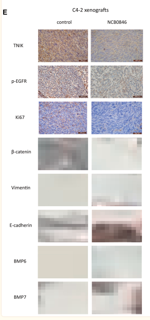
Application: IHC Species: Mouse Sample:

Application: WB Species: human Sample: RPE cell
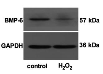
Application: WB Species: human Sample: retinal pigment epithelial (RPE) cells
Restrictive clause
Affinity Biosciences tests all products strictly. Citations are provided as a resource for additional applications that have not been validated by Affinity Biosciences. Please choose the appropriate format for each application and consult Materials and Methods sections for additional details about the use of any product in these publications.
For Research Use Only.
Not for use in diagnostic or therapeutic procedures. Not for resale. Not for distribution without written consent. Affinity Biosciences will not be held responsible for patent infringement or other violations that may occur with the use of our products. Affinity Biosciences, Affinity Biosciences Logo and all other trademarks are the property of Affinity Biosciences LTD.