


")
 0.5μM brusatol was utilized to treat the expression of Nrf2 in PATU-8988 cells for the indicated time points. (b) 0.5μM brusatol lowered the protein levels of Nrf2 and its downstreamgenesafter8-hourtreatments.")
 or NS intraperitoneally every 3 days (n = 6 per group).F. IHC of MARK2, P-gp, and cleaved caspase 3 protein levels in xenograft tumor sections collected from mice treated with cisplatin. Magnification: 400 ×, scale bars: 100 μm. NS,normal saline; IHC, immunohistochemistry.")
 and (B) Protein expression of P-glycoprotein in jejunum; (C) and (D) Protein expression of Mrp2 in ileum. Data are shown as mean ± SD (n = 3). *p < 0.05 compared to without QSYQ.")
. Relative protein expression of PXR (B), MDR1 (C), BCRP (D), and CYP3A4 (E). (x¯±s, n=3), *, P<0.05; **, P<0.01 vs. control group. PECZ, petroleum ether extracts of Curcuma zedoaria; PXR, pregnane X receptor; MDR1, multidrug resistance 1; BCRP, breast cancer resistance protein; CYP3A4, cytochrome P-450.")
. PECZ, petroleum ether extracts of Curcuma zedoaria; PXR, pregnane X receptor; MDR1, multidrug resistance 1; BCRP, breast cancer resistance protein; CYP3A4, cytochrome P-450.")
 or transient silenced by siRNA targeting IL-17RA for 48h (b), and then treated with 1ng/ml rhIL-17A for 24h. B. Up-regulating effects of rhIL-17A on the expression of ABCG2, MDR1 and Gli1 in A2780 and OVCAR3 cells could be blocked by Gant61. OVCA cells were pretreated with 25μM Gant 61 for 1h and then treated with 1ng/ml rhIL-17A for 24h. Protein lysates from OVCA cells were prepared and analyzed by western blotting. Representative result from three independent experiments was showed. C. Gant61 could partially reverse the increase-effect of rhIL-17A on OVCA cell viability. A2780 and OVCAR3 cells were pretreated with 25μM Gant 61 for 1h and then treated with 1ng/ml rhIL-17A and/or DDP (10μM for A2780 cells, 100μM for OVCAR3 cells) for 24h. Cell viability was detected by MTT assay. Data represent means±SD from three independent experiments.")
 Comparison of P-gp in A549 and A549/Taxol cells. Statistical comparisons were performed with unpaired Student’s t-test (n = 3). **p < 0.01 versus A549. (B, C) After treatment of DHW-221 or verapamil for 48 h, Rho-123 (15 µM) was added into the mediums to co-incubate with DHW-221 or verapamil for 3 h in dark. The intracellular accumulation of Rho-123 was evaluated by flow cytometry and confocal microscope, respectively. Verapamil (VRP, 10 μM) was used as a positive control. Green fluorescence indicated Rho-123, and blue fluorescence indicated DAPI. Scale bar = 100 μm. Nuclear mean fluorescence intensity of Rho 123 was measured by ImageJ and analyzed using FlowJo 10 software. Statistical comparisons were performed with one-way ANOVA followed by Dunnett’s post-hoc test for multiple comparisons (n = 3). *p < 0.05, **p < 0.01, ***p < 0.001 versus control. (D) Effect of DHW-221 or verapamil to reverse Taxol resistance for 48 h in A549/Taxol cells. (E) Western blot analysis of DHW-221 inhibited P-gp expression in A549/Taxol cells. (F) Ribbon diagram of human ABCB1 (PDB code: 7A6E, purple spiral) bound to drug (membrane region) and predicted binding modes for DHW-221 (blue stick) and tariquidar (orange stick, a known third-generation P-gp inhibitor) with ABCB1. Hydrogen bonds were shown as green dashed lines. Key residues for DHW-221 and tariquidar interaction were highlighted. (G) Effect of DHW-221 on the thermal stability of ABCB1 was quantitatively detected by a cellular thermal shift assay (CETSA). Statistical comparisons were performed with one-way ANOVA followed by Dunnett’s post-hoc test for multiple comparisons (n = 3). Data are presented as mean ± SD. **p < 0.01, ****p < 0.0001versus control.")
 Intracellular expression of ABCB1, MYCN and CARM1 was detected by western blotting. Expression of ABCB1 was significantly increased in the RPF2 group (P")
 CCK-8 detected cell proliferation ability of GECs after inducing by MRL/lpr Mø Exo or SR-MRL/lpr Mø Exo. (B) Flow cytometry was conducted to detect cell apoptosis. (C) The mRNA levels of MDR1 was measured by PCR. (D) The MDR1 protein expression levels in GECs were confirmed by Western blot. (E) The P-gp levels and accumulation of Rh-123 in GECs were detected by flow cytometry. Values are expressed as mean ± SD (n = 3), ∗p < 0.05, ∗∗p < 0.01, ∗∗∗p < 0.001.")
 Western blot analysis was used to detected MDR-1 and H2AX expression and its gray level in A549 and A549/OS cells. Take Tubulin as the internal parameter. (c, d) MTT was used to detect the cell viability of A549 and A549/OS cells in different concentration gradients of osimertinib. A549/OS cells were incubated with griffithazanone A (2 μM) for 48 h, and the cell viability was observed in different concentrations of osimertinib by MTT compared to the control group. (e) The morphology of A549/OS cells under different conditions were observed under the microscope. (f, i) TUNEL staining shows the number of apoptotic cells, and counted the proportion of apoptotic cells to all cells. (g, h) Colony formation is used to detect the ability of griffithazanone A to reverse the clonal ability of A549/OS cells against osimertinib resistance.")
| Product: | P Glycoprotein 1(MDR1) Antibody |
| Catalog: | AF5185 |
| Description: | Rabbit polyclonal antibody to P Glycoprotein 1(MDR1) |
| Application: | WB IHC |
| Cited expt.: | WB, IHC |
| Reactivity: | Human, Mouse, Rat |
| Prediction: | Pig, Horse, Rabbit, Dog, Chicken, Xenopus |
| Mol.Wt.: | 141 kDa(Observed); 141kD(Calculated). |
| Uniprot: | P08183 |
| RRID: | AB_2837671 |
Control Products

Related Downloads
Protocols
Product Info
*The optimal dilutions should be determined by the end user. For optimal experimental results, antibody reuse is not recommended.
*Tips:
WB: For western blot detection of denatured protein samples. IHC: For immunohistochemical detection of paraffin sections (IHC-p) or frozen sections (IHC-f) of tissue samples. IF/ICC: For immunofluorescence detection of cell samples. ELISA(peptide): For ELISA detection of antigenic peptide.
Cite Format: Affinity Biosciences Cat# AF5185, RRID:AB_2837671.
Fold/Unfold
ABC20; ABCB1; ATP binding cassette, sub family B (MDR/TAP), member 1; ATP-binding cassette sub-family B member 1; CD243; CLCS; Colchicin sensitivity; Doxorubicin resistance; GP170; MDR1; MDR1_HUMAN; Multidrug resistance 1; Multidrug resistance protein 1; P glycoprotein 1; P gp; P-glycoprotein 1; PGY1;
Immunogens
A synthesized peptide derived from human P Glycoprotein 1(MDR1), corresponding to a region within the internal amino acids.
- P08183 MDR1_HUMAN:
- Protein BLAST With
- NCBI/
- ExPASy/
- Uniprot
MDLEGDRNGGAKKKNFFKLNNKSEKDKKEKKPTVSVFSMFRYSNWLDKLYMVVGTLAAIIHGAGLPLMMLVFGEMTDIFANAGNLEDLMSNITNRSDINDTGFFMNLEEDMTRYAYYYSGIGAGVLVAAYIQVSFWCLAAGRQIHKIRKQFFHAIMRQEIGWFDVHDVGELNTRLTDDVSKINEGIGDKIGMFFQSMATFFTGFIVGFTRGWKLTLVILAISPVLGLSAAVWAKILSSFTDKELLAYAKAGAVAEEVLAAIRTVIAFGGQKKELERYNKNLEEAKRIGIKKAITANISIGAAFLLIYASYALAFWYGTTLVLSGEYSIGQVLTVFFSVLIGAFSVGQASPSIEAFANARGAAYEIFKIIDNKPSIDSYSKSGHKPDNIKGNLEFRNVHFSYPSRKEVKILKGLNLKVQSGQTVALVGNSGCGKSTTVQLMQRLYDPTEGMVSVDGQDIRTINVRFLREIIGVVSQEPVLFATTIAENIRYGRENVTMDEIEKAVKEANAYDFIMKLPHKFDTLVGERGAQLSGGQKQRIAIARALVRNPKILLLDEATSALDTESEAVVQVALDKARKGRTTIVIAHRLSTVRNADVIAGFDDGVIVEKGNHDELMKEKGIYFKLVTMQTAGNEVELENAADESKSEIDALEMSSNDSRSSLIRKRSTRRSVRGSQAQDRKLSTKEALDESIPPVSFWRIMKLNLTEWPYFVVGVFCAIINGGLQPAFAIIFSKIIGVFTRIDDPETKRQNSNLFSLLFLALGIISFITFFLQGFTFGKAGEILTKRLRYMVFRSMLRQDVSWFDDPKNTTGALTTRLANDAAQVKGAIGSRLAVITQNIANLGTGIIISFIYGWQLTLLLLAIVPIIAIAGVVEMKMLSGQALKDKKELEGSGKIATEAIENFRTVVSLTQEQKFEHMYAQSLQVPYRNSLRKAHIFGITFSFTQAMMYFSYAGCFRFGAYLVAHKLMSFEDVLLVFSAVVFGAMAVGQVSSFAPDYAKAKISAAHIIMIIEKTPLIDSYSTEGLMPNTLEGNVTFGEVVFNYPTRPDIPVLQGLSLEVKKGQTLALVGSSGCGKSTVVQLLERFYDPLAGKVLLDGKEIKRLNVQWLRAHLGIVSQEPILFDCSIAENIAYGDNSRVVSQEEIVRAAKEANIHAFIESLPNKYSTKVGDKGTQLSGGQKQRIAIARALVRQPHILLLDEATSALDTESEKVVQEALDKAREGRTCIVIAHRLSTIQNADLIVVFQNGRVKEHGTHQQLLAQKGIYFSMVSVQAGTKRQ
Predictions
Score>80(red) has high confidence and is suggested to be used for WB detection. *The prediction model is mainly based on the alignment of immunogen sequences, the results are for reference only, not as the basis of quality assurance.
High(score>80) Medium(80>score>50) Low(score<50) No confidence
Research Backgrounds
Translocates drugs and phospholipids across the membrane. Catalyzes the flop of phospholipids from the cytoplasmic to the exoplasmic leaflet of the apical membrane. Participates mainly to the flop of phosphatidylcholine, phosphatidylethanolamine, beta-D-glucosylceramides and sphingomyelins. Energy-dependent efflux pump responsible for decreased drug accumulation in multidrug-resistant cells.
Cell membrane>Multi-pass membrane protein. Apical cell membrane.
Expressed in liver, kidney, small intestine and brain.
Belongs to the ABC transporter superfamily. ABCB family. Multidrug resistance exporter (TC 3.A.1.201) subfamily.
Research Fields
· Environmental Information Processing > Membrane transport > ABC transporters.
· Human Diseases > Cancers: Overview > MicroRNAs in cancer.
· Human Diseases > Cancers: Specific types > Gastric cancer. (View pathway)
References
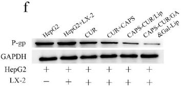
Application: WB Species: Human Sample: HepG2 cells

Application: WB Species: Mouse Sample: BLCA cells
Application: WB Species: human Sample: EOC ascites cells

Application: WB Species: Mouse Sample:
Restrictive clause
Affinity Biosciences tests all products strictly. Citations are provided as a resource for additional applications that have not been validated by Affinity Biosciences. Please choose the appropriate format for each application and consult Materials and Methods sections for additional details about the use of any product in these publications.
For Research Use Only.
Not for use in diagnostic or therapeutic procedures. Not for resale. Not for distribution without written consent. Affinity Biosciences will not be held responsible for patent infringement or other violations that may occur with the use of our products. Affinity Biosciences, Affinity Biosciences Logo and all other trademarks are the property of Affinity Biosciences LTD.