
.
Bands result from membrane strip incubation.")


| Product: | AQP4 Antibody |
| Catalog: | AF5164 |
| Description: | Rabbit polyclonal antibody to AQP4 |
| Application: | WB IHC |
| Cited expt.: | WB, IHC |
| Reactivity: | Human, Mouse, Rat |
| Prediction: | Pig, Bovine, Horse, Rabbit, Dog, Chicken |
| Mol.Wt.: | 34 kDa(Observed); 35kD(Calculated). |
| Uniprot: | P55087 |
| RRID: | AB_2837650 |
Control Products

Related Downloads
Protocols
Product Info
*The optimal dilutions should be determined by the end user. For optimal experimental results, antibody reuse is not recommended.
*Tips:
WB: For western blot detection of denatured protein samples. IHC: For immunohistochemical detection of paraffin sections (IHC-p) or frozen sections (IHC-f) of tissue samples. IF/ICC: For immunofluorescence detection of cell samples. ELISA(peptide): For ELISA detection of antigenic peptide.
Cite Format: Affinity Biosciences Cat# AF5164, RRID:AB_2837650.
Fold/Unfold
AQP 4; AQP-4; AQP4; AQP4_HUMAN; Aquaporin type 4; Aquaporin-4; Aquaporin4; HMIWC 2; HMIWC2; Mercurial insensitive water channel; Mercurial-insensitive water channel; MGC22454; MIWC; WCH 4; WCH4;
Immunogens
A synthesized peptide derived from human AQP4, corresponding to a region within the internal amino acids.
Detected in skeletal muscle (PubMed:29055082). Detected in stomach, along the glandular base region of the fundic gland (at protein level) (PubMed:8601457). Detected in brain, lung and skeletal muscle, and at much lower levels in heart and ovary (PubMed:7559426, PubMed:8601457).
- P55087 AQP4_HUMAN:
- Protein BLAST With
- NCBI/
- ExPASy/
- Uniprot
MSDRPTARRWGKCGPLCTRENIMVAFKGVWTQAFWKAVTAEFLAMLIFVLLSLGSTINWGGTEKPLPVDMVLISLCFGLSIATMVQCFGHISGGHINPAVTVAMVCTRKISIAKSVFYIAAQCLGAIIGAGILYLVTPPSVVGGLGVTMVHGNLTAGHGLLVELIITFQLVFTIFASCDSKRTDVTGSIALAIGFSVAIGHLFAINYTGASMNPARSFGPAVIMGNWENHWIYWVGPIIGAVLAGGLYEYVFCPDVEFKRRFKEAFSKAAQQTKGSYMEVEDNRSQVETDDLILKPGVVHVIDVDRGEEKKGKDQSGEVLSSV
Predictions
Score>80(red) has high confidence and is suggested to be used for WB detection. *The prediction model is mainly based on the alignment of immunogen sequences, the results are for reference only, not as the basis of quality assurance.
High(score>80) Medium(80>score>50) Low(score<50) No confidence
Research Backgrounds
Forms a water-specific channel. Plays an important role in brain water homeostasis and in glymphatic solute transport. Required for a normal rate of water exchange across the blood brain interface. Required for normal levels of cerebrospinal fluid influx into the brain cortex and parenchyma along paravascular spaces that surround penetrating arteries, and for normal drainage of interstitial fluid along paravenous drainage pathways. Thereby, it is required for normal clearance of solutes from the brain interstitial fluid, including soluble beta-amyloid peptides derived from APP. Plays a redundant role in urinary water homeostasis and urinary concentrating ability (By similarity).
Phosphorylation by PKC at Ser-180 reduces conductance by 50%. Phosphorylation by PKG at Ser-111 in response to glutamate increases conductance by 40% (By similarity).
Isoform 2: Palmitoylated on its N-terminal region. Isoform 1: Not palmitoylated.
Cell membrane>Multi-pass membrane protein. Basolateral cell membrane>Multi-pass membrane protein. Endosome membrane. Cell membrane>Sarcolemma>Multi-pass membrane protein. Cell projection.
Note: Activation of the vasopressin receptor AVPR1A triggers AQP4 phosphorylation at Ser-180 and promotes its internalization from the cell membrane. Detected on brain astrocyte processes and astrocyte endfeet close to capillaries.
Detected in skeletal muscle. Detected in stomach, along the glandular base region of the fundic gland (at protein level). Detected in brain, lung and skeletal muscle, and at much lower levels in heart and ovary.
Aquaporins contain two tandem repeats each containing three membrane-spanning domains and a pore-forming loop with the signature motif Asn-Pro-Ala (NPA).
Belongs to the MIP/aquaporin (TC 1.A.8) family.
Research Fields
· Organismal Systems > Excretory system > Vasopressin-regulated water reabsorption.
References
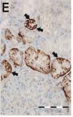
Application: IHC Species: Human Sample: lung tissues

Application: IHC Species: Rat Sample:

Application: WB Species: Rat Sample: kidney tissue
Restrictive clause
Affinity Biosciences tests all products strictly. Citations are provided as a resource for additional applications that have not been validated by Affinity Biosciences. Please choose the appropriate format for each application and consult Materials and Methods sections for additional details about the use of any product in these publications.
For Research Use Only.
Not for use in diagnostic or therapeutic procedures. Not for resale. Not for distribution without written consent. Affinity Biosciences will not be held responsible for patent infringement or other violations that may occur with the use of our products. Affinity Biosciences, Affinity Biosciences Logo and all other trademarks are the property of Affinity Biosciences LTD.