Product: RANTES Antibody
Catalog: AF5151
Description: Rabbit polyclonal antibody to RANTES
Application: WB IHC
Reactivity: Human, Mouse, Rat
Prediction: Pig, Bovine, Horse, Sheep, Rabbit, Dog
Mol.Wt.: 9 kDa; 10kD(Calculated).
Uniprot: P13501
RRID: AB_2837637

View similar products>>

   Size Price Inventory
 100ul $280 In stock
 200ul $350 In stock

Lead Time: Same day delivery

For pricing and ordering contact:
Local distributors

Product Info

Source:
Rabbit
Application:
WB 1:500-1:2000, IHC 1:50-1:200
*The optimal dilutions should be determined by the end user.
*Tips:

WB: For western blot detection of denatured protein samples. IHC: For immunohistochemical detection of paraffin sections (IHC-p) or frozen sections (IHC-f) of tissue samples. IF/ICC: For immunofluorescence detection of cell samples. ELISA(peptide): For ELISA detection of antigenic peptide.

Reactivity:
Human,Mouse,Rat
Prediction:
Pig(100%), Bovine(100%), Horse(100%), Sheep(100%), Rabbit(100%), Dog(89%)
Clonality:
Polyclonal
Specificity:
RANTES Antibody detects endogenous levels of total RANTES.
RRID:
AB_2837637
Cite Format: Affinity Biosciences Cat# AF5151, RRID:AB_2837637.
Conjugate:
Unconjugated.
Purification:
The antiserum was purified by peptide affinity chromatography using SulfoLink™ Coupling Resin (Thermo Fisher Scientific).
Storage:
Rabbit IgG in phosphate buffered saline , pH 7.4, 150mM NaCl, 0.02% sodium azide and 50% glycerol. Store at -20 °C. Stable for 12 months from date of receipt.
Alias:

Fold/Unfold

Beta chemokine RANTES; Beta chemokine RANTES precursor; C C motif chemokine 5; CCL 5; CCL5; CCL5_HUMAN; Chemokine (C C motif) ligand 5; Chemokine CC Motif Ligand 5; D17S136E; EoCP; Eosinophil chemotactic cytokine; MGC17164; RANTES(4-68); Regulated upon activation normally T expressed and presumably secreted; SCYA 5; SCYA5; SIS delta; SIS-delta; SISd; Small inducible cytokine A5 (RANTES); Small inducible cytokine A5; Small inducible cytokine subfamily A (Cys Cys) member 5; Small-inducible cytokine A5; T cell specific protein p288; T cell specific RANTES protein; T cell-specific protein P228; T-cell-specific protein RANTES; TCP 228; TCP228;

Immunogens

Immunogen:
Uniprot:
Gene(ID):
Expression:
P13501 CCL5_HUMAN:

Expressed in the follicular fluid (at protein level). T-cell and macrophage specific.

Description:
Chemoattractant for blood monocytes, memory T-helper cells and eosinophils. Causes the release of histamine from basophils and activates eosinophils. Binds to CCR1, CCR3, CCR4 and CCR5. One of the major HIV-suppressive factors produced by CD8+ T-cells. Recombinant RANTES protein induces a dose-dependent inhibition of different strains of HIV-1, HIV-2, and simian immunodeficiency virus (SIV).
Sequence:
MKVSAAALAVILIATALCAPASASPYSSDTTPCCFAYIARPLPRAHIKEYFYTSGKCSNPAVVFVTRKNRQVCANPEKKWVREYINSLEMS

Predictions

Predictions:

Score>80(red) has high confidence and is suggested to be used for WB detection. *The prediction model is mainly based on the alignment of immunogen sequences, the results are for reference only, not as the basis of quality assurance.

Species
Results
Score
Pig
100
Horse
100
Bovine
100
Sheep
100
Rabbit
100
Dog
89
Xenopus
67
Chicken
56
Zebrafish
0
Model Confidence:
High(score>80) Medium(80>score>50) Low(score<50) No confidence

PTMs - P13501 As Substrate

Site PTM Type Enzyme
S27 O-Glycosylation
S28 O-Glycosylation
K68 Ubiquitination

Research Backgrounds

Function:

Chemoattractant for blood monocytes, memory T-helper cells and eosinophils. Causes the release of histamine from basophils and activates eosinophils. May activate several chemokine receptors including CCR1, CCR3, CCR4 and CCR5. One of the major HIV-suppressive factors produced by CD8+ T-cells. Recombinant RANTES protein induces a dose-dependent inhibition of different strains of HIV-1, HIV-2, and simian immunodeficiency virus (SIV). The processed form RANTES(3-68) acts as a natural chemotaxis inhibitor and is a more potent inhibitor of HIV-1-infection. The second processed form RANTES(4-68) exhibits reduced chemotactic and HIV-suppressive activity compared with RANTES(1-68) and RANTES(3-68) and is generated by an unidentified enzyme associated with monocytes and neutrophils. May also be an agonist of the G protein-coupled receptor GPR75, stimulating inositol trisphosphate production and calcium mobilization through its activation. Together with GPR75, may play a role in neuron survival through activation of a downstream signaling pathway involving the PI3, Akt and MAP kinases. By activating GPR75 may also play a role in insulin secretion by islet cells.

PTMs:

N-terminal processed form RANTES(3-68) is produced by proteolytic cleavage, probably by DPP4, after secretion from peripheral blood leukocytes and cultured sarcoma cells.

The identity of the O-linked saccharides at Ser-27 and Ser-28 are not reported in. They are assigned by similarity.

Subcellular Location:

Secreted.

Extracellular region or secreted Cytosol Plasma membrane Cytoskeleton Lysosome Endosome Peroxisome ER Golgi apparatus Nucleus Mitochondrion Manual annotation Automatic computational assertionSubcellular location
Tissue Specificity:

Expressed in the follicular fluid (at protein level). T-cell and macrophage specific.

Family&Domains:

Belongs to the intercrine beta (chemokine CC) family.

Research Fields

· Environmental Information Processing > Signaling molecules and interaction > Cytokine-cytokine receptor interaction.   (View pathway)

· Environmental Information Processing > Signal transduction > TNF signaling pathway.   (View pathway)

· Human Diseases > Neurodegenerative diseases > Prion diseases.

· Human Diseases > Infectious diseases: Bacterial > Epithelial cell signaling in Helicobacter pylori infection.

· Human Diseases > Infectious diseases: Parasitic > Chagas disease (American trypanosomiasis).

· Human Diseases > Infectious diseases: Viral > Influenza A.

· Human Diseases > Infectious diseases: Viral > Herpes simplex infection.

· Human Diseases > Immune diseases > Rheumatoid arthritis.

· Organismal Systems > Immune system > Chemokine signaling pathway.   (View pathway)

· Organismal Systems > Immune system > Toll-like receptor signaling pathway.   (View pathway)

· Organismal Systems > Immune system > NOD-like receptor signaling pathway.   (View pathway)

· Organismal Systems > Immune system > Cytosolic DNA-sensing pathway.   (View pathway)

References

1). Hyperphosphorylated tau mediates neuronal death by inducing necroptosis and inflammation in Alzheimer’s disease. Journal of Neuroinflammation, 2022 (PubMed: 35971179) [IF=9.3]

Application: WB    Species: Mouse    Sample: HT22 cells

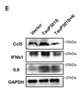
Fig. 2Hyperphosphorylated tau induces cytokine upregulation. A Differentially expressed genes (DEGs) between HT22 cells transfected with vector or TauP301S for 48 h. B Analysis of DEGs in the TauP301S and Tau groups according to the Kyoto Encyclopedia of Genes and Genomes (KEGG) database. C Heat map of cytokine genes differentially expressed in HT22 cells transfected with vector or Tau or TauP301S following treatment with DMSO or Nec-1 (30 μM); low expression is shown in blue and high expression in red. D mRNAs from HT22 cells transfected with vector or TauP301S following treatment with DMSO or Nec-1 (30 μM) were extracted and quantified to determine indicated cytokine levels by qPCR. E Cytokine levels in HT22 cells transfected with vector or TauP301S following treatment with DMSO or Nec-1 (30 μM) were analyzed by western blotting with the indicated antibodies. F ROS levels in HT22 cells transfected with vector or TauP301S. G Chemotaxis of pTau-induced cytokines on BV2 cells was analyzed by transwell assays, Scale bars, 100 μm. Data are presented as the mean ± standard error of the mean (SEM) of three experiments, and statistical analysis was performed using one-way ANOVA with Dunnett’s multiple comparisons test in D and two-tailed unpaired t test in F, G

2). CCL5 derived from tumor-associated macrophages promotes prostate cancer stem cells and metastasis via activating β-catenin/STAT3 signaling. Cell Death & Disease, 2020 (PubMed: 32300100) [IF=9.0]

Application: WB    Species: human    Sample: prostate cancer cells

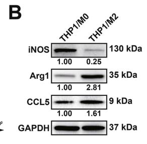
Fig. 3 TAM-secreted CCL5 promoted the invasion of prostate cancer cells and the self-renewal of PCSCs. a, b The reliability of THP1-derived TAMs was validated by analyzing the macrophage surface markers. THP1 monocytes were induced differentiation into macrophages (M0) by 100 ng/ ml PMA treatment for 24 h, which were further transformed into M2 phenotype macrophages (TAMs) by 10 ng/ml IL-4 induction for 72 h. c, d The CM of THP1-derived TAMs significantly promoted EMT, migration and invasion of prostate cancer cells, while CCL5 NA could partly abrogate that. Scale bars represent 200 μm for wound healing assay images and 50 μm for transwell assay images. e The CM of THP1-derived TAMs significantly promoted the self-renewal of PCSCs, while CCL5 NA could partly abrogate that. All values are presented as the mean ± SD. n = 3, *p < 0.05, **p < 0.01.

Application: IF/ICC    Species: human    Sample: prostate cancer cells

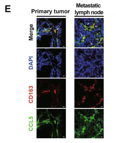
Fig. 1 TAMs-derived CCL5 was elevated in prostate cancer and associated with metastasis. a, b The mRNA and protein expression differences of indicated genes between prostate cancer tumor tissues and para-carcinoma tissues (n = 3). c Elisa assay indicated that the blood samples of prostate cancer patients (n = 30) exhibited elevated expression of CCL5 when compared with that of healthy male participants (n = 30). d Prostate cancer patients with higher Gleason grade exhibited an increased CCL5 accumulation in the blood (n = 30). e Tissue immunofluorescence assay suggested that CCL5 and the macrophage marker CD163 were co-localized in both the primary tumor and metastatic lymph node of prostate cancer patients. Scale bar, 10 μm. f TAMs exhibited increased secretion of CCL5 than immature macrophages or prostate cancer cells. CCL5 concentrations in the cell culture supernatants of THP1-derived macrophages (M0), THP1-derived TAMs (M2), DU145 and PC3 cells were measured using Elisa method (n = 10). All values are presented as the mean ± SD. *p < 0.05, **p < 0.01.

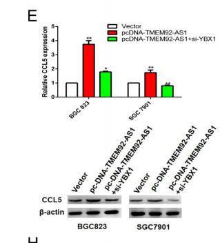
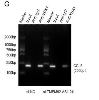
3). A novel long noncoding RNA, TMEM92‐AS1, promotes gastric cancer progression by binding to YBX1 to mediate CCL5. Molecular Oncology, 2021 (PubMed: 33247987) [IF=6.6]

Application: WB    Species: human    Sample: BGC823 and SGC7901 cells

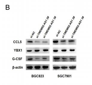
Figure 4 CCL5 was confirmed as a downstream target gene of TMEM92-AS1. (A and B) RT-qPCR and western blot were used to verify changed genes and proteins after TMEM92-AS1 knockdown.

Application: WB    Species: human    Sample: BGC823 cells

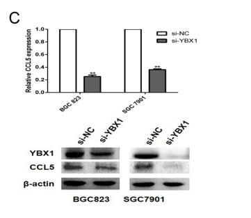
Figure 5(C) RT-qPCR and western blot were performed to detect CCL5 expressions after YBX1 knockdown.

Application: WB    Species: human    Sample: BGC823 cells

Figure 5(E) RT-qPCR and western blot were performed to detect the effect of TMEM92-AS1 overexpression and YBX1 knockdown on CCL5 expression.

Application: WB    Species: human    Sample: BGC823 cells

Figure 5 (G) ChIP results of YBX1 of the promoter region of CCL5 after si-TMEM92-AS1 2# in BGC823 cell

4). IFN-γ Activates the TLR4-CCL5 Signaling Through Reducing Arginine Level, Leading to Enhanced Susceptibility of Bovine Mammary Epithelial Cells to Staphylococcus aureus. INFLAMMATION, 2020 (PubMed: 32725514) [IF=5.1]

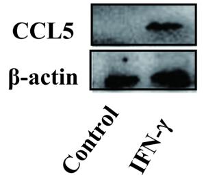
Application: WB    Species: Cows    Sample: bovine mammary epithelial cells (BMECs)

Fig. 2. IFN-γ promoted the expression of CCL5 and CCL5 antibody inhibited the infection of S. aureus. a DEGs associated with inflammation were screened out: DEGs were screened according to false discovery rate (FDR) ≤ 0.05 and |log2 ratio ≥ 1. Among 12 genes associated with inflammation, seven of the genes were increased and five decreased. b Western blot detection of CCL5 expression in BMECs that were stimulated by IFN-γ. c The expression ratio of CCL5 in (b) was quantified by ImageJ software. d The expression of CCL5 and the adhesion of S. aureus were detected by immunofluorescence after IFN-γ treatment. Green indicated S. aureus, blue indicated the nucleus of BMECs, and anti-SPA referred to S. aureus-specific antibody. e The adhesion of S. aureus to BMECs after treatment with CCL5 antibody. f The invasion of S. aureus to BMECs after treatment with CCL5 antibody. RB-IgG means rabbit IgG. The experiment was repeated three times.

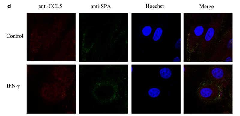
Application: IHC    Species: Cows    Sample: bovine mammary epithelial cells (BMECs)

Fig. 2. IFN-γ promoted the expression of CCL5 and CCL5 antibody inhibited the infection of S. aureus. a DEGs associated with inflammation were screened out: DEGs were screened according to false discovery rate (FDR) ≤ 0.05 and |log2 ratio ≥ 1. Among 12 genes associated with inflammation, seven of the genes were increased and five decreased. b Western blot detection of CCL5 expression in BMECs that were stimulated by IFN-γ. c The expression ratio of CCL5 in (b) was quantified by ImageJ software. d The expression of CCL5 and the adhesion of S. aureus were detected by immunofluorescence after IFN-γ treatment. Green indicated S. aureus, blue indicated the nucleus of BMECs, and anti-SPA referred to S. aureus-specific antibody. e The adhesion of S. aureus to BMECs after treatment with CCL5 antibody. f The invasion of S. aureus to BMECs after treatment with CCL5 antibody. RB-IgG means rabbit IgG. The experiment was repeated three times.

5). RNA demethylase ALKBH5 suppresses tumorigenesis via inhibiting proliferation and invasion and promoting CD8+ T cell infiltration in colorectal cancer. Translational Oncology, 2023 (PubMed: 37224767) [IF=5.0]

Application: WB    Species: Human    Sample: HCT116 and SW620 cells

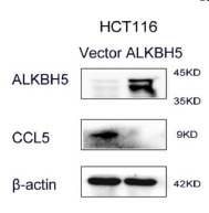
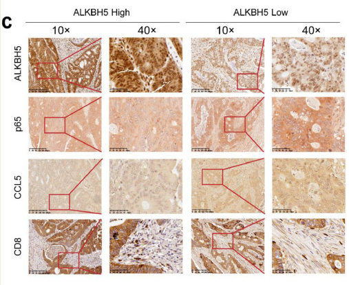
Fig. 6 ALKBH5 disrupting NF-κB-CCL5 signaling. a-b Relative expression of CCL5 in ALKBH5-overexpressed HCT116 and SW620 cells. c Immunohistochemistry was used to detect the expression of ALKBH5, p65, CCL5, and CD8 in human colorectal cancer tissues, the data represented mean ± SD (n=56). d CCL5 expression and the total and phosphorylation level of p65 and IκB-α after ALKBH5 overexpression were examined by western blot analysis. e SW620 were pretreated with 100 and 200 ng/ml of specific NF-κB activator PMA, and western blot analysis was to assess the expression of CCL5 and p-p65.

Application: IHC    Species: Human    Sample: HCT116 and SW620 cells

Fig. 6 ALKBH5 disrupting NF-κB-CCL5 signaling. a-b Relative expression of CCL5 in ALKBH5-overexpressed HCT116 and SW620 cells. c Immunohistochemistry was used to detect the expression of ALKBH5, p65, CCL5, and CD8 in human colorectal cancer tissues, the data represented mean ± SD (n=56). d CCL5 expression and the total and phosphorylation level of p65 and IκB-α after ALKBH5 overexpression were examined by western blot analysis. e SW620 were pretreated with 100 and 200 ng/ml of specific NF-κB activator PMA, and western blot analysis was to assess the expression of CCL5 and p-p65.

6). Radiotherapy Increases 12-LOX and CCL5 Levels in Esophageal Cancer Cells and Promotes Cancer Metastasis via THP-1-Derived Macrophages. OncoTargets and Therapy, 2020 (PubMed: 32801779) [IF=4.0]

Application: WB    Species: Human    Sample: esophageal cancer cells

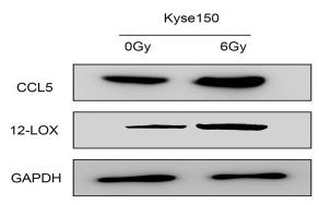
Figure 1 Radiotherapy increases 12-LOX levels in esophageal cancer cells. (A) 12-LOX expression in Eca109 cells irradiated with different X-ray doses (0, 3, and 6 Gy). (B and D) Cytokines (12-LOX, VEGF, TGF-beta1, and IL-4) and chemotactic factors (CCL2, CCL5, CCL22, and CSF1) showed increased mRNA levels after radiotherapy in Eca109 and Kyse150 cells. (C and E) CCL5 and 12-LOX showed increased protein levels after radiotherapy in Eca109 cells. (F and G) Changes in the mRNA levels of the M1- and M2-related genes of THP-1-derived macrophages with CM from irradiated cells. Experimental data were obtained from three independent tests. *P < 0.05; **P < 0.01; ***P < 0.001. Abbreviations: CM, conditioned media; ns, no significance.

Application: IHC    Species: Human    Sample: macrophages

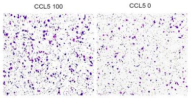
Figure 5 CCL5 attracts macrophages and induces their polarity into the M2 subtype. (A and B) The number of migratory macrophages in the CCL5 100 group increased significantly. (C) The number of migratory THP-1 cells in the CCL5 100 group increased significantly. (D) M1- and M2-related gene expression levels of macrophages changed at the mRNA level with exogenous CCL5 expression. Experimental data were obtained from three independent tests. *P < 0.05; **P < 0.01; ***P < 0.001. Abbreviation: ns, no significance.

7). IGF2BP3 Enhances the Growth of Hepatocellular Carcinoma Tumors by Regulating the Properties of Macrophages and CD8+ T Cells in the Tumor Microenvironment. Journal of Clinical and Translational Hepatology, 2023 (PubMed: 37719968) [IF=3.6]

Application: WB    Species: Mouse    Sample: tumor cells

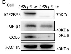
Fig. 4 IGF2BP3 facilitates macrophage infiltration and polarization by promoting the secretion of CCL5 and TGF-β1 from tumor cells. (A–C) qRT-PCR (A) and western blots showing the expression of CCL5 and TGF-β1 in Igf2bp3_wt and Igf2bp3_ko cells (B) and culture supernatants (C). (D) ELISA results showing the levels of CCL5 and TGF-β1 in Hepa1-6 cell culture supernatant. (E) Immunohistochemical (IHC) staining for CCL5 and TGF-β1 expression in Igf2bp3_wt and Igf2bp3_ko tumor tissue (left); the histogram summarizes the staining intensity (right). Scale bar=50 µm. (F, G) Transwell assay performed with Raw264.7 cells. Cells were suspended in serum-free media and seeded into the upper chamber. Cell culture supernatant was added to the lower chamber. CCL5 (100 ng/mL), Anti-CCL5 (200 ng/mL). The histogram summarizes the migration results. The graph shows the number of cells per field of view. Scale bar=100 µm. (H) Igf2bp3_wt cells were cultured with TGF-β1 neutralizing antibody (1 µg/mL). Raw264.7 cells stimulated with tumor supernatant. The H2-Ab1, Tnf-α, Arg1, and Il-10 expression in Raw264.7 cells was determined by qRT-PCR. Igf2bp3_wt Sup., Igf2bp3_wt cell culture supernatants; Igf2bp3_ko Sup., Igf2bp3_ko cell culture supernatants. Data are mean±SD. *p

8). BPIFB2 is highly expressed in “cold” lung adenocarcinoma and decreases T cell chemotaxis via activation of the STAT3 pathway. MOLECULAR AND CELLULAR PROBES, 2022 (PubMed: 35240266) [IF=3.3]

9). Long Noncoding RNA ZFAS1 Promotes Progression of Oral Squamous Cell Carcinoma Through Targeting miR-6499-3p/CCL5 Axis. In Vivo, 2021 (PubMed: 34697152) [IF=2.3]

Application: WB    Species: Mouse    Sample:

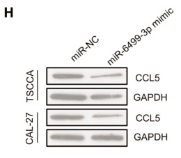
Figure 3 A: Predicted binding sites for long non-coding RNA (lncRNA) zinc finger nuclear transcription factor, X-box binding 1-type containing 1 antisense RNA 1 (ZFAS1), and miR-6499-3p using the DIANA bioinformatics tool (http://diana.imis.athena-innovation.gr/DianaTools/index.php). B: Relative luciferase activity in luciferase reporter plasmids, which included mutant-type (Mut) and wild-type (WT) pGL3-ZFAS1-3'UTR. Activity in pGL3-ZFAS1-Mut-transfected cells was not suppressed by miR-6499-3p. C: There was much more accumulated ZFAS1 binding to Argonaute 2 (AGO2) protein in the miR-6499-3p mimic group. D: Expression of miRNA-6499-3p was up-regulated in si-lncRNA ZFAS1 cells. E: The predicted binding site for C-C motif chemokine ligand 5 (CCL5) and miR-6499-3p by bioinformatics (http://www.targetscan.org). F: Relative luciferase activity in pGL3-CCL5-Mut-transfected cells was not suppressed by miRNA-6499-3p. G: and H: In cells transfected with miRNA-6499-3p mimic, expression of both CCL5 RNA and protein were reduced.

10). Magnolol protects against acute gastrointestinal injury in sepsis by down-regulating regulated on activation, normal T-cell expressed and secreted. World Journal of Clinical Cases, 2021 (PubMed: 35004977) [IF=1.1]

Application: WB    Species: Human    Sample: Caco2 cells

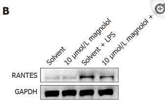
Figure 3 Expression of regulated on activation, normal T-cell expressed and secreted protein in Caco2 cells in each group. A: The cells were pretreated with magnolol or solvent for 8 h, followed by lipopolysaccharide (LPS) (100 μg/mL) for 24 h. The content of RANTES in the supernatant of the four groups of cells was determined by ELISA. Data are presented as the mean ± SD; the comparison between different groups was performed using the LSD method in one-way ANOVA (bP < 0.01); B: The cells were pretreated with magnolol or solvent for 8 h and then with LPS (100 μg/mL) for 24 h. The expression of RANTES protein in Caco2 cells in each group was detected by Western blot analysis. 0 or Solvent: Treated with solvent only; 10 μmol/L magnolol: Treated with 10 μmol/L magnolol; LPS: Treated with solvent and LPS (100 μg/mL); LPS + magnolol 10 μmol/L: Treated with magnolol 10 μmol/L and with LPS (100 μg/mL); GAPDH: Glyceraldehyde-3-phosphate dehydrogenase; RANTES: Regulated on activation, normal T-cell expressed and secreted.

Load more

Restrictive clause


Affinity Biosciences tests all products strictly. Citations are provided as a resource for additional applications that have not been validated by Affinity Biosciences. Please choose the appropriate format for each application and consult Materials and Methods sections for additional details about the use of any product in these publications.

For Research Use Only.
Not for use in diagnostic or therapeutic procedures. Not for resale. Not for distribution without written consent. Affinity Biosciences will not be held responsible for patent infringement or other violations that may occur with the use of our products. Affinity Biosciences, Affinity Biosciences Logo and all other trademarks are the property of Affinity Biosciences LTD.