Product: CD34 Antibody
Catalog: AF5149
Description: Rabbit polyclonal antibody to CD34
Application: WB IF/ICC
Reactivity: Human, Mouse, Rat
Prediction: Rabbit, Dog
Mol.Wt.: 120 kDa; 41kD(Calculated).
Uniprot: P28906
RRID: AB_2837635

View similar products>>

   Size Price Inventory
 100ul $280 In stock
 200ul $350 In stock

Lead Time: Same day delivery

For pricing and ordering contact:
Local distributors

Product Info

Source:
Rabbit
Application:
WB 1:500-1:2000, IF/ICC 1:0-1:500
*The optimal dilutions should be determined by the end user.
*Tips:

WB: For western blot detection of denatured protein samples. IHC: For immunohistochemical detection of paraffin sections (IHC-p) or frozen sections (IHC-f) of tissue samples. IF/ICC: For immunofluorescence detection of cell samples. ELISA(peptide): For ELISA detection of antigenic peptide.

Reactivity:
Human,Mouse,Rat
Prediction:
Rabbit(82%), Dog(82%)
Clonality:
Polyclonal
Specificity:
CD34 Antibody detects endogenous levels of total CD34.
RRID:
AB_2837635
Cite Format: Affinity Biosciences Cat# AF5149, RRID:AB_2837635.
Conjugate:
Unconjugated.
Purification:
The antiserum was purified by peptide affinity chromatography using SulfoLink™ Coupling Resin (Thermo Fisher Scientific).
Storage:
Rabbit IgG in phosphate buffered saline , pH 7.4, 150mM NaCl, 0.02% sodium azide and 50% glycerol. Store at -20 °C. Stable for 12 months from date of receipt.
Alias:

Fold/Unfold

CD34; CD34 antigen; CD34 molecule; CD34_HUMAN; Cluster designation 34; Hematopoietic progenitor cell antigen CD34; HPCA1; Mucosialin; OTTHUMP00000034733; OTTHUMP00000034734;

Immunogens

Immunogen:
Uniprot:
Gene(ID):
Expression:
P28906 CD34_HUMAN:

Selectively expressed on hematopoietic progenitor cells and the small vessel endothelium of a variety of tissues.

Description:
Possible adhesion molecule with a role in early hematopoiesis by mediating the attachment of stem cells to the bone marrow extracellular matrix or directly to stromal cells. Could act as a scaffold for the attachment of lineage specific glycans, allowing stem cells to bind to lectins expressed by stromal cells or other marrow components. Presents carbohydrate ligands to selectins.
Sequence:
MLVRRGARAGPRMPRGWTALCLLSLLPSGFMSLDNNGTATPELPTQGTFSNVSTNVSYQETTTPSTLGSTSLHPVSQHGNEATTNITETTVKFTSTSVITSVYGNTNSSVQSQTSVISTVFTTPANVSTPETTLKPSLSPGNVSDLSTTSTSLATSPTKPYTSSSPILSDIKAEIKCSGIREVKLTQGICLEQNKTSSCAEFKKDRGEGLARVLCGEEQADADAGAQVCSLLLAQSEVRPQCLLLVLANRTEISSKLQLMKKHQSDLKKLGILDFTEQDVASHQSYSQKTLIALVTSGALLAVLGITGYFLMNRRSWSPTGERLGEDPYYTENGGGQGYSSGPGTSPEAQGKASVNRGAQENGTGQATSRNGHSARQHVVADTEL

Predictions

Predictions:

Score>80(red) has high confidence and is suggested to be used for WB detection. *The prediction model is mainly based on the alignment of immunogen sequences, the results are for reference only, not as the basis of quality assurance.

Species
Results
Score
Rabbit
82
Dog
82
Horse
70
Pig
60
Sheep
55
Bovine
45
Xenopus
0
Zebrafish
0
Chicken
0
Model Confidence:
High(score>80) Medium(80>score>50) Low(score<50) No confidence

PTMs - P28906 As Substrate

Site PTM Type Enzyme
S198 Phosphorylation
K268 Acetylation
S282 Phosphorylation
Y286 Phosphorylation
Y329 Phosphorylation
Y330 Phosphorylation
T331 Phosphorylation
Y339 Phosphorylation
S341 Phosphorylation
S346 Phosphorylation P31751 (AKT2)
S354 Phosphorylation

Research Backgrounds

Function:

Possible adhesion molecule with a role in early hematopoiesis by mediating the attachment of stem cells to the bone marrow extracellular matrix or directly to stromal cells. Could act as a scaffold for the attachment of lineage specific glycans, allowing stem cells to bind to lectins expressed by stromal cells or other marrow components. Presents carbohydrate ligands to selectins.

PTMs:

Highly glycosylated.

Phosphorylated on serine residues by PKC.

Subcellular Location:

Membrane>Single-pass type I membrane protein.

Extracellular region or secreted Cytosol Plasma membrane Cytoskeleton Lysosome Endosome Peroxisome ER Golgi apparatus Nucleus Mitochondrion Manual annotation Automatic computational assertionSubcellular location
Tissue Specificity:

Selectively expressed on hematopoietic progenitor cells and the small vessel endothelium of a variety of tissues.

Family&Domains:

Belongs to the CD34 family.

Research Fields

· Environmental Information Processing > Signaling molecules and interaction > Cell adhesion molecules (CAMs).   (View pathway)

· Organismal Systems > Immune system > Hematopoietic cell lineage.   (View pathway)

References

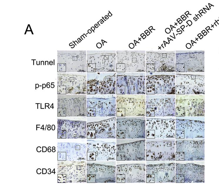
1). Berberine-mediated up-regulation of surfactant protein D facilitates cartilage repair by modulating immune responses via the inhibition of TLR4/NF-ĸB signaling. Pharmacological Research, 2020 (PubMed: 32057894) [IF=9.3]

Application: IHC    Species: rat    Sample: knee joints

Fig. 5. Chondrocyte apoptosis and immune molecular biomarkers were inhibited by BBR binding to SP-D in vivo in OA. OA model rats (n = 5/group) were injected using 25 μl of 200 μM BBR, followed by 25 μl rhSP-D (40 μg/mL) 1 day later. Sham-operated and OA-induction groups received an injection of 50 μl PBS into the right knee joint. For rats receiving rAAV-GFP or rAAV-SP-D shRNA, injections were performed for 10 consecutive days beginning 1 day after the initial BBR injection. At 10 weeks post-surgery, animals were sacrificed and the knee joints were collected for TUNEL and IHC analysis. (A) TUNEL staining (400 ×) was used to assess apoptosis. The p-p65, TLR4, F4/80, CD68, and CD34 expression was assessed via IHC (400 × with higher 800 × magnification inset). (B) Quantification of the indicated proteins in IHC images. Each column represents the mean ± SEM (n = 5). ***P < 0.001 versus the sham-operated group; ##P < 0.01 and ###P < 0.001 versus the OA-induction group; &P < 0.05 and &&P < 0.01 versus the OA + BBR group.

2). Mangiferin alleviates diabetic pulmonary fibrosis in mice via inhibiting endothelial-mesenchymal transition through AMPK/FoxO3/SIRT3 axis. Acta pharmacologica Sinica, 2024 (PubMed: 38225395) [IF=8.2]

3). Resolvin D1 ameliorates Inflammation-Mediated Blood-Brain Barrier Disruption After Subarachnoid Hemorrhage in rats by Modulating A20 and NLRP3 Inflammasome. Frontiers in Pharmacology, 2021 (PubMed: 33732145) [IF=5.6]

4). Radiation-induced FAP + fibroblasts are involved in keloid recurrence after radiotherapy. Frontiers in Cell and Developmental Biology, 2022 (PubMed: 36092734) [IF=5.5]

5). BCL2L10 inhibits growth and metastasis of hepatocellular carcinoma both in vitro and in vivo. MOLECULAR CARCINOGENESIS, 2017 (PubMed: 27770580) [IF=4.6]

Application: IHC    Species: mouse    Sample:

(D) Representative microscopic feature of subtaneous tumor in H&E-stained sections of nude mice (left). CD34 protein expression was higher in tumors with low expression of BCL2L10 by IHC (middle and right).

6). Organoid models derived from patients with malignant phyllodes tumor of the breast. Breast Cancer Research and Treatment, 2023 (PubMed: 37204665) [IF=3.8]

7). Upregulated VEGF and Robo4 correlate with the reduction of miR-15a in the development of diabetic retinopathy. ENDOCRINE, 2019 (PubMed: 30980286) [IF=3.7]

8). Andrographolide improves the dysfunction of endothelial progenitor cells from angiotensin II-induced hypertensive mice through SIRT1 signaling. BIOCHEMICAL AND BIOPHYSICAL RESEARCH COMMUNICATIONS, 2023 (PubMed: 36543019) [IF=3.1]

9). Sesquiterpene binding Gly-Leu-Ser/Lys-"co-adaptation pocket" to inhibit lung cancer cell epithelial-mesenchymal transition. Oncotarget, 2017 (PubMed: 29050271)

10). PU.1 alleviates the inhibitory effects of cigarette smoke on endothelial progenitor cell function and lung-homing through Wnt/β-catenin and CXCL12/CXCR4 pathways. Tobacco Induced Diseases, 2024 (PubMed: 38274000)

Restrictive clause


Affinity Biosciences tests all products strictly. Citations are provided as a resource for additional applications that have not been validated by Affinity Biosciences. Please choose the appropriate format for each application and consult Materials and Methods sections for additional details about the use of any product in these publications.

For Research Use Only.
Not for use in diagnostic or therapeutic procedures. Not for resale. Not for distribution without written consent. Affinity Biosciences will not be held responsible for patent infringement or other violations that may occur with the use of our products. Affinity Biosciences, Affinity Biosciences Logo and all other trademarks are the property of Affinity Biosciences LTD.