Caspase 4 Antibody - #AF5130
| Product: | Caspase 4 Antibody |
| Catalog: | AF5130 |
| Description: | Rabbit polyclonal antibody to Caspase 4 |
| Application: | WB IHC |
| Cited expt.: | WB |
| Reactivity: | Human, Mouse, Rat |
| Prediction: | Bovine, Dog |
| Mol.Wt.: | 35~50 kD; 43kD(Calculated). |
| Uniprot: | P49662 |
| RRID: | AB_2837616 |
Related Downloads
Protocols
Product Info
*The optimal dilutions should be determined by the end user. For optimal experimental results, antibody reuse is not recommended.
*Tips:
WB: For western blot detection of denatured protein samples. IHC: For immunohistochemical detection of paraffin sections (IHC-p) or frozen sections (IHC-f) of tissue samples. IF/ICC: For immunofluorescence detection of cell samples. ELISA(peptide): For ELISA detection of antigenic peptide.
Cite Format: Affinity Biosciences Cat# AF5130, RRID:AB_2837616.
Fold/Unfold
Apoptotic cysteine protease Mih1/TX; CASP-4; CASP4; CASP4_HUMAN; Caspase 4 apoptosis related cysteine peptidase; Caspase-4 subunit 2; Caspase4; ICE(rel)-II; ICE(rel)II; ICEREL II; ICH2; Mih1/TX; Protease ICH-2; Protease TX; TX; CASP11;caspase 11;
Immunogens
A synthesized peptide derived from human Caspase 4, corresponding to a region within the internal amino acids.
Widely expressed, including in keratinocytes and colonic and small intestinal epithelial cells (at protein level). Not detected in brain.
- P49662 CASP4_HUMAN:
- Protein BLAST With
- NCBI/
- ExPASy/
- Uniprot
MAEGNHRKKPLKVLESLGKDFLTGVLDNLVEQNVLNWKEEEKKKYYDAKTEDKVRVMADSMQEKQRMAGQMLLQTFFNIDQISPNKKAHPNMEAGPPESGESTDALKLCPHEEFLRLCKERAEEIYPIKERNNRTRLALIICNTEFDHLPPRNGADFDITGMKELLEGLDYSVDVEENLTARDMESALRAFATRPEHKSSDSTFLVLMSHGILEGICGTVHDEKKPDVLLYDTIFQIFNNRNCLSLKDKPKVIIVQACRGANRGELWVRDSPASLEVASSQSSENLEEDAVYKTHVEKDFIAFCSSTPHNVSWRDSTMGSIFITQLITCFQKYSWCCHLEEVFRKVQQSFETPRAKAQMPTIERLSMTRYFYLFPGN
Predictions
Score>80(red) has high confidence and is suggested to be used for WB detection. *The prediction model is mainly based on the alignment of immunogen sequences, the results are for reference only, not as the basis of quality assurance.
High(score>80) Medium(80>score>50) Low(score<50) No confidence
Research Backgrounds
Inflammatory caspase. Essential effector of NLRP3 inflammasome-dependent CASP1 activation and IL1B and IL18 secretion in response to non-canonical activators, such as UVB radiation, cholera enterotoxin subunit B and cytosolic LPS. Independently of NLRP3 inflammasome and CASP1, promotes pyroptosis, through GSDMD cleavage and activation, and IL1A, IL18 and HMGB1 release in response to non-canonical inflammasome activators. Plays a crucial role in the restriction of Salmonella typhimurium replication in colonic epithelial cells during infection. In later stages of the infection, LPS from cytosolic Salmonella triggers CASP4 activation, which ultimately results in pyroptosis of infected cells and their extrusion into the gut lumen, as well as in IL18 secretion. Pyroptosis limits bacterial replication, while cytokine secretion promotes the recruitment and activation of immune cells and triggers mucosal inflammation. Involved in LPS-induced IL6 secretion; this activity may not require caspase enzymatic activity. Involved in cell death induced by endoplasmic reticulum stress and by treatment with cytotoxic APP peptides found Alzheimer's patient brains. Activated by direct binding to LPS without the need of an upstream sensor. Does not directly process IL1B. During non-canonical inflammasome activation, cuts CGAS and may play a role in the regulation of antiviral innate immune activation.
In response to activation signals, including endoplasmic reticulum stress or treatment with amyloid-beta A4 protein fragments (such as amyloid-beta protein 40), undergoes autoproteolytic cleavage.
Cytoplasm>Cytosol. Endoplasmic reticulum membrane>Peripheral membrane protein>Cytoplasmic side. Mitochondrion. Inflammasome. Secreted.
Note: Predominantly localizes to the endoplasmic reticulum (ER). Association with the ER membrane requires TMEM214 (PubMed:15123740). Released in the extracellular milieu by keratinocytes following UVB irradiation (PubMed:22246630).
Widely expressed, including in keratinocytes and colonic and small intestinal epithelial cells (at protein level). Not detected in brain.
The CARD domain mediates LPS recognition and homooligomerization.
Belongs to the peptidase C14A family.
Research Fields
· Organismal Systems > Immune system > NOD-like receptor signaling pathway. (View pathway)
References
Application: IF/ICC Species: Mouse Sample: kidney
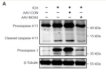
Application: WB Species: Rat Sample:
Application: WB Species: Mice Sample: liver tissues
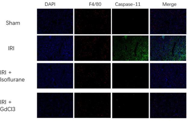
Application: IF/ICC Species: Mice Sample: liver tissues
Restrictive clause
Affinity Biosciences tests all products strictly. Citations are provided as a resource for additional applications that have not been validated by Affinity Biosciences. Please choose the appropriate format for each application and consult Materials and Methods sections for additional details about the use of any product in these publications.
For Research Use Only.
Not for use in diagnostic or therapeutic procedures. Not for resale. Not for distribution without written consent. Affinity Biosciences will not be held responsible for patent infringement or other violations that may occur with the use of our products. Affinity Biosciences, Affinity Biosciences Logo and all other trademarks are the property of Affinity Biosciences LTD.


