
.
Bands result from membrane strip incubation.")


 and mouse anti-beta tubulin Ab(T0023 1:200) for 1 hour at 37°C. An AlexaFluor594 conjugated goat anti-rabbit IgG(H+L) Ab(Red) and an AlexaFluor488 conjugated goat anti-mouse IgG(H+L) Ab(Green) were used as the secondary antibody.
The nuclear counter stain is DAPI(blue).")
 and mouse anti-beta tubulin Ab(#T0023) for 1 hour at 37°C. An AlexaFluor594 conjugated goat anti-rabbit IgG Ab(Red) and an AlexaFluor488 conjugated goat anti-mouse IgG Ab(Green) were used as the secondary antibody.
The nuclear counter stain is DAPI (blue).")

 were used as negative control. (A) Immunoblot analysis to assess the expression of autophagic flux. GAPDH served as protein loading control. Semiquantitative analyses of protein expression in following histogram (mean ± S.E.M., three independent replicates per groups). * p < 0.05, ** p < 0.01.")
 The expression and quantification of HMGB1, RAGE, LC3, and Beclin1 in the A498 cell line were analyzed by Western blot analysis.")
 Mitochondria dynamics in control and BHPF-exposed oocytes. HSP60, red; α-Tubulin, green; DNA, blue. Arrow, mitochondria aggregation. Scale bar, 10 μm. (B) Relative mtDNA copy numbers in control and BHPF-exposed oocytes. (C) Immunoblot analysis to assess the effects of BHPF on the expression of PINK1, Beclin1, LC3B, TOMM20,and TIMM17A proteins.")
 for 48 hours. Cell apoptosis was detected via flow cytometry analysis. The apoptotic ratio was calculated as the percentage of early apoptotic (Annexin V+/PI-) cells plus late apoptotic (Annexin V+/PI+) cells (A,B).The expression of apoptosis-related proteins and autophagy markers was measured via Western blot analysis (C,D).")
 HUVECs were pretreated for 2h with 10 mM 3-MA, 20μM Z-VAD-FMK, or 10
mM NAC, and then treated with 5μg/ml HMME and light dose of 2 J/cm2
. Cell viability
was detected by CCK8 assay 24h after HMME-PDT.
** p<0.01. (B) Autophagosome
formation was observed in HUVECs 6h after HMME-PDT. Scale bar= 25μm. (C) The
number of early autophagosomes (yellow puncta) per cell was calculated, and more
than 10 cells were quantified for each condition.
** p<0.01. (D) TUNEL staining of
HUVECs was performed 24h after HMME-PDT. Scale bar=100μm. (E) Apoptosis ratio
Journal Pre-proof
was calculated according to the TUNEL staining of HUVECs.
** p<0.01. (F) Western
blotting analysis was used to measure the expression levels of Cytochrome C, Cleaved
Caspase 3, PARP, Cleaved PARP, Beclin 1, LC3, and β-actin in HUVECs 24h after
HMME-PDT")
 Autophagy related proteins LC3-II/LC3-I, Beclin-1, and P62 were detected and quantified by Western blot in THP-1, THP-1/ADM, K562 and K562/ADM cells.")
for 6 h. B. Results were detected by western blot when cells were pretreated with CHX with or without 17β-E2. C–F. After chondrocyte were pretreated with lysosome inhibitors CQ, 3-MA, ALLN, MG-312 with or without 17β-E2, ASIC1a protein were analyzed by Western blot. G. Western blot analysis of LC3-II, Beclin-1 levels.")
: Immunofluorescent staining of
beclin1 and cleaved-caspase3 in lung tissue. (B): Bar graph of levels of beclin1 and cleaved-caspase3 expression in lung tissue. (C):
Western blot bands of beclin1 and cleaved-caspase3 protein in lung tissue. (D): Bar graph of Western blot bands in Figure 5 (C).
Data are representative of at least three separate experiments. (*p < 0.05, **p < 0.01 vs. control group; #
p < 0.05, ##p < 0.01 vs. model
group).")
 Representative immunoblots and relative protein levels of Beclin1, and LC3. (C, D) Hepatic
expression and distribution of autophagy protein (Beclin1 and LC3) by immunofluorescent staining, and relative flourescent intensity of Beclin1 and LC3 in the liver.
(E, F) Representative immunoblots and relative protein levels of Beclin1 and LC3. ^p < 0.05* vs WT-ND; p < 0.05; **p < 0.01 vs WT-HFD; ##p < 0.01;
###p < 0.005 vs ApoE−/−-HFD. n = 3 for each group.")
 Representative images of Beclin1, LC3 and P62 (immunohistochemical staining; magnification, ×400). Quantitative analysis of (B) Beclin1, (C) LC3 and (D) P62 were normalized to the normal control group. (E) Protein expression levels of Beclin1, LC3 and P62 in the liver tissues were detected via western blotting. Semi-quantitative analysis of (F) Beclin1, (G) LC3 and (H) P62 was normalised to β-actin. Data are presented as the mean ± SD, n=3. *P<0.05, ***P<0.001 vs. control group; #P<0.05, ##P<0.01, ###P<0.001 vs. model group. Con, Control; Mod, Model; AS-IV, Astragaloside IV (80 mg/kg); Met, Metformin (200 mg/kg).")
 Representative images of Beclin1, LC3 and P62 (immunohistochemical staining; magnification, ×400). Quantitative analysis of (B) Beclin1, (C) LC3 and (D) P62 were normalized to the normal control group. (E) Protein expression levels of Beclin1, LC3 and P62 in the liver tissues were detected via western blotting. Semi-quantitative analysis of (F) Beclin1, (G) LC3 and (H) P62 was normalised to β-actin. Data are presented as the mean ± SD, n=3. *P<0.05, ***P<0.001 vs. control group; #P<0.05, ##P<0.01, ###P<0.001 vs. model group. Con, Control; Mod, Model; AS-IV, Astragaloside IV (80 mg/kg); Met, Metformin (200 mg/kg).")
.")

 Following treatment of HEC-1A and Ishikawa cells with various concentrations of chrysin (0, 10, 20, 40 and 80 µM) for 48 h, the levels of LC3II, Beclin 1 and p62 were examined by western blotting. (C and D) HEC-1A and Ishikawa cells were transfected with si-negative control or siATG5 for 24 h, and then treated with chrysin at 40 µM for 24 h. The expression of ATG5 and LC3 was detected by western blotting. #P<0.05, ##P<0.01, ###P<0.001 vs. siNC. (E and F) HEC-1A and Ishikawa cells were untreated or treated with 5 µM CQ for 1 h, followed by chrysin treatment at 40 µM for 24 h. The expression levels of LC3II were then examined by western blotting. Values are reported as the mean ± standard deviation (n=3). *P<0.05, **P<0.01, ***P<0.001 vs. control group; #P<0.05, ##P<0.01, ###P<0.001 vs. chrysin group. si, small interfering RNA; NC, negative control; ATG5, autophagy-related gene 5; CQ, chloroquinone; LC3, Microtubule-associated proteins 1A/1B light chain 3B.")
 Expression levels of beclin 1, LC3 II, and p62 proteins in the LV of mice. n = 6, ∗P < 0.05, ∗∗P < 0.01 vs. control group; #P < 0.05, ##P < 0.01 vs. Ang II group. (b) Representative images of the ultrastructure of H9c2 cells showing the changes of autophagy under a transmission electron microscope. Scale bar in (i), 2 μm and magnification, ×10000; in (ii), 1 μm, ×20000. Yellow arrow refers to the autophagosome, which contains dense electronic content such as autolysosomes or mitochondria. (c) Expression of beclin 1 and LC3 mRNA in H9c2 cells. (d, e) Expression of beclin 1, LC3II, and P62 proteins in H9c2 cells. In cell experiments, (1) control, (2) Ang II, (3) NC, (4) NC + Ang II, (5) miR-128 antag, (6) miR-128 antag + Ang II, (7) Ang II + CQ (chloroquine), and (8) miR-128 antag + Ang II + CQ. n = 3 independent batches, repeated 2 ~ 3 wells/each batch. Data are expressed as mean ± SD. Statistical significance was assessed by using one-way ANOVA followed by LSD. ∗P < 0.05, ∗∗P < 0.01 vs. control group; #P < 0.05, ##P < 0.01 vs. NC + Ang II group; ∆P < 0.05, P < 0.01 vs. Ang II group; ▴P < 0.05 vs. Ang II + CQ group.")
. b Confocal microscopy images of cells treated with or without PS-T (5 μg/mL) for 24 h after transfection with mRFP-GFP-LC3 plasmid (scale bars: 10 μm). Quantification of LC3-GFP and LC3-RFP puncta/cells from three independent experiments (***P < 0.001). c MDA-MB-231 cells are treated with PS-T (5 μg/mL) for 24 h with or without LY294002 (10 µM). The levels of each EMT marker in MDA-MB-231cells were quantified using NIH ImageJ software. (mean ± SD, *P < 0.05 and **P < 0.01)")
. B, Detection of the autophagy-related proteins LC3II/I, Beclin1, AKT, PTEN, mTOR and STX3 by Western blotting.")
 Expression levels of beclin 1, LC3 II, and p62 proteins in the LV of mice. n = 6, ∗P < 0:05, ∗∗P < 0:01 vs. control group; #P < 0:05, ##P < 0:01 vs. Ang II group.")
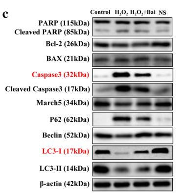
 To confrm that the efect of baicalin is mediated by regulating autophagy, 3-MA was used with baicalin treatment, and the levels of the autophagy-related proteins LC3II/I, Beclin1, P62, p-AMPK, AMPK, and mTOR were measured by Western blotting.")


 Representative protein blots of p62, LC3, and Beclin 1. (b) Quantitative analysis of p62, LC3, and Beclin 1 proteins. (c) Representative protein blots of PI3K/AKT pathway-related proteins. (d) Quantitative analysis of PI3K pathway-related targets in each group. ∗P < 0.05 and ∗∗P < 0.01 vs. sham group. ▲P < 0.05 and ▲▲P < 0.01 vs. IR group.")
 Protein levels of beclin-1, p62, and LC3II as measured by western blotting. (B–D) Relative density values showing P62, beclin1, LC3- II/LC3- I expression. (E,F) Representative images of TEM and immunohistochemical staining for P62. The white arrows indicate autophagosomes and autolysosomes, the yellow arrowheads indicate abnormal mitochondria. Data are expressed as *p < 0.05, **p < 0.01, compared with sham group; #p < 0.05, ##p < 0.01, compared with model group.")
 post‐oxygen–glucose deprivation (OGD). (A, B) Apoptosis of BMSCs in different groups was stained by Annexin V‐FITC/PI, and then assessed by flow cytometry (FCM). (C–E) Western blot images and relative quantification of apoptosis‐related proteins (Bcl‐2, Bax) and Gapdh (n = 4). (F–I) Western blot analysis of P62, Beclin‐1, LC3, and Gapdh and quantitative analysis (n = 4). * means p value <0.05, ** means p value <0.01, and *** means p value <0.001, p values were calculated with one‐way ANOVA, followed by Bonferroni's multiple comparison test")
 Immunofluorescence staining of LC3 and F4/80 in the lung tissues of WT and Nrf2−/− mice before or after CLP treatment. (C,D) Western blot analyses of autophagy-associated proteins including LC3 I, LC3 II, Beclin1, and p62 in lung tissues of each group. ** p < 0.01, *** p < 0.001. (E) Transmission electron microscopy assay detected the numbers of double-membrane autophagosomes and autolysosomes (red arrows in the pictures) in lung tissues of each group.")
 treated VSMCs. The relative protein expressions in Ang II-treated VSMCs with presence of C/EBPα overexpression or knockdown were analyzed by Western blot. GAPDH was used as an internal control. Data are presented as means ± SEM. (a, b) Evaluation of the influence of C/EBPα on the expression of α-SMA, SM-MHC, and OPN proteins. (c, d) Evaluation of the influence of C/EBPα on the expression of PIK3C2A protein. (e, f) Evaluation of the influence of C/EBPα on the expression of Beclin-1, p62, LC3II, and LC3I proteins. (g, h) Evaluation of the influence of C/EBPα on the expression of MMP-2 and MMP-9 proteins. (i) Intracellular ROS was measured by flow cytometry using DCFH-DA probe with presence of C/EBPα overexpression or knockdown. Control: control group; model: treated with 1 μM Ang II; vector: treated with 1 μM Ang II in vector transfected VSMCs; pcC/EBPα: treated with 1 μM Ang II in C/EBPα-overexpressed VSMCs; siNC: treated with 1 μM Ang II in siRNA NC-transfected VSMCs; siC/EBPα: treated with 1 μM Ang II in C/EBPα-knockdown VSMCs. ∗P < 0.05 vs. control,")
 of P62 (A, B), LC3-II (A, C), and Beclin-1 (A, D) were analyzed. The proteins expression of P62 (E, F), LC3-II (E, G), and Beclin-1 (E, H) in DEX-treated MC3T3-E1 cells were determined by western blot. * *P˂0.01. NC, negative control group; DEX, model rat group treated with DEX (5 mg/kg).")
 of P62 (A, B), LC3-II (A, C), and Beclin-1 (A, D) were analyzed. The proteins expression of P62 (E, F), LC3-II (E, G), and Beclin-1 (E, H) in DEX-treated MC3T3-E1 cells were determined by western blot. * *P˂0.01. NC, negative control group; DEX, model rat group treated with DEX (5 mg/kg).")
, p62 (B), and LC3(C) were captured under fluorescence microscopy, and the relative fluorescence intensity was calculated. All images were obtained at identical magnification, ×200, scale bar = 50 μm. Data are represented as mean ± SEM (n = 3). **P")


. Western blot detection of proteins in the NLRP3 inflammatory body: NLRP3, cleaved-CASP1, and cleaved-IL-1β (B). Western blot detection of proteins in the autophagy pathway: Beclin1, p-VPS34, and p62 (C). The expression of NLRP3 inflammatory bodies and autophagic vesicles in uterine tissues was observed by IF: NLRP3 was labeled with red fluorescence, VPS34 was labeled with green fluorescence, and nuclei were labeled with DAPI with blue fluorescence (D). Western blot detection of proteins in the lysosomal pathway: LAMP2, Ubiquitin and LC3 Ⅱ. (E). Results are presented as the mean ± S.E.M.")
 Western blot analysis of apoptosis-related protein expression levels. (B–D) Quantification of cleaved-caspase-3 (B), Bax (C), and Bcl-2 (D) protein levels from the western blot data. (E) Western blot analysis of autophagy-related protein expression levels. (F–H) Quantification of LC3II/LC3I (F), Beclin-1 (G), and P62 (H) protein levels from the western blot data. (I) Western blot analysis of oxidative stress-related protein expression levels. β-Actin was used as an internal reference for cleaved-caspase-3, Bax, and Bcl-2. GAPDH was used as an internal reference for LC3I, LC3II, Beclin-1, P62, Nrf2, and HO-1. (J, K) Western blot analysis of Nrf2 (J) and HO-1 (K) protein expression levels. All western blot data were normalized to the sham group. Data are expressed as mean ± SD (n = 3). **P < 0.01, vs. sham group; #P < 0.05, ##P < 0.01, vs. model group (one-way analysis of variance followed by Tukey's post hoc test). BA: Biochanin A; HO-1: heme oxygenase-1; LC3: microtube-associated protein 1 light-chain 3; Nrf2: nuclear factor erythroid2-related factor 2; P62: sequestosome-1; PC: positive control; SCI: spinal cord injury.")
 Hepatic LC3B, Beclin1, and p62 proteins expressions. (f–h) Hepatic Beclin1, LC3B, and p62 genes expressions. Results were expressed as the mean ± SD, n = 3 in (a–e) and n = 6 in (f–h). ∗P < 0.05 and ∗∗P < 0.01, compared with the model group.")
. According to the integrity of the lung tissues and the degree of inflammatory cell aggregation, the degree of lung damage was scored, being proportional to the score. C-H. The NLRP3 inflammasome and autophagy-related protein levels of the lung were measured by Western blot (n=3). The levels of ASC, pro-caspase 1, and NLRP3 protein in the lungs of CLP rats were reduced by Rap treatment, and the Beclin-1 and ratio of LC3Ⅱ to Ⅰ levels were increased by Rap treatment, while 3-MA treatment had the contrary effect. Data are presented as the mean ± standard deviation. ##P")
 BECLIN1, (C,D) LC3 were observed at the protein level and photographed under a microscope at 200× magnification. Scale bars represent 100 μm. typical areas were magnified at 400×. Scale bar represents 50 μm. Compared with Sham group: *p")
 Hematoxylin and Eosin (H&E) staining showed the nucleus in blue-purple and the cytoplasm in pink. Masson staining highlighted collagen fibers in light blue, cytoplasm, and red blood cells in red, with blue-purple nuclei. Sirius Red staining revealed brownish-brown nuclei and red collagen fibers. Magnification: ×200; Scale = 100 μm. (B–D) Analysis of the number of renal tubules and collagen volume in cattle and yaks. (E) Localization and distribution of P-AMPK, P-mTOR, Parkin, PINK1, LC3 and BECN1 proteins in cattle and yak kidneys. Magnification: ×40, Bar = 50 µm. (F,G) Western blot analysis was used to assess the expression levels of P-AMPK/AMPK, P-mTOR/mTOR, Parkin, PINK1, LC3 Ⅱ/Ⅰ and BECN1 proteins in the kidney. The original WB images are shown in Figure S1. All data are presented as means ± SEM, ns = not significant, * p < 0.05; ** p < 0.01; *** p < 0.001.")
 Hematoxylin and Eosin (H&E) staining showed the nucleus in blue-purple and the cytoplasm in pink. Masson staining highlighted collagen fibers in light blue, cytoplasm, and red blood cells in red, with blue-purple nuclei. Sirius Red staining revealed brownish-brown nuclei and red collagen fibers. Magnification: ×200; Scale = 100 μm. (B–D) Analysis of the number of renal tubules and collagen volume in cattle and yaks. (E) Localization and distribution of P-AMPK, P-mTOR, Parkin, PINK1, LC3 and BECN1 proteins in cattle and yak kidneys. Magnification: ×40, Bar = 50 µm. (F,G) Western blot analysis was used to assess the expression levels of P-AMPK/AMPK, P-mTOR/mTOR, Parkin, PINK1, LC3 Ⅱ/Ⅰ and BECN1 proteins in the kidney. The original WB images are shown in Figure S1. All data are presented as means ± SEM, ns = not significant, * p < 0.05; ** p < 0.01; *** p < 0.001.")
 Western blotting of LC3, Beclin 1, ATG5, and p62 levels in chondrocytes stimulated by IL-1β or/and transfected with miR-103-3p inhibitor. (F-G) Immunofluorescence staining of LC3 in chondrocytes. (H-L) Western blotting of LC3, Beclin 1, ATG5, and p62 protein levels in chondrocytes stimulated by IL-1β or/and transfected with miR-103-3p mimic. (M-N) Flow cytometry evaluated apoptosis in IL-1β-stimulated chondrocytes treated with miR-103-3p mimic or 3-MA. (O-S) Western blotting of Bax, Bcl-2, MMP13, and collagen II protein levels in chondrocytes. N = 3. ***p")
. The red arrow indicates a double membrane autophagosome. Scale bar, 1 μm. B The mRNA expression levels of Beclin1 and LC3 were detected by RT-qPCR. C The protein expression levels of Beclin1, p62, and LC3I/II were detected by western blot. D The LC3 puncta was detected by immunofluorescence staining (IF). Scale bar, 10 μm. Data represented mean ± SD (n = 3 biological replicates). *P")

. A. The CD4+ T cells in spleens of CLP rats by flow cytometry. B. The CD8+ T cells in spleens of CLP rats by flow cytometry. C. Bands of proteins; D-H. The statistical analysis results of NLRP3, Sirt1, Pro-IL-1β, Beclin-1, and LC3 Ⅱ/LC3 Ⅰ proteins bands by Western blot. Sham means the group underwent sham operation; CLP meats the group underwent CLP operation; HNK means the group was injected intraperitoneally with HNK 24h after the operation; And all rats were injected intraperitoneally with HNK (5 mg/kg twice) or PBS in the same volume. HNK: honokiol; CLP: cecal ligation and puncture; PBS: Phosphate buffer saline; NLRP3: NOD-like receptor thermal protein domain associated protein 3; Sirt1: Sirtuin1; ProIL-1β: Pro-Interleukin-1β; LC3: MAP1LC3.")
. C Quantitative results of TNF-α, IL-1β and IL-18 assays in serum by ELISA (TNF-α: t6 = 3.757, p = 0.0094; IL-1β: t6 = 3.507, p = 0.0127; IL-18: t6 = 2.663, p = 0.0374; n = 4 mice per group). D Representative immunoblotting bands of Atg7, p62, Atg5 and Beclin1. E Quantitative results of Atg7, p62, Atg5 and Beclin1 immunoblotting assays (Atg7: t10 = 2.460, p = 0.0337; p62: t10 = 7.415, p")
 MLg cells were exposed to 20 μM chloroquine and/or oleuropein for 24 h, after which samples were collected for Western blot analysis to assess the protein expression levels of p62 (original images can be found in Figure S1). The results are presented as Mean ± SD, n = 3. # indicates differences between the control group and the CQ group, with # p < 0.05. * indicates differences between the CQ group and the oleuropein treatment group, with * p < 0.05, and *** p < 0.001. NIH-3T3 (B) and MLg (C) cells were exposed to oleuropein and/or TGF-β1 (5 ng/mL) for 24 h, followed by sample collection for Western blot analysis to evaluate the protein expression levels of Beclin1, LC3, and p62 (original images can be found in Figure S1). The results are presented as Mean ± SD, n = 3. # indicates differences between the control group and the TGF-β1 group, with ## p < 0.01, and #### p < 0.0001. * indicates differences between the TGF-β1 group and the oleuropein treatment group, with * p < 0.05, *** p < 0.001, and **** p < 0.0001.")
—alignments of the edited loci, guide RNA’s sequences are in blue, PAM sites—in red, inserts—in green and in violet. Direct repeats are marked by underlining. Coding strands are shown as plus strands for all loci. The sequence of the plasmid px458N is present in Supporting File S1, Genbank entry NC_048598.1 is the CHO chromosome 7; NZ_CP080620.1—E. coli TOP10 strain. (B)—Western blotting of cell lysates with anti-Bak1 and anti-Bax antibodies, day 3 samples from Figure 2D are used. (C)—Western blotting of cell lysates with anti-Bcl-2 and anti-Beclin-1 antibodies, untransfected cells, day 3 samples were used. Three replicate samples for each cell line were tested. (D)—change in Bcl-2 and Beclin-1 expression levels determined by Western blotting. (E)—change in target protein titers upon target gene amplification by increasing the MTX concentration in culture medium from 50 nM to 500 nM. Product titers are measured on day 3 of cultures. Cells were seeded as 3 × 10⁵ cells/mL. (F)—induction of apoptosis in the 4BGD cells by various agents, measured by flow cytometry, propidium iodide staining. (D)—Error bars indicate the standard deviation, n = 3 for panel (D), n = 2 for panel (E), n = 3–4 for panel (F). Statistical significance of differences by the two-tailed t-test, *—p < 0.05, **—p < 0.01.")
 Representative western blot results of placental tissues. (B) Representative western blot results showing HTR8/SVeno and TEV-1 expression. Data shown in the bar chart are presented as mean ± SD. Statistical analysis between two groups in (A) was performed by the student t-test, inter-group statistical analysis in (B) was performed by one-way ANOVA. ** in (A) represent p 0.05, and ** represent p")
, intermediate (50 μg/kg body weight), or high (100 μg/kg body weight) doses of melittin via intraperitoneal injection. Four weeks after the administration, kidney tissues were collected from the rats for assessment. (A) Podocin and (B) nephrin expression levels in the kidney tissues were assessed by qRT-PCR. Expression levels of (C, D) podocin, (C, E) nephrin, (C, F) Beclin 1, (C, G) LC3II/LC3I, and (C, H) p-mTOR/mTOR in the kidney tissues assessed by Western blot.")
| Product: | Beclin 1 Antibody |
| Catalog: | AF5128 |
| Description: | Rabbit polyclonal antibody to Beclin 1 |
| Application: | WB IHC IF/ICC |
| Cited expt.: | WB, IHC, IF/ICC |
| Reactivity: | Human, Mouse, Rat |
| Prediction: | Pig, Zebrafish, Bovine, Horse, Sheep, Rabbit, Dog, Chicken, Xenopus |
| Mol.Wt.: | 52 kDa(Observed); 52kD(Calculated). |
| Uniprot: | Q14457 |
| RRID: | AB_2837614 |
Control Products

Related Downloads
Protocols
Product Info
*The optimal dilutions should be determined by the end user. For optimal experimental results, antibody reuse is not recommended.
*Tips:
WB: For western blot detection of denatured protein samples. IHC: For immunohistochemical detection of paraffin sections (IHC-p) or frozen sections (IHC-f) of tissue samples. IF/ICC: For immunofluorescence detection of cell samples. ELISA(peptide): For ELISA detection of antigenic peptide.
Cite Format: Affinity Biosciences Cat# AF5128, RRID:AB_2837614.
Fold/Unfold
APG6; ATG 6; ATG6; ATG6 autophagy related 6 homolog; Bcl-2-interacting protein beclin; Beclin 1 (coiled coil moesin like BCL2 interacting protein); Beclin 1 autophagy related; Beclin-1; Beclin1; BECN 1; Becn1; BECN1_HUMAN; Coiled coil myosin like BCL2 interacting protein; Coiled-coil myosin-like BCL2-interacting protein; GT197; Protein GT197; VPS 30; VPS30;
Immunogens
A synthesized peptide derived from human Beclin 1, corresponding to a region within the internal amino acids.
- Q14457 BECN1_HUMAN:
- Protein BLAST With
- NCBI/
- ExPASy/
- Uniprot
MEGSKTSNNSTMQVSFVCQRCSQPLKLDTSFKILDRVTIQELTAPLLTTAQAKPGETQEEETNSGEEPFIETPRQDGVSRRFIPPARMMSTESANSFTLIGEASDGGTMENLSRRLKVTGDLFDIMSGQTDVDHPLCEECTDTLLDQLDTQLNVTENECQNYKRCLEILEQMNEDDSEQLQMELKELALEEERLIQELEDVEKNRKIVAENLEKVQAEAERLDQEEAQYQREYSEFKRQQLELDDELKSVENQMRYAQTQLDKLKKTNVFNATFHIWHSGQFGTINNFRLGRLPSVPVEWNEINAAWGQTVLLLHALANKMGLKFQRYRLVPYGNHSYLESLTDKSKELPLYCSGGLRFFWDNKFDHAMVAFLDCVQQFKEEVEKGETRFCLPYRMDVEKGKIEDTGGSGGSYSIKTQFNSEEQWTKALKFMLTNLKWGLAWVSSQFYNK
Predictions
Score>80(red) has high confidence and is suggested to be used for WB detection. *The prediction model is mainly based on the alignment of immunogen sequences, the results are for reference only, not as the basis of quality assurance.
High(score>80) Medium(80>score>50) Low(score<50) No confidence
Research Backgrounds
Plays a central role in autophagy. Acts as core subunit of the PI3K complex that mediates formation of phosphatidylinositol 3-phosphate; different complex forms are believed to play a role in multiple membrane trafficking pathways: PI3KC3-C1 is involved in initiation of autophagosomes and PI3KC3-C2 in maturation of autophagosomes and endocytosis. Involved in regulation of degradative endocytic trafficking and required for the abcission step in cytokinesis, probably in the context of PI3KC3-C2. Essential for the formation of PI3KC3-C2 but not PI3KC3-C1 PI3K complex forms. Involved in endocytosis. Protects against infection by a neurovirulent strain of Sindbis virus. May play a role in antiviral host defense.
Beclin-1-C 35 kDa localized to mitochondria can promote apoptosis; it induces the mitochondrial translocation of BAX and the release of proapoptotic factors.
Phosphorylation at Thr-119 by DAPK1 reduces its interaction with BCL2 and BCL2L1 and promotes induction of autophagy. In response to autophagic stimuli, phosphorylated at serine residues by AMPK in an ATG14-dependent manner, and this phosphorylation is critical for maximally efficient autophagy.
Polyubiquitinated by NEDD4, both with 'Lys-11'- and 'Lys-63'-linkages. 'Lys-11'-linked polyubiquitination leads to degradation and is enhanced when the stabilizing interaction partner VPS34 is depleted. Deubiquitinated by USP10 and USP13, leading to stabilize the PIK3C3/VPS34-containing complexes. Polyubiquitinated at Lys-402 with 'Lys-48'-linkages. 'Lys-48'-linked polyubiquitination of Lys-402 leads to degradation. Deubiquitinated by ATXN3, leading to stabilization.
Proteolytically processed by caspases including CASP8 and CASP3; the C-terminal fragments lack autophagy-inducing capacity and are proposed to induce apoptosis. Thus the cleavage is proposed to be an determinant to switch from autophagy to apoptosis pathways affecting cellular homeostasis including viral infections and survival of tumor cells.
Cytoplasm. Golgi apparatus>trans-Golgi network membrane>Peripheral membrane protein. Endosome membrane>Peripheral membrane protein. Endoplasmic reticulum membrane>Peripheral membrane protein. Mitochondrion membrane>Peripheral membrane protein. Endosome. Cytoplasmic vesicle>Autophagosome.
Note: Interaction with ATG14 promotes translocation to autophagosomes. Expressed in dendrites and cell bodies of cerebellar Purkinje cells (By similarity).
Mitochondrion. Nucleus. Cytoplasm.
Mitochondrion.
Ubiquitous.
The coiled coil domain can form antiparallel homodimers and mediates dimerization with the coiled coil domains of ATG14 or UVRAG involved in the formation of PI3K complexes.
The C-terminal evolutionary conserved domain (ECD) contains poly-Gln-binding domains such as the ATXN3 poly-Gln motif, consistent with structural docking models revealing two highly scored poly-Gln-binding pockets in the ECD (PubMed:28445460). As some binding is observed with BECN1 lacking the ECD, other domains of BECN1 may also interact with ATXN3 (PubMed:28445460).
Belongs to the beclin family.
Research Fields
· Cellular Processes > Transport and catabolism > Autophagy - other. (View pathway)
· Cellular Processes > Transport and catabolism > Autophagy - animal. (View pathway)
· Cellular Processes > Cell growth and death > Apoptosis - multiple species. (View pathway)
· Environmental Information Processing > Signal transduction > Apelin signaling pathway. (View pathway)
References

Application: WB Species: Mouse Sample: retinal tissue
Application: WB Species: Human Sample: CRC cells
Restrictive clause
Affinity Biosciences tests all products strictly. Citations are provided as a resource for additional applications that have not been validated by Affinity Biosciences. Please choose the appropriate format for each application and consult Materials and Methods sections for additional details about the use of any product in these publications.
For Research Use Only.
Not for use in diagnostic or therapeutic procedures. Not for resale. Not for distribution without written consent. Affinity Biosciences will not be held responsible for patent infringement or other violations that may occur with the use of our products. Affinity Biosciences, Affinity Biosciences Logo and all other trademarks are the property of Affinity Biosciences LTD.