Product: CD63 Antibody
Catalog: AF5117
Description: Rabbit polyclonal antibody to CD63
Application: WB IHC
Reactivity: Human, Mouse, Rat
Mol.Wt.: 25kDa,47kDa; 26kD(Calculated).
Uniprot: P08962
RRID: AB_2837603

View similar products>>

   Size Price Inventory
 100ul $280 In stock
 200ul $350 In stock

Lead Time: Same day delivery

For pricing and ordering contact:
Local distributors

Product Info

Source:
Rabbit
Application:
IHC 1:50-1:200, WB 1:1000
*The optimal dilutions should be determined by the end user.
*Tips:

WB: For western blot detection of denatured protein samples. IHC: For immunohistochemical detection of paraffin sections (IHC-p) or frozen sections (IHC-f) of tissue samples. IF/ICC: For immunofluorescence detection of cell samples. ELISA(peptide): For ELISA detection of antigenic peptide.

Reactivity:
Human,Mouse,Rat
Clonality:
Polyclonal
Specificity:
CD63 Antibody detects endogenous levels of total CD63.
RRID:
AB_2837603
Cite Format: Affinity Biosciences Cat# AF5117, RRID:AB_2837603.
Conjugate:
Unconjugated.
Purification:
The antiserum was purified by peptide affinity chromatography using SulfoLink™ Coupling Resin (Thermo Fisher Scientific).
Storage:
Rabbit IgG in phosphate buffered saline , pH 7.4, 150mM NaCl, 0.02% sodium azide and 50% glycerol. Store at -20 °C. Stable for 12 months from date of receipt.
Alias:

Fold/Unfold

Lysosomal associated membrane protein 3; CD 63; CD63; CD63 antigen (melanoma 1 antigen); CD63 antigen; CD63 antigen melanoma 1 antigen; CD63 molecule; CD63_HUMAN; gp55; Granulophysin; LAMP 3; LAMP-3; LAMP3; LIMP; Lysosomal-associated membrane protein 3; Lysosome associated membrane glycoprotein 3; Mast cell antigen AD1; ME491; Melanoma 1 antigen; Melanoma associated antigen ME491; Melanoma associated antigen MLA1; Melanoma-associated antigen ME491; MGC72893; MLA 1; MLA1; NGA; Ocular melanoma associated antigen; Ocular melanoma-associated antigen; OMA81H; PTLGP40; Tetraspanin 30; Tetraspanin-30; Tspan 30; Tspan-30; TSPAN30;

Immunogens

Immunogen:
Uniprot:
Gene(ID):
Expression:
P08962 CD63_HUMAN:

Detected in platelets (at protein level). Dysplastic nevi, radial growth phase primary melanomas, hematopoietic cells, tissue macrophages.

Description:
Functions as cell surface receptor for TIMP1 and plays a role in the activation of cellular signaling cascades. Plays a role in the activation of ITGB1 and integrin signaling, leading to the activation of AKT, FAK/PTK2 and MAP kinases. Promotes cell survival, reorganization of the actin cytoskeleton, cell adhesion, spreading and migration, via its role in the activation of AKT and FAK/PTK2. Plays a role in VEGFA signaling via its role in regulating the internalization of KDR/VEGF
Sequence:
MAVEGGMKCVKFLLYVLLLAFCACAVGLIAVGVGAQLVLSQTIIQGATPGSLLPVVIIAVGVFLFLVAFVGCCGACKENYCLMITFAIFLSLIMLVEVAAAIAGYVFRDKVMSEFNNNFRQQMENYPKNNHTASILDRMQADFKCCGAANYTDWEKIPSMSKNRVPDSCCINVTVGCGINFNEKAIHKEGCVEKIGGWLRKNVLVVAAAALGIAFVEVLGIVFACCLVKSIRSGYEVM

PTMs - P08962 As Substrate

Site PTM Type Enzyme
K110 Ubiquitination
K128 Ubiquitination
N130 N-Glycosylation
S159 Phosphorylation
K162 Ubiquitination
K188 Ubiquitination
K194 Ubiquitination

Research Backgrounds

Function:

Functions as cell surface receptor for TIMP1 and plays a role in the activation of cellular signaling cascades. Plays a role in the activation of ITGB1 and integrin signaling, leading to the activation of AKT, FAK/PTK2 and MAP kinases. Promotes cell survival, reorganization of the actin cytoskeleton, cell adhesion, spreading and migration, via its role in the activation of AKT and FAK/PTK2. Plays a role in VEGFA signaling via its role in regulating the internalization of KDR/VEGFR2. Plays a role in intracellular vesicular transport processes, and is required for normal trafficking of the PMEL luminal domain that is essential for the development and maturation of melanocytes. Plays a role in the adhesion of leukocytes onto endothelial cells via its role in the regulation of SELP trafficking. May play a role in mast cell degranulation in response to Ms4a2/FceRI stimulation, but not in mast cell degranulation in response to other stimuli.

PTMs:

Palmitoylated at a low, basal level in unstimulated platelets. The level of palmitoylation increases when platelets are activated by thrombin (in vitro).

Subcellular Location:

Cell membrane>Multi-pass membrane protein. Lysosome membrane>Multi-pass membrane protein. Late endosome membrane>Multi-pass membrane protein. Endosome>Multivesicular body. Melanosome. Secreted>Extracellular exosome. Cell surface.
Note: Also found in Weibel-Palade bodies of endothelial cells (PubMed:10793155). Located in platelet dense granules (PubMed:7682577). Detected in a subset of pre-melanosomes. Detected on intralumenal vesicles (ILVs) within multivesicular bodies (PubMed:21962903).

Extracellular region or secreted Cytosol Plasma membrane Cytoskeleton Lysosome Endosome Peroxisome ER Golgi apparatus Nucleus Mitochondrion Manual annotation Automatic computational assertionSubcellular location
Tissue Specificity:

Detected in platelets (at protein level). Dysplastic nevi, radial growth phase primary melanomas, hematopoietic cells, tissue macrophages.

Subunit Structure:

Interacts with TIMP1 and ITGB1 and recruits TIMP1 to ITGB1. Interacts with CD9. Identified in a complex with CD9 and ITGB3. Interacts with PMEL. Interacts with KDR/VEGFR2; identified in a complex with ITGB1 and KDR/VEGFR2 and is required to recruit KDR to ITGB1 complexes. Interacts with SYT7 (By similarity).

Family&Domains:

Belongs to the tetraspanin (TM4SF) family.

Research Fields

· Cellular Processes > Transport and catabolism > Lysosome.   (View pathway)

· Human Diseases > Cancers: Overview > Proteoglycans in cancer.

References

1). Exosomes-loaded electroconductive nerve dressing for nerve regeneration and pain relief against diabetic peripheral nerve injury. Bioactive Materials (PubMed: 36923267) [IF=18.9]

2). Iron Oxide Nanoparticles Engineered Macrophage-Derived Exosomes for Targeted Pathological Angiogenesis Therapy. ACS nano (PubMed: 38412252) [IF=17.1]

Application: WB    Species: Mouse    Sample:

Figure 2 Characterization of ESIONPs@EXO derived from ESIONPs engineered macrophages. (A) The morphology of EXO and ESIONPs@EXO determined by TEM. Scale bar: 200 nm (left) and 100 nm (right). (B) The size distribution of EXO and ESIONPs@EXO evaluated by NTA. (C) Western blot analysis of CD9, CD63, CD81, TSG101, and calnexin. (D) Relaxation properties of ESIONPs@EXO. (E) T1 and T2 weighted MR images of ESIONPs@EXO at different concentrations (measured on a 3 T MR scanner). 1/T1 (F) and 1/T2 (G) relaxation rates of ESIONPs@EXO at different concentrations.

3). Exosomal miR-21 from tubular cells contributes to renal fibrosis by activating fibroblasts via targeting PTEN in obstructed kidneys. Theranostics (PubMed: 34522205) [IF=12.4]

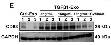
Application: WB    Species: Mice    Sample: NRK-52E cells

Figure 2 TGF-β1 promotes the secretion of exosomes by renal tubular epithelial cells and activates fibroblasts in vitro. (A) Schematic diagram of experimental process. Exosomes from NRK-52E cells treated without (Ctrl-Exos) or with TGF-β1 (TGFβ1-Exos) were extracted and incubated with NRK-49F cells. (B,C) DLS and NTA of exosomes from NRK-52E cells. (D) TEM image of exosomes isolated from NRK-52E cells. Scale bar = 100 nm. (E, F) Representative western blot (E) and quantitative data (F) of CD63 as an exosome marker in exosomes from TGF-β1- or GW4869-treated NRK-52E cells. Numbers (1 to 3) indicate each independent treatment in the given group. *p < 0.05 versus Ctrl-Exos, #p < 0.05 versus 5 ng/ml TGFβ1-Exos, &p < 0.05 versus 15 ng/ml TGFβ1-Exos (n = 3). (G) Fluorescent staining image of PKH-67-labeled NRK-52E cells. Scales bars=50 μm. (H) Fluorescent staining image of NRK-52E cell-derived exosomes taken up by NRK-49F cells. Scales bars=10 μm. (I, K) Representative western blot (I) and quantitative data (K) of α-SMA and PCNA in NRK-49F cells incubated with exosomes from TGF-β1- or GW4869-treated NRK-52E cells. Numbers (1 to 3) indicate each independent treatment in the giving group. *p < 0.05 versus Ctrl-Exos, #p < 0.05 versus 5 ng/ml TGFβ1-Exos, &p < 0.05 versus 15 ng/ml TGFβ1-Exos (n = 3). (J) Proliferation rate of NRK-49F cells incubated with NRK-52E cell-derived exosomes measured by CCK-8. *p < 0.05 versus Ctrl-Exos, #p < 0.05 versus 5 ng/ml TGFβ1-Exos, &p < 0.05 versus 15 ng/mL TGFβ1-Exos (n = 3). (L-N) Double immunofluorescence staining (green for Col-I and red for fibronectin) demonstrates the expression of Col-I and fibronectin in NRK-49F cells incubated with NRK-52E cell-derived exosomes. Scales bars=50 μm. *p < 0.05 versus Ctrl-Exos, #p < 0.05 versus 5 ng/ml TGFβ1-Exos, &p < 0.05 versus 15 ng/mL TGFβ1-Exos.

4). Small extracellular vesicles encapsulating lefty1 mRNA inhibit hepatic fibrosis. Asian Journal of Pharmaceutical Sciences (PubMed: 36382306) [IF=10.2]

5). Tubular cell-derived exosomal miR-150-5p contributes to renal fibrosis following unilateral ischemia-reperfusion injury by activating fibroblast in vitro and in vivo. International Journal of Biological Sciences (PubMed: 34671216) [IF=9.2]

Application: IF/ICC    Species: Mice    Sample: kidney

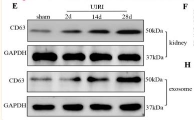
Figure 2 Increased secretion of exosomes in the kidney following UIRI. (A) Immunofluorescence staining for CD63 and TSG-101 (red). (B) Integrated optical density (IOD) of TSG-101. (C) Integrated optical density (IOD) of CD63. (D) Double immunofluorescence staining for CD63 (red) and AQP-1 (green), respectively. (E) Western blotting of CD63 in kidney tissue and exosome at different time points after UIRI (n=6). (F) Quantitative of CD63 in kidney. (G) Quantitative of CD63 in exosome. (H) TEM image of the exosomes in the kidney tissue after UIRI. *P < 0.05 versus sham.

Application: WB    Species: Mice    Sample: kidney

Figure 2 Increased secretion of exosomes in the kidney following UIRI. (A) Immunofluorescence staining for CD63 and TSG-101 (red). (B) Integrated optical density (IOD) of TSG-101. (C) Integrated optical density (IOD) of CD63. (D) Double immunofluorescence staining for CD63 (red) and AQP-1 (green), respectively. (E) Western blotting of CD63 in kidney tissue and exosome at different time points after UIRI (n=6). (F) Quantitative of CD63 in kidney. (G) Quantitative of CD63 in exosome. (H) TEM image of the exosomes in the kidney tissue after UIRI. *P < 0.05 versus sham.

6). MicroRNA-enriched small extracellular vesicles possess odonto-immunomodulatory properties for modulating the immune response of macrophages and promoting odontogenesis. Stem Cell Research & Therapy (PubMed: 33256846) [IF=7.5]

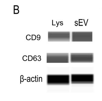
Application: WB    Species: Human    Sample: DPSCs-sEV

Fig. 1 Identification and characterization of DPSCs-sEV. a The morphology of DPSCs-sEV was determined by TEM, scale bar = 50 nm. b Expression of CD9 and CD63 in the DPSCs-sEV (sEV represents DPSCs-sEV and Lys represents DPSCs lysate, 尾-actin is a control for the lysate). c Nano-flow cytometry showed DPSCs-sEV ranged between 30 and 150 nm in diameter. d PKH26-labeled DPSCs-sEV were found in macrophage cytosol

7). Autophagy and Exosome Coordinately Enhance Macrophage M1 Polarization and Recruitment in Influenza A Virus Infection. Frontiers in Immunology (PubMed: 35371077) [IF=7.3]

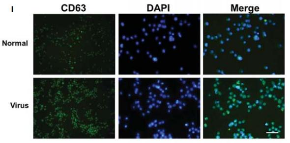
Application: IF/ICC    Species: mouse    Sample: lung

FIGURE 3 | M1 polarization and LC3/CD63/IL-1b analysis of infected ANA-1 macrophages. Total RNA was isolated from infected ANA-1 cells at 24-h post-infection and used for transcriptional analysis of the related genes.(I) CD63 cellular immunofluorescence

Application: WB    Species: mouse    Sample: macrophages

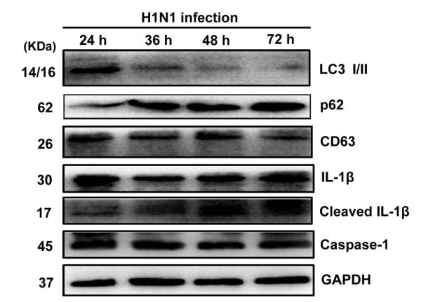
FIGURE s4 |The total proteins of infected primary peritoneal macrophages were extracted at 24, 36, 48, and 72 h post-infection and then subjected for Western blotting. The corresponding antibodies were used to analyze autophagic protein (LC3, p62), exosome marker (CD63), and IL-1β activation pathway (IL-1β, cleaved IL-1β, and caspase-1) normalized to GAPDH.

8). Focal ischemic stroke modifies microglia-derived exosomal miRNAs: potential role of mir-212-5p in neuronal protection and functional recovery. Biological Research (PubMed: 37789455) [IF=6.7]

Application: WB    Species: Rat    Sample:

Fig. 2 MiRNA sequencing of microglial exosomes 3 days after MCAO/R and qRT–PCR validation. A, B Characterisation of microglial exosomes using nanoparticle tracking analysis and transmission electron microscopy scanning. Scale bar = 200 nm. C The exosome markers CD9, CD63, and CD81 were detected using western blot analysis. D Microglia-derived exosomes were detected by western blot analysis using the microglia marker CD11b. E Heatmap showing the levels of miRNAs in microglial exosomes at 3 days after MCAO/R. F Expression of miR-30c-5p, miR-126a-5p, miR-128-3p, miR-212-5p and miR-1949 in the ischemic penumbra of the cortex at 3 days after MCAO/R was determined using qRT–PCR. G Expression levels of the miRNA-targeting genes PLXNA2, PTEN and FOXO3 in the ischemic penumbra of the cortex at 3 days after MCAO/R. H The target sites of miR-212-5p in PLXNA2 mRNA 3’ untranslated region (3’UTR). I Map of the pmirGLO luciferase reporter vector. J Dual luciferase assays revealed the binding of miR-212-5p to the 3’UTR of PLXNA2. The data are presented as the means ± SEM (n = 5 per group). *P 

9). Chemerin-Induced Down-Regulation of Placenta-Derived Exosomal miR-140-3p and miR-574-3p Promotes Umbilical Vein Endothelial Cells Proliferation, Migration, and Tube Formation in Gestational Diabetes Mellitus. Cells (PubMed: 36359855) [IF=6.0]

10). OSCC cell-secreted exosomal CMTM6 induced M2-like macrophages polarization via ERK1/2 signaling pathway. Cancer immunology, immunotherapy : CII (PubMed: 33104837) [IF=5.8]

Load more

Restrictive clause


Affinity Biosciences tests all products strictly. Citations are provided as a resource for additional applications that have not been validated by Affinity Biosciences. Please choose the appropriate format for each application and consult Materials and Methods sections for additional details about the use of any product in these publications.

For Research Use Only.
Not for use in diagnostic or therapeutic procedures. Not for resale. Not for distribution without written consent. Affinity Biosciences will not be held responsible for patent infringement or other violations that may occur with the use of our products. Affinity Biosciences, Affinity Biosciences Logo and all other trademarks are the property of Affinity Biosciences LTD.