Product: LHX1 Antibody
Catalog: DF4823
Description: Rabbit polyclonal antibody to LHX1
Application: WB IHC IF/ICC
Reactivity: Human, Mouse, Rat
Prediction: Pig, Zebrafish, Bovine, Horse, Sheep, Rabbit, Dog, Chicken, Xenopus
Mol.Wt.: 44 KD; 45kD(Calculated).
Uniprot: P48742
RRID: AB_2837188

View similar products>>

   Size Price Inventory
 100ul $280 In stock
 200ul $350 In stock

Lead Time: Same day delivery

For pricing and ordering contact:
Local distributors

Product Info

Source:
Rabbit
Application:
WB 1:500-1:1000, IHC 1:50-1:200, IF/ICC 1:100-1:500
*The optimal dilutions should be determined by the end user.
*Tips:

WB: For western blot detection of denatured protein samples. IHC: For immunohistochemical detection of paraffin sections (IHC-p) or frozen sections (IHC-f) of tissue samples. IF/ICC: For immunofluorescence detection of cell samples. ELISA(peptide): For ELISA detection of antigenic peptide.

Reactivity:
Human,Mouse,Rat
Prediction:
Pig(91%), Zebrafish(82%), Bovine(91%), Horse(91%), Sheep(91%), Rabbit(91%), Dog(91%), Chicken(91%), Xenopus(91%)
Clonality:
Polyclonal
Specificity:
LHX1 Antibody detects endogenous levels of total LHX1.
RRID:
AB_2837188
Cite Format: Affinity Biosciences Cat# DF4823, RRID:AB_2837188.
Conjugate:
Unconjugated.
Purification:
The antiserum was purified by peptide affinity chromatography using SulfoLink™ Coupling Resin (Thermo Fisher Scientific).
Storage:
Rabbit IgG in phosphate buffered saline , pH 7.4, 150mM NaCl, 0.02% sodium azide and 50% glycerol. Store at -20 °C. Stable for 12 months from date of receipt.
Alias:

Fold/Unfold

hLim-1; Homeo box protein Lim 1; Homeo box protein Lim1; Homeobox protein Lim 1; Homeobox protein Lim-1; Homeobox protein Lim1; LHX 1; LHX1; LHX1_HUMAN; LIM 1; LIM homeo box 1; LIM homeo box protein 1; LIM homeobox 1; LIM homeobox protein 1; LIM-1; LIM/homeobox protein Lhx 1; LIM/homeobox protein Lhx1; MGC126723; MGC138141;

Immunogens

Immunogen:
Uniprot:
Gene(ID):
Expression:
P48742 LHX1_HUMAN:

Expressed in the brain, thymus, and tonsils. Expressed in samples from patients with chronic myeloid leukemia (CML) and in 58% of acute myeloid leukemia (AML) cell lines.

Sequence:
MVHCAGCKRPILDRFLLNVLDRAWHVKCVQCCECKCNLTEKCFSREGKLYCKNDFFRCFGTKCAGCAQGISPSDLVRRARSKVFHLNCFTCMMCNKQLSTGEELYIIDENKFVCKEDYLSNSSVAKENSLHSATTGSDPSLSPDSQDPSQDDAKDSESANVSDKEAGSNENDDQNLGAKRRGPRTTIKAKQLETLKAAFAATPKPTRHIREQLAQETGLNMRVIQVWFQNRRSKERRMKQLSALGARRHAFFRSPRRMRPLVDRLEPGELIPNGPFSFYGDYQSEYYGPGGNYDFFPQGPPSSQAQTPVDLPFVPSSGPSGTPLGGLEHPLPGHHPSSEAQRFTDILAHPPGDSPSPEPSLPGPLHSMSAEVFGPSPPFSSLSVNGGASYGNHLSHPPEMNEAAVW

Predictions

Predictions:

Score>80(red) has high confidence and is suggested to be used for WB detection. *The prediction model is mainly based on the alignment of immunogen sequences, the results are for reference only, not as the basis of quality assurance.

Species
Results
Score
Pig
91
Horse
91
Bovine
91
Sheep
91
Dog
91
Xenopus
91
Chicken
91
Rabbit
91
Zebrafish
82
Model Confidence:
High(score>80) Medium(80>score>50) Low(score<50) No confidence

PTMs - P48742 As Substrate

Site PTM Type Enzyme
K48 Sumoylation
S71 Phosphorylation
S162 Phosphorylation

Research Backgrounds

Function:

Potential transcription factor. May play a role in early mesoderm formation and later in lateral mesoderm differentiation and neurogenesis.

Subcellular Location:

Nucleus.

Extracellular region or secreted Cytosol Plasma membrane Cytoskeleton Lysosome Endosome Peroxisome ER Golgi apparatus Nucleus Mitochondrion Manual annotation Automatic computational assertionSubcellular location
Tissue Specificity:

Expressed in the brain, thymus, and tonsils. Expressed in samples from patients with chronic myeloid leukemia (CML) and in 58% of acute myeloid leukemia (AML) cell lines.

Subunit Structure:

Interacts with LDB1 via the tandem LIM domains.

Family&Domains:

The LIM domains exert a negative regulatory function and disruption of the LIM domains produces an activated form. In addition, two activation domains and a negative regulatory domain exist C-terminally to the homeobox (By similarity).

References

1). BUB1 drives the occurrence and development of bladder cancer by mediating the STAT3 signaling pathway. JOURNAL OF EXPERIMENTAL & CLINICAL CANCER RESEARCH, 2021 (PubMed: 34852826) [IF=11.3]

2). Degradation of Different Molecular Weight Fucoidans and Their Inhibition of TGF-β1 Induced Epithelial-Mesenchymal Transition in Mouse Renal Tubular Epithelial Cells. International Journal of Biological Macromolecules, 2020 (PubMed: 32057857) [IF=7.7]

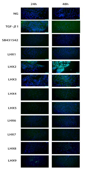
Application: IF/ICC    Species: Mouse    Sample: MTEC cells

Fig. 7. The result of cell immunofluorescence assay after LHXs and TGF-β1 treated MTEC for 24 and 48 h. Representative images (3 visual fields for each tissue analyzed) of immunolabeling for Fn and nuclear staining with DAPI. Scale bar, 50 ~μm.

3). Long non-coding RNA LHX1-DT regulates cardiomyocyte differentiation through H2A.Z-mediated LHX1 transcriptional activation. iScience, 2023 (PubMed: 37942009)

Application: WB    Species: Human    Sample:

Figure 4 LHX1-DT enhances LHX1 expression to promote cardiac differentiation (A) Schematic representation of the LHX1-DT and LHX1 gene locus and their sequence conservation analysis from UCSC genome browser. Red and blue arrows indicate the direction of transcription of LHX1-DT and LHX1 respectively. (B) Genome browser track of the selected genomic regions around LHX1-DT and LHX1. The gray shadow rectangle showing the expression levels and ATAC signals of LHX1-DT and LHX1 at the four stages of cardiac development. (C) qRT-PCR analysis of LHX1-DT and LHX1 expression during cardiac differentiation, n = 4. (D) Localization of LHX1-DT and LHX1 in mesodermal cells derived from hESCs by FISH study. Scale bars, 20 μm, n = 4. (E) Western blot analysis of LHX1 in undifferentiated and in mesoderm differentiating cells with LHX1-DT knockdown. ∗∗p < 0.01, n = 3. (F) qRT-PCR analysis of LHX1 expression in differentiating hESCs 2 days following siRNA transfection of cardiac differentiation. ∗∗∗p < 0.001, n = 4. (G and H) qRT-PCR analysis of the expression levels of LHX1-DT and mesoderm markers (T and Mesp1) on Day 2 and cardiomyocyte markers (cTnT, α-actinin, NKX2.5, and MYL7) on Day 10 in LHX1-DT depleted hESCs. ∗∗∗p < 0.001, n = 4. (I) Immunostaining of cTnT (green) and α-actinin (red) in LHX1 knockdown and siNC cells on Day 10 of CM differentiation. Scale bar, 50 μm, n = 4. (J) Representative flow cytometry analysis of the percentage of cTnT-positive cardiomyocytes between siLHX1 and siNC group. ∗∗∗p < 0.001, n = 3. (K) qRT-PCR analysis of cardiac marker genes expression on Day 10 in LHX1-DT+/+, LHX1-DT−/−, and LHX1-DT−/− + LHX1 cells. ∗p < 0.05, ∗∗p < 0.01, ∗∗∗p < 0.001 vs. LHX1-DT+/+, n = 4. ##p < 0.01, ###p < 0.001 vs. LHX1-DT−/−, n = 4. (L) Immunostaining of cTnT (green) and α-actinin (red) in LHX1-DT+/+, LHX1-DT−/−, and LHX1-DT−/− + LHX1 cells on Day 10 of CM differentiation. Scale bar, 50 μm. Scale bars, 50 μm, n = 4. (M) Representative flow cytometry analysis of the percentage of cTnT-positive cells in LHX1-DT+/+, LHX1-DT−/−, and LHX1-DT−/− + LHX1 cardiomyocytes. ∗∗∗p < 0.001 vs. LHX1-DT+/+, ###p < 0.001 vs. LHX1-DT−/−, n = 3. Data are presented as mean ± SEM.

Application: IF/ICC    Species: Human    Sample:

Figure 4 LHX1-DT enhances LHX1 expression to promote cardiac differentiation (A) Schematic representation of the LHX1-DT and LHX1 gene locus and their sequence conservation analysis from UCSC genome browser. Red and blue arrows indicate the direction of transcription of LHX1-DT and LHX1 respectively. (B) Genome browser track of the selected genomic regions around LHX1-DT and LHX1. The gray shadow rectangle showing the expression levels and ATAC signals of LHX1-DT and LHX1 at the four stages of cardiac development. (C) qRT-PCR analysis of LHX1-DT and LHX1 expression during cardiac differentiation, n = 4. (D) Localization of LHX1-DT and LHX1 in mesodermal cells derived from hESCs by FISH study. Scale bars, 20 μm, n = 4. (E) Western blot analysis of LHX1 in undifferentiated and in mesoderm differentiating cells with LHX1-DT knockdown. ∗∗p < 0.01, n = 3. (F) qRT-PCR analysis of LHX1 expression in differentiating hESCs 2 days following siRNA transfection of cardiac differentiation. ∗∗∗p < 0.001, n = 4. (G and H) qRT-PCR analysis of the expression levels of LHX1-DT and mesoderm markers (T and Mesp1) on Day 2 and cardiomyocyte markers (cTnT, α-actinin, NKX2.5, and MYL7) on Day 10 in LHX1-DT depleted hESCs. ∗∗∗p < 0.001, n = 4. (I) Immunostaining of cTnT (green) and α-actinin (red) in LHX1 knockdown and siNC cells on Day 10 of CM differentiation. Scale bar, 50 μm, n = 4. (J) Representative flow cytometry analysis of the percentage of cTnT-positive cardiomyocytes between siLHX1 and siNC group. ∗∗∗p < 0.001, n = 3. (K) qRT-PCR analysis of cardiac marker genes expression on Day 10 in LHX1-DT+/+, LHX1-DT−/−, and LHX1-DT−/− + LHX1 cells. ∗p < 0.05, ∗∗p < 0.01, ∗∗∗p < 0.001 vs. LHX1-DT+/+, n = 4. ##p < 0.01, ###p < 0.001 vs. LHX1-DT−/−, n = 4. (L) Immunostaining of cTnT (green) and α-actinin (red) in LHX1-DT+/+, LHX1-DT−/−, and LHX1-DT−/− + LHX1 cells on Day 10 of CM differentiation. Scale bar, 50 μm. Scale bars, 50 μm, n = 4. (M) Representative flow cytometry analysis of the percentage of cTnT-positive cells in LHX1-DT+/+, LHX1-DT−/−, and LHX1-DT−/− + LHX1 cardiomyocytes. ∗∗∗p < 0.001 vs. LHX1-DT+/+, ###p < 0.001 vs. LHX1-DT−/−, n = 3. Data are presented as mean ± SEM.

Restrictive clause


Affinity Biosciences tests all products strictly. Citations are provided as a resource for additional applications that have not been validated by Affinity Biosciences. Please choose the appropriate format for each application and consult Materials and Methods sections for additional details about the use of any product in these publications.

For Research Use Only.
Not for use in diagnostic or therapeutic procedures. Not for resale. Not for distribution without written consent. Affinity Biosciences will not be held responsible for patent infringement or other violations that may occur with the use of our products. Affinity Biosciences, Affinity Biosciences Logo and all other trademarks are the property of Affinity Biosciences LTD.