CAMKK2 Antibody - #DF4793
| Product: | CAMKK2 Antibody |
| Catalog: | DF4793 |
| Description: | Rabbit polyclonal antibody to CAMKK2 |
| Application: | WB IHC IF/ICC |
| Cited expt.: | WB |
| Reactivity: | Human, Mouse, Rat |
| Prediction: | Pig, Bovine, Horse, Sheep, Rabbit, Dog, Chicken |
| Mol.Wt.: | 65 KD; 65kD(Calculated). |
| Uniprot: | Q96RR4 |
| RRID: | AB_2837147 |
Product Info
*The optimal dilutions should be determined by the end user. For optimal experimental results, antibody reuse is not recommended.
*Tips:
WB: For western blot detection of denatured protein samples. IHC: For immunohistochemical detection of paraffin sections (IHC-p) or frozen sections (IHC-f) of tissue samples. IF/ICC: For immunofluorescence detection of cell samples. ELISA(peptide): For ELISA detection of antigenic peptide.
Cite Format: Affinity Biosciences Cat# DF4793, RRID:AB_2837147.
Fold/Unfold
Calcium/calmodulin dependent protein kinase beta; Calcium/calmodulin dependent protein kinase kinase 2; Calcium/calmodulin dependent protein kinase kinase 2 beta; Calcium/calmodulin dependent protein kinase kinase beta; Calcium/calmodulin-dependent protein kinase kinase 2; Calcium/calmodulin-dependent protein kinase kinase beta; CaM kinase kinase beta; CaM KK beta; CaM-kinase kinase 2; CaM-kinase kinase beta; CaM-KK 2; CaM-KK beta; CaMKK 2; CAMKK; CaMKK beta; CAMKK beta protein; Camkk2; CAMKKB; KKCC2_HUMAN;
Immunogens
A synthesized peptide derived from human CAMKK2, corresponding to a region within C-terminal amino acids.
Ubiquitously expressed with higher levels in the brain. Intermediate levels are detected in spleen, prostate, thyroid and leukocytes. The lowest level is in lung.
- Q96RR4 KKCC2_HUMAN:
- Protein BLAST With
- NCBI/
- ExPASy/
- Uniprot
MSSCVSSQPSSNRAAPQDELGGRGSSSSESQKPCEALRGLSSLSIHLGMESFIVVTECEPGCAVDLGLARDRPLEADGQEVPLDTSGSQARPHLSGRKLSLQERSQGGLAAGGSLDMNGRCICPSLPYSPVSSPQSSPRLPRRPTVESHHVSITGMQDCVQLNQYTLKDEIGKGSYGVVKLAYNENDNTYYAMKVLSKKKLIRQAGFPRRPPPRGTRPAPGGCIQPRGPIEQVYQEIAILKKLDHPNVVKLVEVLDDPNEDHLYMVFELVNQGPVMEVPTLKPLSEDQARFYFQDLIKGIEYLHYQKIIHRDIKPSNLLVGEDGHIKIADFGVSNEFKGSDALLSNTVGTPAFMAPESLSETRKIFSGKALDVWAMGVTLYCFVFGQCPFMDERIMCLHSKIKSQALEFPDQPDIAEDLKDLITRMLDKNPESRIVVPEIKLHPWVTRHGAEPLPSEDENCTLVEVTEEEVENSVKHIPSLATVILVKTMIRKRSFGNPFEGSRREERSLSAPGNLLTKKPTRECESLSELKEARQRRQPPGHRPAPRGGGGSALVRGSPCVESCWAPAPGSPARMHPLRPEEAMEPE
Predictions
Score>80(red) has high confidence and is suggested to be used for WB detection. *The prediction model is mainly based on the alignment of immunogen sequences, the results are for reference only, not as the basis of quality assurance.
High(score>80) Medium(80>score>50) Low(score<50) No confidence
Research Backgrounds
Calcium/calmodulin-dependent protein kinase belonging to a proposed calcium-triggered signaling cascade involved in a number of cellular processes. Isoform 1, isoform 2 and isoform 3 phosphorylate CAMK1 and CAMK4. Isoform 3 phosphorylates CAMK1D. Isoform 4, isoform 5 and isoform 6 lacking part of the calmodulin-binding domain are inactive. Efficiently phosphorylates 5'-AMP-activated protein kinase (AMPK) trimer, including that consisting of PRKAA1, PRKAB1 and PRKAG1. This phosphorylation is stimulated in response to Ca(2+) signals (By similarity). Seems to be involved in hippocampal activation of CREB1 (By similarity). May play a role in neurite growth. Isoform 3 may promote neurite elongation, while isoform 1 may promoter neurite branching.
Autophosphorylated and phosphorylated by PKA. Each isoform may show a different pattern of phosphorylation.
Nucleus. Cytoplasm. Cell projection>Neuron projection.
Note: Predominantly nuclear in unstimulated cells, relocalizes into cytoplasm and neurites after forskolin induction.
Ubiquitously expressed with higher levels in the brain. Intermediate levels are detected in spleen, prostate, thyroid and leukocytes. The lowest level is in lung.
The autoinhibitory domain overlaps with the calmodulin binding region and may be involved in intrasteric autoinhibition.
The RP domain (arginine/proline-rich) is involved in the recognition of CAMKI and CAMK4 as substrates.
Belongs to the protein kinase superfamily. Ser/Thr protein kinase family.
Research Fields
· Cellular Processes > Transport and catabolism > Autophagy - animal. (View pathway)
· Environmental Information Processing > Signal transduction > AMPK signaling pathway. (View pathway)
· Human Diseases > Substance dependence > Alcoholism.
· Organismal Systems > Aging > Longevity regulating pathway. (View pathway)
· Organismal Systems > Endocrine system > Adipocytokine signaling pathway.
· Organismal Systems > Endocrine system > Oxytocin signaling pathway.
References
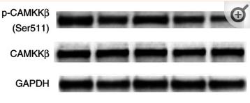
Application: WB Species: Mice Sample: CRC cells
Application: WB Species: Human Sample: HepG2 cells
Application: WB Species: Human Sample: L-02 cells
Application: WB Species: mouse Sample: livers
Application: WB Species: Rat Sample: myocardial tissue
Restrictive clause
Affinity Biosciences tests all products strictly. Citations are provided as a resource for additional applications that have not been validated by Affinity Biosciences. Please choose the appropriate format for each application and consult Materials and Methods sections for additional details about the use of any product in these publications.
For Research Use Only.
Not for use in diagnostic or therapeutic procedures. Not for resale. Not for distribution without written consent. Affinity Biosciences will not be held responsible for patent infringement or other violations that may occur with the use of our products. Affinity Biosciences, Affinity Biosciences Logo and all other trademarks are the property of Affinity Biosciences LTD.


![Fig. 7
L-theanine regulates hepatocyte lipid metabolic pathways via the CaMKKβ-AMPK signaling pathway. HepG2 cells were treated with 500 μM OA for 24 h with or without pretreated L-theanine for 2 h. A Western blot analysis showing protein expression of p-CaMKKβ and p-AMPK in HepG2 cells. HepG2 cells were pretreated with 10 μM STO-609 for 2 h, then added L-theanine co-incubated for 2 h, finally, OA were added co-incubated for 24 h. B Abrogating effect of CaMKKβ inhibitor on L-theanine-facilitated phosphorylation of CaMKKβ and AMPKα in HepG2 hepatocytes. HepG2 cells were pretreated with 10 μM BAPTA-AM for 1 h, then added L-theanine co-incubated for 2 h, finally, OA were added co-incubated for 24 h. C Intracellular Ca2+ levels were assessed in HepG2 cells with Fluo-4 AM using a fluorescence microscope. Scale bar: 20 μm (20 ×). D [Ca2+]i was detected by a multifunctional microplate reader using Fluo-4 AM. under the condition of 488 nm excitation wavelength and 520 nm emission wavelength. E Western blots of indicated proteins in HepG2 cells treated with an intracellular Ca2+ chelator BAPTA-AM. Band intensity was quantified by densitometry analysis. Values are expressed as mean ± SEM of three independent experiments. *p < 0.05, **p < 0.01, ***p < 0.001, versus control group; #p < 0.05, ##p < 0.01, versus OA group; &p < 0.05 versus OA + STO-609 group, $p < 0.05 versus OA + BAPTA-AM group. n.s.: not significant (p > 0.05) versus control group CAMKK2 Antibody - Fig.](http://img.affbiotech.cn/images/cited_image/202212/cite-wx-50-1669972653.jpg)
