Product: MRC1/CD206 Antibody
Catalog: DF4149
Description: Rabbit polyclonal antibody to MRC1/CD206
Application: WB IHC IF/ICC
Reactivity: Human, Mouse, Rat
Prediction: Pig, Bovine, Horse, Sheep, Rabbit, Dog
Mol.Wt.: 166 KD; 166kD(Calculated).
Uniprot: P22897
RRID: AB_2836514

View similar products>>

   Size Price Inventory
 100ul $280 In stock
 200ul $350 In stock

Lead Time: Same day delivery

For pricing and ordering contact:
Local distributors

Product Info

Source:
Rabbit
Application:
WB 1:500-1:1000, IHC 1:50-1:100, IF/ICC 1:100-1:500
*The optimal dilutions should be determined by the end user.
*Tips:

WB: For western blot detection of denatured protein samples. IHC: For immunohistochemical detection of paraffin sections (IHC-p) or frozen sections (IHC-f) of tissue samples. IF/ICC: For immunofluorescence detection of cell samples. ELISA(peptide): For ELISA detection of antigenic peptide.

Reactivity:
Human,Mouse,Rat
Prediction:
Pig(91%), Bovine(91%), Horse(100%), Sheep(91%), Rabbit(91%), Dog(91%)
Clonality:
Polyclonal
Specificity:
MRC1 Antibody detects endogenous levels of total MRC1.
RRID:
AB_2836514
Cite Format: Affinity Biosciences Cat# DF4149, RRID:AB_2836514.
Conjugate:
Unconjugated.
Purification:
The antiserum was purified by peptide affinity chromatography using SulfoLink™ Coupling Resin (Thermo Fisher Scientific).
Storage:
Rabbit IgG in phosphate buffered saline , pH 7.4, 150mM NaCl, 0.02% sodium azide and 50% glycerol. Store at -20 °C. Stable for 12 months from date of receipt.
Alias:

Fold/Unfold

bA541I19.1; C type lectin domain family 13 member D; C-type lectin domain family 13 member D; CD 206; CD206; CD206 antigen; CLEC13D; CLEC13DL; Macrophage mannose receptor 1; Macrophage mannose receptor 1 like protein 1; Macrophage mannose receptor; Mannose receptor C type 1; Mannose receptor C type 1 like 1; MMR; MRC 1; MRC1; MRC1_HUMAN; MRC1L1; OTTHUMP00000045206;

Immunogens

Immunogen:
Uniprot:
Gene(ID):
Sequence:
MRLPLLLVFASVIPGAVLLLDTRQFLIYNEDHKRCVDAVSPSAVQTAACNQDAESQKFRWVSESQIMSVAFKLCLGVPSKTDWVAITLYACDSKSEFQKWECKNDTLLGIKGEDLFFNYGNRQEKNIMLYKGSGLWSRWKIYGTTDNLCSRGYEAMYTLLGNANGATCAFPFKFENKWYADCTSAGRSDGWLWCGTTTDYDTDKLFGYCPLKFEGSESLWNKDPLTSVSYQINSKSALTWHQARKSCQQQNAELLSITEIHEQTYLTGLTSSLTSGLWIGLNSLSFNSGWQWSDRSPFRYLNWLPGSPSAEPGKSCVSLNPGKNAKWENLECVQKLGYICKKGNTTLNSFVIPSESDVPTHCPSQWWPYAGHCYKIHRDEKKIQRDALTTCRKEGGDLTSIHTIEELDFIISQLGYEPNDELWIGLNDIKIQMYFEWSDGTPVTFTKWLRGEPSHENNRQEDCVVMKGKDGYWADRGCEWPLGYICKMKSRSQGPEIVEVEKGCRKGWKKHHFYCYMIGHTLSTFAEANQTCNNENAYLTTIEDRYEQAFLTSFVGLRPEKYFWTGLSDIQTKGTFQWTIEEEVRFTHWNSDMPGRKPGCVAMRTGIAGGLWDVLKCDEKAKFVCKHWAEGVTHPPKPTTTPEPKCPEDWGASSRTSLCFKLYAKGKHEKKTWFESRDFCRALGGDLASINNKEEQQTIWRLITASGSYHKLFWLGLTYGSPSEGFTWSDGSPVSYENWAYGEPNNYQNVEYCGELKGDPTMSWNDINCEHLNNWICQIQKGQTPKPEPTPAPQDNPPVTEDGWVIYKDYQYYFSKEKETMDNARAFCKRNFGDLVSIQSESEKKFLWKYVNRNDAQSAYFIGLLISLDKKFAWMDGSKVDYVSWATGEPNFANEDENCVTMYSNSGFWNDINCGYPNAFICQRHNSSINATTVMPTMPSVPSGCKEGWNFYSNKCFKIFGFMEEERKNWQEARKACIGFGGNLVSIQNEKEQAFLTYHMKDSTFSAWTGLNDVNSEHTFLWTDGRGVHYTNWGKGYPGGRRSSLSYEDADCVVIIGGASNEAGKWMDDTCDSKRGYICQTRSDPSLTNPPATIQTDGFVKYGKSSYSLMRQKFQWHEAETYCKLHNSLIASILDPYSNAFAWLQMETSNERVWIALNSNLTDNQYTWTDKWRVRYTNWAADEPKLKSACVYLDLDGYWKTAHCNESFYFLCKRSDEIPATEPPQLPGRCPESDHTAWIPFHGHCYYIESSYTRNWGQASLECLRMGSSLVSIESAAESSFLSYRVEPLKSKTNFWIGLFRNVEGTWLWINNSPVSFVNWNTGDPSGERNDCVALHASSGFWSNIHCSSYKGYICKRPKIIDAKPTHELLTTKADTRKMDPSKPSSNVAGVVIIVILLILTGAGLAAYFFYKKRRVHLPQEGAFENTLYFNSQSSPGTSDMKDLVGNIEQNEHSVI

Predictions

Predictions:

Score>80(red) has high confidence and is suggested to be used for WB detection. *The prediction model is mainly based on the alignment of immunogen sequences, the results are for reference only, not as the basis of quality assurance.

Species
Results
Score
Horse
100
Pig
91
Bovine
91
Sheep
91
Dog
91
Rabbit
91
Xenopus
0
Zebrafish
0
Chicken
0
Model Confidence:
High(score>80) Medium(80>score>50) Low(score<50) No confidence

PTMs - P22897 As Substrate

Site PTM Type Enzyme
S62 Phosphorylation
S64 Phosphorylation
Y200 Phosphorylation
T389 Phosphorylation
T390 Phosphorylation
T633 Phosphorylation
K670 Acetylation
T820 Phosphorylation
Y850 Phosphorylation
Y860 Phosphorylation
T1081 Phosphorylation
S1128 Phosphorylation
N1205 N-Glycosylation
S1268 Phosphorylation
S1279 Phosphorylation
S1280 Phosphorylation
S1454 Phosphorylation

Research Backgrounds

Function:

Mediates the endocytosis of glycoproteins by macrophages. Binds both sulfated and non-sulfated polysaccharide chains.

(Microbial infection) Acts as phagocytic receptor for bacteria, fungi and other pathogens.

(Microbial infection) Acts as a receptor for Dengue virus envelope protein E.

(Microbial infection) Interacts with Hepatitis B virus envelope protein.

Subcellular Location:

Endosome membrane>Single-pass type I membrane protein. Cell membrane>Single-pass type I membrane protein.

Extracellular region or secreted Cytosol Plasma membrane Cytoskeleton Lysosome Endosome Peroxisome ER Golgi apparatus Nucleus Mitochondrion Manual annotation Automatic computational assertionSubcellular location
Subunit Structure:

(Microbial infection) Interacts with Dengue virus.

(Microbial infection) May act as a receptor for hepatitis B virus, enabling uptake of the virus in hepatic dendritic cells.

Research Fields

· Cellular Processes > Transport and catabolism > Phagosome.   (View pathway)

· Human Diseases > Infectious diseases: Bacterial > Tuberculosis.

References

1). Plasma Membrane-Derived Biomimetic Apoptotic Nanovesicles Targeting Inflammation and Cartilage Degeneration for Osteoarthritis. Small methods, 2024 (PubMed: 39036830) [IF=12.4]

2). Honokiol@ PF127 crosslinked hyaluronate-based hydrogel for promoting wound healing by regulating macrophage polarization. Carbohydrate Polymers, 2023 (PubMed: 36657865) [IF=11.2]

3). Dually Crosslinked Copper‐Poly (Tannic Acid) Nanoparticles with Microenvironment‐Responsiveness for Infected Wound Treatment. Advanced Healthcare Materials, 2023 (PubMed: 36842067) [IF=10.0]

4). Soft apoptotic-cell-inspired nanoparticles persistently bind to macrophage membranes and promote anti-inflammatory and pro-healing effects. Acta Biomaterialia, 2021 (PubMed: 34245890) [IF=9.7]

5). Reprogramming of TAMs via the STAT3/CD47-SIRPα axis promotes acquired resistance to EGFR-TKIs in lung cancer. Cancer Letters, 2023 (PubMed: 37146936) [IF=9.7]

6). Oroxin A alleviates early brain injury after subarachnoid hemorrhage by regulating ferroptosis and neuroinflammation. Journal of neuroinflammation, 2024 (PubMed: 38702778) [IF=9.3]

Application: WB    Species: Mouse    Sample: hippocampal tissues

Fig. 5 OA treatment inhibited microglial activation and ameliorated neuroinflammation following SAH. A: qPCR assay indicating the mRNA expression levels of TNF-a, IL-6, IL-1β, and IL-10 in hippocampal tissues following SAH B: ELISA analysis indicates the protein secretion levels of TNF-a, IL-6, IL-1β, and IL-10 in hippocampal tissues following SAH. C: Immunofluorescence staining for Iba-1 in hippocampal tissues following surgery/anesthesia. D: qPCR assay indicating the mRNA expression levels of CD-32 and CD-206 in hippocampal tissues following SAH. E: Representative Western blot bands (as normalized to GAPDH) illustrate the protein levels of CD 32 and CD206 in hippocampal tissues following SAH. The data are summarized as mean ± SD (n = 3). Scale bar = 20 μm

7). Oesophageal squamous cell carcinoma–associated IL‐33 rewires macrophage polarization towards M2 via activating ornithine decarboxylase. CELL PROLIFERATION, 2021 (PubMed: 33305406) [IF=8.5]

Application: IHC    Species: human    Sample: non-tumour and tumour

FIGURE 1|M2 macrophage infiltration and IL-33 production are enhanced with close correlation in oesophageal squamous cell carcinoma (ESCC). A, Representative images of IL-33+ cell, CD206+ cell and CD68+ cell in non-tumour and tumour tissue (scale bar = 50 μm).

Application: WB    Species: human    Sample: non-tumour and tumour

FIGURE 1|M2 macrophage infiltration and IL-33 production are enhanced with close correlation in oesophageal squamous cell carcinoma (ESCC). A, Representative images of IL-33+ cell, CD206+ cell and CD68+ cell in non-tumour and tumour tissue (scale bar = 50 μm).B, Western blot analysis of IL-33 and CD206 expression in non-tumour and tumour tissues, and GAPDH was used as a reference control.

8). Facile preparation of polyphenol-crosslinked chitosan-based hydrogels for cutaneous wound repair. International Journal of Biological Macromolecules, 2023 (PubMed: 36565830) [IF=8.2]

9). Role of transient receptor potential ankyrin 1 in idiopathic pulmonary fibrosis: modulation of M2 macrophage polarization. Cellular and molecular life sciences : CMLS, 2024 (PubMed: 38635081) [IF=8.0]

Application: IHC    Species: Mouse    Sample:

Fig. 6 Inhibition of TRPA1 diminishes macrophage polarization to the M2 phenotype by disrupting the phosphorylation of the TGF-β1-suppressor of Smad2 pathway. A Western blot analysis of TGF-β1-Smad2 pathway activation and the expression of macrophage polarization markers CD206, IL-4, IL-10, and IL-13. B Immunohistochemical staining to detect the expression of CD206, IL-4, IL-10, and IL-13 proteins. C Schematic representation of cellular localization and quantification of CD206, IL-4, IL-10, and IL-13. Statistical significance was denoted as *P (Bleomycin group vs. Control group, *P 

Application: WB    Species: Mouse    Sample:

Fig. 6 Inhibition of TRPA1 diminishes macrophage polarization to the M2 phenotype by disrupting the phosphorylation of the TGF-β1-suppressor of Smad2 pathway. A Western blot analysis of TGF-β1-Smad2 pathway activation and the expression of macrophage polarization markers CD206, IL-4, IL-10, and IL-13. B Immunohistochemical staining to detect the expression of CD206, IL-4, IL-10, and IL-13 proteins. C Schematic representation of cellular localization and quantification of CD206, IL-4, IL-10, and IL-13. Statistical significance was denoted as *P (Bleomycin group vs. Control group, *P 

10). Huoluo Xiaoling Pellet promotes microglia M2 polarization through increasing MCPIP1 expression for ischemia stroke alleviation. Biomedicine & Pharmacotherapy, 2023 (PubMed: 37236023) [IF=7.5]

Application: IF/ICC    Species: Rat    Sample:

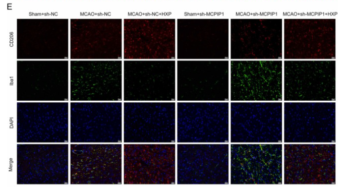
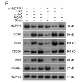
Fig. 3. HXP reduced neuroinflammation in MCAO rats by promoting MCPIP1-mediated microglia M2 polarization. A. After reperfusion for 7 d, brain water contents were examined. B. Brain infract volume was detected by 2,3,5-Triphentltetrazolium chloride staining. C. Changes in IL-1β, IL-6, iNOS, and TNF-α were measured via ELISA assy. D. Representative images of histopathological analysis. Scale bar, 20 µm. E. Representative immunofluorescence staining for CD206 (red) and Iba1 (green) in brain tissues. Scale bar, 20 µm. F. The expression of MCPIP1, CD16, iNOS, CD206, Arg1, and PPARγ was detected using Western blotting assay. Data are represented as mean ± standard deviation (N = 5). * * P 

Application: WB    Species: Rat    Sample:

Fig. 3. HXP reduced neuroinflammation in MCAO rats by promoting MCPIP1-mediated microglia M2 polarization. A. After reperfusion for 7 d, brain water contents were examined. B. Brain infract volume was detected by 2,3,5-Triphentltetrazolium chloride staining. C. Changes in IL-1β, IL-6, iNOS, and TNF-α were measured via ELISA assy. D. Representative images of histopathological analysis. Scale bar, 20 µm. E. Representative immunofluorescence staining for CD206 (red) and Iba1 (green) in brain tissues. Scale bar, 20 µm. F. The expression of MCPIP1, CD16, iNOS, CD206, Arg1, and PPARγ was detected using Western blotting assay. Data are represented as mean ± standard deviation (N = 5). * * P 

Load more

Restrictive clause


Affinity Biosciences tests all products strictly. Citations are provided as a resource for additional applications that have not been validated by Affinity Biosciences. Please choose the appropriate format for each application and consult Materials and Methods sections for additional details about the use of any product in these publications.

For Research Use Only.
Not for use in diagnostic or therapeutic procedures. Not for resale. Not for distribution without written consent. Affinity Biosciences will not be held responsible for patent infringement or other violations that may occur with the use of our products. Affinity Biosciences, Affinity Biosciences Logo and all other trademarks are the property of Affinity Biosciences LTD.