FOLR1 Antibody - #DF4058
| Product: | FOLR1 Antibody |
| Catalog: | DF4058 |
| Description: | Rabbit polyclonal antibody to FOLR1 |
| Application: | WB IHC IF/ICC |
| Cited expt.: | WB, IF/ICC |
| Reactivity: | Human, Mouse, Rat |
| Prediction: | Pig, Bovine, Horse, Sheep, Rabbit, Dog, Xenopus |
| Mol.Wt.: | 34 KD; 30kD(Calculated). |
| Uniprot: | P15328 |
| RRID: | AB_2836432 |
Related Downloads
Protocols
Product Info
*The optimal dilutions should be determined by the end user. For optimal experimental results, antibody reuse is not recommended.
*Tips:
WB: For western blot detection of denatured protein samples. IHC: For immunohistochemical detection of paraffin sections (IHC-p) or frozen sections (IHC-f) of tissue samples. IF/ICC: For immunofluorescence detection of cell samples. ELISA(peptide): For ELISA detection of antigenic peptide.
Cite Format: Affinity Biosciences Cat# DF4058, RRID:AB_2836432.
Fold/Unfold
adult; Adult folate binding protein; Adult folate-binding protein; FBP; Folate Receptor 1 Adult; Folate receptor 1; Folate Receptor 1 Precursor; Folate receptor adult; Folate receptor alpha; Folate receptor; FOLR; FOLR1; FOLR1_HUMAN; FR alpha; FR-alpha; FRalpha; KB cells FBP; MOV18; Ovarian cancer associated antigen; Ovarian tumor associated antigen; Ovarian tumor associated antigen MOv18; Ovarian tumor-associated antigen MOv18;
Immunogens
A synthesized peptide derived from human FOLR1, corresponding to a region within the internal amino acids.
Primarily expressed in tissues of epithelial origin. Expression is increased in malignant tissues. Expressed in kidney, lung and cerebellum. Detected in placenta and thymus epithelium.
- P15328 FOLR1_HUMAN:
- Protein BLAST With
- NCBI/
- ExPASy/
- Uniprot
MAQRMTTQLLLLLVWVAVVGEAQTRIAWARTELLNVCMNAKHHKEKPGPEDKLHEQCRPWRKNACCSTNTSQEAHKDVSYLYRFNWNHCGEMAPACKRHFIQDTCLYECSPNLGPWIQQVDQSWRKERVLNVPLCKEDCEQWWEDCRTSYTCKSNWHKGWNWTSGFNKCAVGAACQPFHFYFPTPTVLCNEIWTHSYKVSNYSRGSGRCIQMWFDPAQGNPNEEVARFYAAAMSGAGPWAAWPFLLSLALMLLWLLS
Predictions
Score>80(red) has high confidence and is suggested to be used for WB detection. *The prediction model is mainly based on the alignment of immunogen sequences, the results are for reference only, not as the basis of quality assurance.
High(score>80) Medium(80>score>50) Low(score<50) No confidence
Research Backgrounds
Binds to folate and reduced folic acid derivatives and mediates delivery of 5-methyltetrahydrofolate and folate analogs into the interior of cells. Has high affinity for folate and folic acid analogs at neutral pH. Exposure to slightly acidic pH after receptor endocytosis triggers a conformation change that strongly reduces its affinity for folates and mediates their release. Required for normal embryonic development and normal cell proliferation.
The secreted form is derived from the membrane-bound form either by cleavage of the GPI anchor, or/and by proteolysis catalyzed by a metalloprotease.
Cell membrane>Lipid-anchor. Secreted. Cytoplasmic vesicle. Cytoplasmic vesicle>Clathrin-coated vesicle. Endosome. Apical cell membrane.
Note: Endocytosed into cytoplasmic vesicles and then recycled to the cell membrane.
Primarily expressed in tissues of epithelial origin. Expression is increased in malignant tissues. Expressed in kidney, lung and cerebellum. Detected in placenta and thymus epithelium.
Belongs to the folate receptor family.
Research Fields
· Cellular Processes > Transport and catabolism > Endocytosis. (View pathway)
· Human Diseases > Drug resistance: Antineoplastic > Antifolate resistance.
References
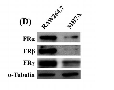
Application: WB Species: Mouse Sample: Raw264.7 and MH7A cells
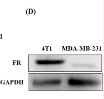
Application: WB Species: Human Sample: 4T1 cells and MDA-MB-231 cells
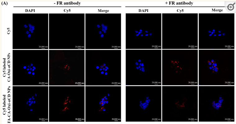
Application: IF/ICC Species: Human Sample: 4T1 cells and MDA-MB-231 cells
Restrictive clause
Affinity Biosciences tests all products strictly. Citations are provided as a resource for additional applications that have not been validated by Affinity Biosciences. Please choose the appropriate format for each application and consult Materials and Methods sections for additional details about the use of any product in these publications.
For Research Use Only.
Not for use in diagnostic or therapeutic procedures. Not for resale. Not for distribution without written consent. Affinity Biosciences will not be held responsible for patent infringement or other violations that may occur with the use of our products. Affinity Biosciences, Affinity Biosciences Logo and all other trademarks are the property of Affinity Biosciences LTD.


