, using ENTPD1/CD39 Antibody at 1/1000 dilution.
5ug/NC membrane strip.
Exposure for 10s with Affinity™ ECL Kit(#KF8001).
Bands result from membrane strip incubation.")
Control Products

Related Downloads
Protocols
Product Info
*The optimal dilutions should be determined by the end user. For optimal experimental results, antibody reuse is not recommended.
*Tips:
WB: For western blot detection of denatured protein samples. IHC: For immunohistochemical detection of paraffin sections (IHC-p) or frozen sections (IHC-f) of tissue samples. IF/ICC: For immunofluorescence detection of cell samples. ELISA(peptide): For ELISA detection of antigenic peptide.
Cite Format: Affinity Biosciences Cat# DF4031, RRID:AB_2836391.
Fold/Unfold
ATPDase; CD 39; CD39; CD39 antigen; DKFZp686D194; DKFZp686I093; Ecto apyrase; Ecto ATP diphosphohydrolase; Ecto-apyrase; Ecto-ATP diphosphohydrolase 1; Ecto-ATPase 1; Ecto-ATPDase 1; Ectonucleoside triphosphate diphosphohydrolase 1; ENTP1_HUMAN; ENTPD 1; ENTPD1; FLJ40921; FLJ40959; Lymphoid cell activation antigen; NTPDase 1; NTPDase1; SPG64;
Immunogens
A synthesized peptide derived from human ENTPD1/CD39, corresponding to a region within the internal amino acids.
Expressed primarily on activated lymphoid cells. Also expressed in endothelial tissues. Isoform 1 and isoform 3 are present in both placenta and umbilical vein, whereas isoform 2 is present in placenta only.
- P49961 ENTP1_HUMAN:
- Protein BLAST With
- NCBI/
- ExPASy/
- Uniprot
MEDTKESNVKTFCSKNILAILGFSSIIAVIALLAVGLTQNKALPENVKYGIVLDAGSSHTSLYIYKWPAEKENDTGVVHQVEECRVKGPGISKFVQKVNEIGIYLTDCMERAREVIPRSQHQETPVYLGATAGMRLLRMESEELADRVLDVVERSLSNYPFDFQGARIITGQEEGAYGWITINYLLGKFSQKTRWFSIVPYETNNQETFGALDLGGASTQVTFVPQNQTIESPDNALQFRLYGKDYNVYTHSFLCYGKDQALWQKLAKDIQVASNEILRDPCFHPGYKKVVNVSDLYKTPCTKRFEMTLPFQQFEIQGIGNYQQCHQSILELFNTSYCPYSQCAFNGIFLPPLQGDFGAFSAFYFVMKFLNLTSEKVSQEKVTEMMKKFCAQPWEEIKTSYAGVKEKYLSEYCFSGTYILSLLLQGYHFTADSWEHIHFIGKIQGSDAGWTLGYMLNLTNMIPAEQPLSTPLSHSTYVFLMVLFSLVLFTVAIIGLLIFHKPSYFWKDMV
Research Backgrounds
In the nervous system, could hydrolyze ATP and other nucleotides to regulate purinergic neurotransmission. Could also be implicated in the prevention of platelet aggregation by hydrolyzing platelet-activating ADP to AMP. Hydrolyzes ATP and ADP equally well.
The N-terminus is blocked.
Palmitoylated in the N-terminal part.
Membrane>Multi-pass membrane protein.
Expressed primarily on activated lymphoid cells. Also expressed in endothelial tissues. Isoform 1 and isoform 3 are present in both placenta and umbilical vein, whereas isoform 2 is present in placenta only.
Belongs to the GDA1/CD39 NTPase family.
Research Fields
· Human Diseases > Infectious diseases: Viral > Epstein-Barr virus infection.
· Metabolism > Nucleotide metabolism > Purine metabolism.
· Metabolism > Nucleotide metabolism > Pyrimidine metabolism.
References
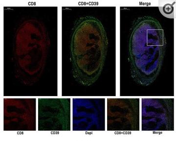
Application: IF/ICC Species: Mice Sample: tumors tissues
Restrictive clause
Affinity Biosciences tests all products strictly. Citations are provided as a resource for additional applications that have not been validated by Affinity Biosciences. Please choose the appropriate format for each application and consult Materials and Methods sections for additional details about the use of any product in these publications.
For Research Use Only.
Not for use in diagnostic or therapeutic procedures. Not for resale. Not for distribution without written consent. Affinity Biosciences will not be held responsible for patent infringement or other violations that may occur with the use of our products. Affinity Biosciences, Affinity Biosciences Logo and all other trademarks are the property of Affinity Biosciences LTD.