
.
Bands result from membrane strip incubation.")


| Product: | CPT1B Antibody |
| Catalog: | DF3904 |
| Description: | Rabbit polyclonal antibody to CPT1B |
| Application: | WB IHC IF/ICC |
| Cited expt.: | WB |
| Reactivity: | Human, Mouse, Rat |
| Prediction: | Pig, Bovine, Horse, Sheep, Dog |
| Mol.Wt.: | 88 KD(Observed); 88kD(Calculated). |
| Uniprot: | Q92523 |
| RRID: | AB_2836257 |
Control Products

Related Downloads
Protocols
Product Info
*The optimal dilutions should be determined by the end user. For optimal experimental results, antibody reuse is not recommended.
*Tips:
WB: For western blot detection of denatured protein samples. IHC: For immunohistochemical detection of paraffin sections (IHC-p) or frozen sections (IHC-f) of tissue samples. IF/ICC: For immunofluorescence detection of cell samples. ELISA(peptide): For ELISA detection of antigenic peptide.
Cite Format: Affinity Biosciences Cat# DF3904, RRID:AB_2836257.
Fold/Unfold
muscle isoform; Carnitine O palmitoyltransferase 1B; Carnitine O palmitoyltransferase I mitochondrial muscle isoform; Carnitine O palmitoyltransferase I muscle isoform; Carnitine O-palmitoyltransferase 1, muscle isoform; Carnitine O-palmitoyltransferase I; Carnitine palmitoyltransferase 1A (muscle); Carnitine palmitoyltransferase 1B (muscle); Carnitine palmitoyltransferase 1B; Carnitine palmitoyltransferase I like protein; Carnitine palmitoyltransferase I muscle; Carnitine palmitoyltransferase I-like protein; CPT 1B; CPT I; CPT1 M; CPT1 muscle; CPT1-M; Cpt1b; CPT1B_HUMAN; CPT1M; CPTI; CPTI M; CPTI muscle; CPTI-M; CPTIM; FLJ55729; FLJ58750; KIAA1670; M CPT1; M-CPT1; MCCPT1; MCPT1;
Immunogens
A synthesized peptide derived from human CPT1B, corresponding to a region within the internal amino acids.
Strong expression in heart and skeletal muscle. No expression in liver and kidney.
- Q92523 CPT1B_HUMAN:
- Protein BLAST With
- NCBI/
- ExPASy/
- Uniprot
MAEAHQAVAFQFTVTPDGVDFRLSREALKHVYLSGINSWKKRLIRIKNGILRGVYPGSPTSWLVVIMATVGSSFCNVDISLGLVSCIQRCLPQGCGPYQTPQTRALLSMAIFSTGVWVTGIFFFRQTLKLLLCYHGWMFEMHGKTSNLTRIWAMCIRLLSSRHPMLYSFQTSLPKLPVPRVSATIQRYLESVRPLLDDEEYYRMELLAKEFQDKTAPRLQKYLVLKSWWASNYVSDWWEEYIYLRGRSPLMVNSNYYVMDLVLIKNTDVQAARLGNIIHAMIMYRRKLDREEIKPVMALGIVPMCSYQMERMFNTTRIPGKDTDVLQHLSDSRHVAVYHKGRFFKLWLYEGARLLKPQDLEMQFQRILDDPSPPQPGEEKLAALTAGGRVEWAQARQAFFSSGKNKAALEAIERAAFFVALDEESYSYDPEDEASLSLYGKALLHGNCYNRWFDKSFTLISFKNGQLGLNAEHAWADAPIIGHLWEFVLGTDSFHLGYTETGHCLGKPNPALAPPTRLQWDIPKQCQAVIESSYQVAKALADDVELYCFQFLPFGKGLIKKCRTSPDAFVQIALQLAHFRDRGKFCLTYEASMTRMFREGRTETVRSCTSESTAFVQAMMEGSHTKADLRDLFQKAAKKHQNMYRLAMTGAGIDRHLFCLYLVSKYLGVSSPFLAEVLSEPWRLSTSQIPQSQIRMFDPEQHPNHLGAGGGFGPVADDGYGVSYMIAGENTIFFHISSKFSSSETNAQRFGNHIRKALLDIADLFQVPKAYS
Predictions
Score>80(red) has high confidence and is suggested to be used for WB detection. *The prediction model is mainly based on the alignment of immunogen sequences, the results are for reference only, not as the basis of quality assurance.
High(score>80) Medium(80>score>50) Low(score<50) No confidence
Research Backgrounds
Mitochondrion outer membrane>Multi-pass membrane protein.
Strong expression in heart and skeletal muscle. No expression in liver and kidney.
Belongs to the carnitine/choline acetyltransferase family.
Research Fields
· Environmental Information Processing > Signal transduction > AMPK signaling pathway. (View pathway)
· Human Diseases > Endocrine and metabolic diseases > Insulin resistance.
· Metabolism > Lipid metabolism > Fatty acid degradation.
· Metabolism > Global and overview maps > Fatty acid metabolism.
· Organismal Systems > Endocrine system > PPAR signaling pathway.
· Organismal Systems > Endocrine system > Adipocytokine signaling pathway.
· Organismal Systems > Endocrine system > Glucagon signaling pathway.
References
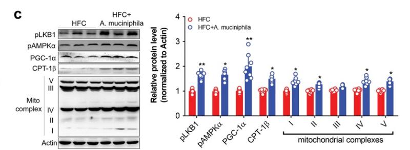
Application: WB Species: Mice Sample: Liver
Restrictive clause
Affinity Biosciences tests all products strictly. Citations are provided as a resource for additional applications that have not been validated by Affinity Biosciences. Please choose the appropriate format for each application and consult Materials and Methods sections for additional details about the use of any product in these publications.
For Research Use Only.
Not for use in diagnostic or therapeutic procedures. Not for resale. Not for distribution without written consent. Affinity Biosciences will not be held responsible for patent infringement or other violations that may occur with the use of our products. Affinity Biosciences, Affinity Biosciences Logo and all other trademarks are the property of Affinity Biosciences LTD.