Product: PSMD3 Antibody
Catalog: DF3645
Description: Rabbit polyclonal antibody to PSMD3
Application: WB IHC IF/ICC
Reactivity: Human, Mouse, Rat
Prediction: Zebrafish, Bovine, Horse, Sheep, Rabbit, Dog, Chicken, Xenopus
Mol.Wt.: 61 KD; 61kD(Calculated).
Uniprot: O43242
RRID: AB_2836017

View similar products>>

   Size Price Inventory
 100ul $280 In stock
 200ul $350 In stock

Lead Time: Same day delivery

For pricing and ordering contact:
Local distributors

Product Info

Source:
Rabbit
Application:
WB 1:500-1:1000, IHC 1:50-1:200, IF/ICC 1:100-1:500
*The optimal dilutions should be determined by the end user.
*Tips:

WB: For western blot detection of denatured protein samples. IHC: For immunohistochemical detection of paraffin sections (IHC-p) or frozen sections (IHC-f) of tissue samples. IF/ICC: For immunofluorescence detection of cell samples. ELISA(peptide): For ELISA detection of antigenic peptide.

Reactivity:
Human,Mouse,Rat
Prediction:
Zebrafish(91%), Bovine(100%), Horse(100%), Sheep(100%), Rabbit(100%), Dog(100%), Chicken(100%), Xenopus(100%)
Clonality:
Polyclonal
Specificity:
PSMD3 Antibody detects endogenous levels of total PSMD3.
RRID:
AB_2836017
Cite Format: Affinity Biosciences Cat# DF3645, RRID:AB_2836017.
Conjugate:
Unconjugated.
Purification:
The antiserum was purified by peptide affinity chromatography using SulfoLink™ Coupling Resin (Thermo Fisher Scientific).
Storage:
Rabbit IgG in phosphate buffered saline , pH 7.4, 150mM NaCl, 0.02% sodium azide and 50% glycerol. Store at -20 °C. Stable for 12 months from date of receipt.
Alias:

Fold/Unfold

26S proteasome non ATPase regulatory subunit 3; 26S proteasome non-ATPase regulatory subunit 3; 26S proteasome regulatory subunit RPN3; 26S proteasome regulatory subunit S3; OTTHUMP00000164347; P58; Proteasome (prosome, macropain) 26S subunit non ATPase 3; Proteasome subunit p58; PSMD3; PSMD3_HUMAN; RPN3; S3; Tissue specific transplantation antigen 2; TSTA2;

Immunogens

Immunogen:
Uniprot:
Gene(ID):
Sequence:
MKQEGSARRRGADKAKPPPGGGEQEPPPPPAPQDVEMKEEAATGGGSTGEADGKTAAAAAEHSQRELDTVTLEDIKEHVKQLEKAVSGKEPRFVLRALRMLPSTSRRLNHYVLYKAVQGFFTSNNATRDFLLPFLEEPMDTEADLQFRPRTGKAASTPLLPEVEAYLQLLVVIFMMNSKRYKEAQKISDDLMQKISTQNRRALDLVAAKCYYYHARVYEFLDKLDVVRSFLHARLRTATLRHDADGQATLLNLLLRNYLHYSLYDQAEKLVSKSVFPEQANNNEWARYLYYTGRIKAIQLEYSEARRTMTNALRKAPQHTAVGFKQTVHKLLIVVELLLGEIPDRLQFRQPSLKRSLMPYFLLTQAVRTGNLAKFNQVLDQFGEKFQADGTYTLIIRLRHNVIKTGVRMISLSYSRISLADIAQKLQLDSPEDAEFIVAKAIRDGVIEASINHEKGYVQSKEMIDIYSTREPQLAFHQRISFCLDIHNMSVKAMRFPPKSYNKDLESAEERREREQQDLEFAKEMAEDDDDSFP

Predictions

Predictions:

Score>80(red) has high confidence and is suggested to be used for WB detection. *The prediction model is mainly based on the alignment of immunogen sequences, the results are for reference only, not as the basis of quality assurance.

Species
Results
Score
Horse
100
Bovine
100
Sheep
100
Dog
100
Xenopus
100
Chicken
100
Rabbit
100
Zebrafish
91
Pig
0
Model Confidence:
High(score>80) Medium(80>score>50) Low(score<50) No confidence

PTMs - O43242 As Substrate

Site PTM Type Enzyme
K2 Sumoylation
S6 Phosphorylation
K14 Acetylation
K16 Ubiquitination
K38 Sumoylation
K38 Ubiquitination
T43 Phosphorylation
S47 Phosphorylation
K54 Acetylation
K54 Ubiquitination
K76 Ubiquitination
K80 Ubiquitination
K84 Ubiquitination
K89 Ubiquitination
S105 Phosphorylation
K186 Ubiquitination
K194 Ubiquitination
K209 Ubiquitination
K223 Ubiquitination
Y264 Phosphorylation
K269 Ubiquitination
K273 Ubiquitination
Y290 Phosphorylation
K296 Ubiquitination
Y302 Phosphorylation
S303 Phosphorylation
K315 Ubiquitination
K325 Ubiquitination
K374 Ubiquitination
Y392 Phosphorylation
T393 Phosphorylation
K404 Ubiquitination
T405 Phosphorylation
S413 Phosphorylation
Y414 Phosphorylation
S418 Phosphorylation
K425 Ubiquitination
S430 Phosphorylation
K440 Ubiquitination
S450 Phosphorylation
K455 Ubiquitination
K461 Ubiquitination
S481 Phosphorylation
K492 Ubiquitination
K499 Ubiquitination
K503 Ubiquitination
S507 Phosphorylation
K523 Methylation
K523 Ubiquitination
S532 Phosphorylation

Research Backgrounds

Function:

Component of the 26S proteasome, a multiprotein complex involved in the ATP-dependent degradation of ubiquitinated proteins. This complex plays a key role in the maintenance of protein homeostasis by removing misfolded or damaged proteins, which could impair cellular functions, and by removing proteins whose functions are no longer required. Therefore, the proteasome participates in numerous cellular processes, including cell cycle progression, apoptosis, or DNA damage repair.

Subunit Structure:

Component of the 19S proteasome regulatory particle complex. The 26S proteasome consists of a 20S core particle (CP) and two 19S regulatory subunits (RP). The regulatory particle is made of a lid composed of 9 subunits including PSMD3, a base containing 6 ATPases and few additional components. Interacts with UBQLN1 (via ubiquitin-like domain). Interacts with ERCC6.

Family&Domains:

Belongs to the proteasome subunit S3 family.

Research Fields

· Genetic Information Processing > Folding, sorting and degradation > Proteasome.

· Human Diseases > Infectious diseases: Viral > Epstein-Barr virus infection.

References

1). PSMD3-ILF3 signaling cascade drives lung cancer cell proliferation and migration. Biology Direct (PubMed: 37337223) [IF=5.5]

Application: WB    Species: Mouse    Sample:

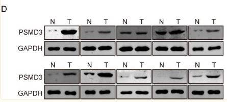
Fig. 1 PSMD3 was upregulated in lung cancer and high PSMD3 indicates poor prognosis. (A) The expression of PSMD3 in LUAD and LUSC used samples from TCGA. (B) Kaplan-Meier analysis showed the low OS in high-PSMD3 TPM. (C) Representative images of IHC on PSMD3 expression in paired tumor and nearby noncancerous tissues from two typical patients. The scale bar stands for 100 μm. (D) The protein level of PSMD3 in 10 pairs normal versus LC tissue samples using western blot. GADPH served as internal control. N: Normal; T: tumor. (E) The protein level of PSMD3 in five LC cell lines and a normal lung cell. GADPH was internal control.

Application: IHC    Species: Mouse    Sample:

Fig. 2 PSMD3 promotes cell proliferation in LC. (A-B) The protein levels of PSMD3 in two LC cell lines transfected with siPSMD3#1, siPSMD3#2, and siCtrl or pcDNA3.1-PSMD3, and Ctrl were determined using western blot. (C-D) Cell viability after PSMD3 knockdown of overexpression at 24, 48, 72 and 96 h were assessed by CCK-8 assay. (E-F) Cell colonies at 14 days in LC cells. (G-H) Cell apoptosis at 24 h after being treated with the suggested siRNAs and plasmids were assessed by flow cytometry. (I) Tumor size in nude mice between shCtrl and shPSMD3 groups were sacrificed at day 35. Images of the xenograft mice with tumors are displayed. (J) The measurement of tumor volumes of nude mice among three groups was made with digital calipers at day 7, 14, 21, 28, and 35. (K) The weight of xenograft tumors was measured among three mice groups at day 35. (L) Images of IHC staining of PSMD3 between two group tumor tissues.

2). TMT-based quantitative proteomics analysis reveals the attenuated replication mechanism of Newcastle disease virus caused by nuclear localization signal mutation in viral matrix protein. Virulence (PubMed: 32420802) [IF=5.2]

Application: WB    Species: Mouse    Sample: BSR-T7/5 cells

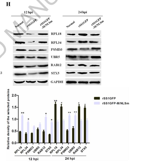
Figure 8. rSS1GFP infection affects the expression of cellular translation, posttranslational modification and trafficking-associated proteins. (A) The heatmap of representative 20 DEPs related to “Translation, ribosomal structure and biogenesis” during rSS1GFP and rSS1GFP-M/NLSm infection at 12 and 24 h. (B) The protein-protein interactions of the DEPs related to “Translation, ribosomal structure and biogenesis” are analyzed by the STRING software. A red line indicates the presence of fusion evidence; a blue line indicates co-occurrence evidence; a light blue line indicates database evidence; a purple line indicates experimental evidence; a green line indicates neighborhood evidence; a black line indicates co-expression evidence. (C) The heatmap of representative 20 DEPs related to “Posttranslational modification, protein turnover, chaperones” during rSS1GFP and rSS1GFP-M/NLSm infection at 12 and 24 h. (D) The protein-protein interactions of the DEPs related to “Posttranslational modification, protein turnover, chaperones” are analyzed by the STRING software. (E) The heatmap of representative 20 DEPs related to “Intracellular trafficking, secretion, and vesicular transport” during rSS1GFP and rSS1GFP-M/NLSm infection at 12 and 24 h. (F) The protein-protein interactions of the DEPs related to “Intracellular trafficking, secretion, and vesicular transport” are analyzed by the STRING software. (G) The mRNA expression levels of six selected DEP genes in BSR-T7/5 cells infected with rSS1GFP and rSS1GFP-M/NLSm were verified by qRT-PCR. (H) The protein expression levels of six DEPs in BSR-T7/5 cells infected with rSS1GFP and rSS1GFP-M/NLSm were examined by Western blotting. The relative expression levels of six DEPs were compared with the control GAPDH expression.

Restrictive clause


Affinity Biosciences tests all products strictly. Citations are provided as a resource for additional applications that have not been validated by Affinity Biosciences. Please choose the appropriate format for each application and consult Materials and Methods sections for additional details about the use of any product in these publications.

For Research Use Only.
Not for use in diagnostic or therapeutic procedures. Not for resale. Not for distribution without written consent. Affinity Biosciences will not be held responsible for patent infringement or other violations that may occur with the use of our products. Affinity Biosciences, Affinity Biosciences Logo and all other trademarks are the property of Affinity Biosciences LTD.