

.
Bands result from membrane strip incubation.")

Control Products

Related Downloads
Protocols
Product Info
*The optimal dilutions should be determined by the end user. For optimal experimental results, antibody reuse is not recommended.
*Tips:
WB: For western blot detection of denatured protein samples. IHC: For immunohistochemical detection of paraffin sections (IHC-p) or frozen sections (IHC-f) of tissue samples. IF/ICC: For immunofluorescence detection of cell samples. ELISA(peptide): For ELISA detection of antigenic peptide.
Cite Format: Affinity Biosciences Cat# DF3641, RRID:AB_2836013.
Fold/Unfold
1 acyl sn glycerol 3 phosphate acyltransferase epsilon; 1 acylglycerol 3 phosphate O acyltransferase 5; 1 AGP acyltransferase 5; 1 AGPAT 5; 1 AGPAT5; 1-acyl-sn-glycerol-3-phosphate acyltransferase epsilon; 1-acylglycerol-3-phosphate O-acyltransferase 5 (lysophosphatidic acid acyltransferase, epsilon); 1-acylglycerol-3-phosphate O-acyltransferase 5; 1-AGP acyltransferase 5; 1-AGPAT 5; 1acylglycerol3phosphate Oacyltransferase 5 (lysophosphatidic acid acyltransferase, epsilon); 1acylglycerol3phosphate Oacyltransferase 5; 1acylglycerol3phosphate Oacyltransferase5 (lysophosphatidic acid acyltransferase, epsilon); 1AGP acyltransferase 5; 1AGPAT5; AGPAT5; LPAAT e; LPAAT epsilon; LPAAT-epsilon; LPAATE; Lysophosphatidic acid acyltransferase epsilon; PLCE_HUMAN;
Immunogens
A synthesized peptide derived from human AGPAT5, corresponding to a region within the internal amino acids.
- Q9NUQ2 PLCE_HUMAN:
- Protein BLAST With
- NCBI/
- ExPASy/
- Uniprot
MLLSLVLHTYSMRYLLPSVVLLGTAPTYVLAWGVWRLLSAFLPARFYQALDDRLYCVYQSMVLFFFENYTGVQILLYGDLPKNKENIIYLANHQSTVDWIVADILAIRQNALGHVRYVLKEGLKWLPLYGCYFAQHGGIYVKRSAKFNEKEMRNKLQSYVDAGTPMYLVIFPEGTRYNPEQTKVLSASQAFAAQRGLAVLKHVLTPRIKATHVAFDCMKNYLDAIYDVTVVYEGKDDGGQRRESPTMTEFLCKECPKIHIHIDRIDKKDVPEEQEHMRRWLHERFEIKDKMLIEFYESPDPERRKRFPGKSVNSKLSIKKTLPSMLILSGLTAGMLMTDAGRKLYVNTWIYGTLLGCLWVTIKA
Predictions
Score>80(red) has high confidence and is suggested to be used for WB detection. *The prediction model is mainly based on the alignment of immunogen sequences, the results are for reference only, not as the basis of quality assurance.
High(score>80) Medium(80>score>50) Low(score<50) No confidence
Research Backgrounds
Converts 1-acyl-sn-glycerol-3-phosphate (lysophosphatidic acid or LPA) into 1,2-diacyl-sn-glycerol-3-phosphate (phosphatidic acid or PA) by incorporating an acyl moiety at the sn-2 position of the glycerol backbone. Acts on LPA containing saturated or unsaturated fatty acids C15:0-C20:4 at the sn-1 position using C18:1-CoA as the acyl donor. Also acts on lysophosphatidylethanolamine using oleoyl-CoA, but not arachidonoyl-CoA, and lysophosphatidylinositol using arachidonoyl-CoA, but not oleoyl-CoA. Activity toward lysophosphatidylglycerol not detectable.
Endoplasmic reticulum membrane>Multi-pass membrane protein. Nucleus envelope. Mitochondrion.
Widely expressed.
The HXXXXD motif is essential for acyltransferase activity and may constitute the binding site for the phosphate moiety of the glycerol-3-phosphate.
Belongs to the 1-acyl-sn-glycerol-3-phosphate acyltransferase family.
Research Fields
· Environmental Information Processing > Signal transduction > Phospholipase D signaling pathway. (View pathway)
· Metabolism > Lipid metabolism > Glycerolipid metabolism.
· Metabolism > Lipid metabolism > Glycerophospholipid metabolism.
· Metabolism > Global and overview maps > Metabolic pathways.
References
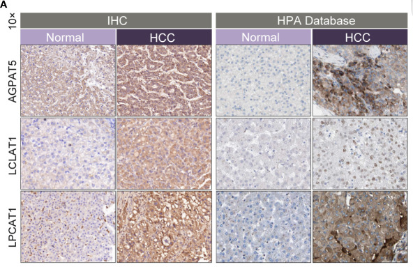
Application: IHC Species: Human Sample: tumor tissues
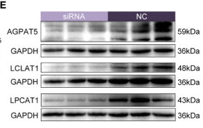
Application: WB Species: Human Sample: tumor tissues
Restrictive clause
Affinity Biosciences tests all products strictly. Citations are provided as a resource for additional applications that have not been validated by Affinity Biosciences. Please choose the appropriate format for each application and consult Materials and Methods sections for additional details about the use of any product in these publications.
For Research Use Only.
Not for use in diagnostic or therapeutic procedures. Not for resale. Not for distribution without written consent. Affinity Biosciences will not be held responsible for patent infringement or other violations that may occur with the use of our products. Affinity Biosciences, Affinity Biosciences Logo and all other trademarks are the property of Affinity Biosciences LTD.