
, using Cytochrome P450 19A1 Antibody at 1/1000 dilution.")
, using Cytochrome P450 19A1 Antibody at 1/1000 dilution.
5ug/NC membrane strip.
Exposure for 20s with Affinity™ ECL Kit(#KF8001).
Bands result from membrane strip incubation.")


 and mouse anti-beta tubulin Ab(T0023 1:200) for 1 hour at 37°C. An AlexaFluor594 conjugated goat anti-rabbit IgG(H+L) Ab(Red) and an AlexaFluor488 conjugated goat anti-mouse IgG(H+L) Ab(Green) were used as the secondary antibody.
The nuclear counter stain is DAPI(blue).")
 and mouse anti-beta tubulin Ab(T0023) for 1 hour at 37°C. An AlexaFluor594 conjugated goat anti-rabbit IgG(H+L) Ab(Red) and an AlexaFluor488 conjugated goat anti-mouse IgG(H+L) Ab(Green) were used as the secondary antibody.
The nuclear counter stain is DAPI (blue).")

| Product: | Cytochrome P450 19A1 Antibody |
| Catalog: | DF3564 |
| Description: | Rabbit polyclonal antibody to Cytochrome P450 19A1 |
| Application: | WB IHC IF/ICC |
| Cited expt.: | WB, IHC |
| Reactivity: | Human, Mouse, Rat |
| Prediction: | Pig, Bovine, Sheep, Rabbit, Dog |
| Mol.Wt.: | 53 KD(Observed); 58kD(Calculated). |
| Uniprot: | P11511 |
| RRID: | AB_2835936 |
Control Products

Related Downloads
Protocols
Product Info
*The optimal dilutions should be determined by the end user. For optimal experimental results, antibody reuse is not recommended.
*Tips:
WB: For western blot detection of denatured protein samples. IHC: For immunohistochemical detection of paraffin sections (IHC-p) or frozen sections (IHC-f) of tissue samples. IF/ICC: For immunofluorescence detection of cell samples. ELISA(peptide): For ELISA detection of antigenic peptide.
Cite Format: Affinity Biosciences Cat# DF3564, RRID:AB_2835936.
Fold/Unfold
ARO; ARO1; Aromatase; CP19A_HUMAN; CPV1; CYAR; CYP19; Cyp19a1; CYPXIX; Cytochrome P-450AROM; Cytochrome P450 19A1; Cytochrome P450, family 19, subfamily A, polypeptide 1; Cytochrome P450, subfamily XIX (aromatization of androgens); Estrogen synthase; Estrogen synthetase; Flavoprotein linked monooxygenase; MGC104309; Microsomal monooxygenase; OTTHUMP00000162543; OTTHUMP00000198350; P 450AROM;
Immunogens
A synthesized peptide derived from human Cytochrome P450 19A1, corresponding to a region within the internal amino acids.
Widely expressed, including in adult and fetal brain, placenta, skin fibroblasts, adipose tissue and gonads.
- P11511 CP19A_HUMAN:
- Protein BLAST With
- NCBI/
- ExPASy/
- Uniprot
MVLEMLNPIHYNITSIVPEAMPAATMPVLLLTGLFLLVWNYEGTSSIPGPGYCMGIGPLISHGRFLWMGIGSACNYYNRVYGEFMRVWISGEETLIISKSSSMFHIMKHNHYSSRFGSKLGLQCIGMHEKGIIFNNNPELWKTTRPFFMKALSGPGLVRMVTVCAESLKTHLDRLEEVTNESGYVDVLTLLRRVMLDTSNTLFLRIPLDESAIVVKIQGYFDAWQALLIKPDIFFKISWLYKKYEKSVKDLKDAIEVLIAEKRRRISTEEKLEECMDFATELILAEKRGDLTRENVNQCILEMLIAAPDTMSVSLFFMLFLIAKHPNVEEAIIKEIQTVIGERDIKIDDIQKLKVMENFIYESMRYQPVVDLVMRKALEDDVIDGYPVKKGTNIILNIGRMHRLEFFPKPNEFTLENFAKNVPYRYFQPFGFGPRGCAGKYIAMVMMKAILVTLLRRFHVKTLQGQCVESIQKIHDLSLHPDETKNMLEMIFTPRNSDRCLEH
Predictions
Score>80(red) has high confidence and is suggested to be used for WB detection. *The prediction model is mainly based on the alignment of immunogen sequences, the results are for reference only, not as the basis of quality assurance.
High(score>80) Medium(80>score>50) Low(score<50) No confidence
Research Backgrounds
A cytochrome P450 monooxygenase that catalyzes the conversion of C19 androgens, androst-4-ene-3,17-dione (androstenedione) and testosterone to the C18 estrogens, estrone and estradiol, respectively. Catalyzes three successive oxidations of C19 androgens: two conventional oxidations at C19 yielding 19-hydroxy and 19-oxo/19-aldehyde derivatives, followed by a third oxidative aromatization step that involves C1-beta hydrogen abstraction combined with cleavage of the C10-C19 bond to yield a phenolic A ring and formic acid. Alternatively, the third oxidative reaction yields a 19-norsteroid and formic acid. Converts dihydrotestosterone to delta1,10-dehydro 19-nordihydrotestosterone and may play a role in homeostasis of this potent androgen. Also displays 2-hydroxylase activity toward estrone. Mechanistically, uses molecular oxygen inserting one oxygen atom into a substrate, and reducing the second into a water molecule, with two electrons provided by NADPH via cytochrome P450 reductase (CPR; NADPH-ferrihemoprotein reductase).
Phosphorylated in vitro by PKA and PKG/PRKG1. These phosphorylations inhibit the catalytic activity as measured by estrone synthesis from androstenedione (36% decrease for PKA and 30% for PKG/PRKG1).
Endoplasmic reticulum membrane>Multi-pass membrane protein. Microsome membrane>Multi-pass membrane protein.
Widely expressed, including in adult and fetal brain, placenta, skin fibroblasts, adipose tissue and gonads.
Belongs to the cytochrome P450 family.
Research Fields
· Metabolism > Lipid metabolism > Steroid hormone biosynthesis.
· Metabolism > Global and overview maps > Metabolic pathways.
· Organismal Systems > Endocrine system > Ovarian steroidogenesis.
References
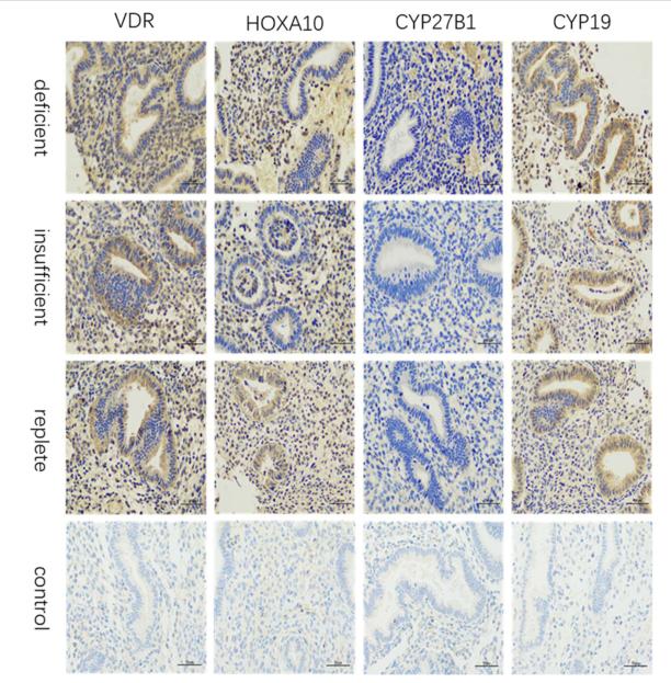
Application: IHC Species: human Sample: endometrial
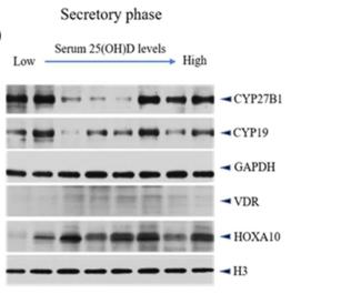
Application: WB Species: human Sample: endometrial
Application: WB Species: porcine Sample: GCs
Application: WB Species: Human Sample: granulosa cells
Restrictive clause
Affinity Biosciences tests all products strictly. Citations are provided as a resource for additional applications that have not been validated by Affinity Biosciences. Please choose the appropriate format for each application and consult Materials and Methods sections for additional details about the use of any product in these publications.
For Research Use Only.
Not for use in diagnostic or therapeutic procedures. Not for resale. Not for distribution without written consent. Affinity Biosciences will not be held responsible for patent infringement or other violations that may occur with the use of our products. Affinity Biosciences, Affinity Biosciences Logo and all other trademarks are the property of Affinity Biosciences LTD.