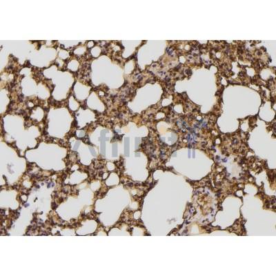
JIP2 Antibody - #DF3260
| Product: | JIP2 Antibody |
| Catalog: | DF3260 |
| Description: | Rabbit polyclonal antibody to JIP2 |
| Application: | WB IHC IF/ICC |
| Cited expt.: | WB |
| Reactivity: | Human, Mouse, Rat, Zebrafish |
| Prediction: | Pig, Bovine, Sheep, Dog, Xenopus |
| Mol.Wt.: | 87 KD; 88kD(Calculated). |
| Uniprot: | Q13387 |
| RRID: | AB_2835558 |
Related Downloads
Protocols
Product Info
*The optimal dilutions should be determined by the end user. For optimal experimental results, antibody reuse is not recommended.
*Tips:
WB: For western blot detection of denatured protein samples. IHC: For immunohistochemical detection of paraffin sections (IHC-p) or frozen sections (IHC-f) of tissue samples. IF/ICC: For immunofluorescence detection of cell samples. ELISA(peptide): For ELISA detection of antigenic peptide.
Cite Format: Affinity Biosciences Cat# DF3260, RRID:AB_2835558.
Fold/Unfold
C jun amino terminal kinase interacting protein 2; C-jun-amino-terminal kinase-interacting protein 2; Homologous to mouse JIP 1; IB 2; IB-2; IB2; Islet brain 2; Islet-brain-2; JIP 2; JIP-2; JIP2; JIP2_HUMAN; JNK interacting protein 2; JNK MAP kinase scaffold protein 2; JNK MAP kinase scaffold protein JIP2; JNK-interacting protein 2; MAPK8IP2; Mitogen activated protein kinase 8 interacting protein 2; Mitogen-activated protein kinase 8-interacting protein 2; PRKM8 interacting protein like; PRKM8IPL;
Immunogens
A synthesized peptide derived from human JIP2, corresponding to a region within the internal amino acids.
Expressed mainly in the brain and pancreas, including insulin-secreting cells. In the nervous system, more abundantly expressed in the cerebellum, pituitary gland, occipital lobe and the amygdala. Also expressed in fetal brain. Very low levels found in uterus, ovary, prostate, colon, testis, adrenal gland, thyroid gland and salivary gland.
- Q13387 JIP2_HUMAN:
- Protein BLAST With
- NCBI/
- ExPASy/
- Uniprot
MADRAEMFSLSTFHSLSPPGCRPPQDISLEEFDDEDLSEITDDCGLGLSYDSDHCEKDSLSLGRSEQPHPICSFQDDFQEFEMIDDNEEEDDEDEEEEEEEEEGDGEGQEGGDPGSEAPAPGPLIPSPSVEEPHKHRPTTLRLTTLGAQDSLNNNGGFDLVRPASWQETALCSPAPEALRELPGPLPATDTGPGGAQSPVRPGCDCEGNRPAEPPAPGGTSPSSDPGIEADLRSRSSGGRGGRRSSQELSSPGSDSEDAGGARLGRMISSISETELELSSDGGSSSSGRSSHLTNSIEEASSPASEPEPPREPPRRPAFLPVGPDDTNSEYESGSESEPDLSEDADSPWLLSNLVSRMISEGSSPIRCPGQCLSPAPRPPGEPVSPAGGAAQDSQDPEAAAGPGGVELVDMETLCAPPPPAPAAPRPGPAQPGPCLFLSNPTRDTITPLWAAPGRAARPGRACSAACSEEEDEEDDEEEEDAEDSAGSPGGRGTGPSAPRDASLVYDAVKYTLVVDEHTQLELVSLRRCAGLGHDSEEDSGGEASEEEAGAALLGGGQVSGDTSPDSPDLTFSKKFLNVFVNSTSRSSSTESFGLFSCLVNGEEREQTHRAVFRFIPRHPDELELDVDDPVLVEAEEDDFWFRGFNMRTGERGVFPAFYAHAVPGPAKDLLGSKRSPCWVERFDVQFLGSVEVPCHQGNGILCAAMQKIATARKLTVHLRPPASCDLEISLRGVKLSLSGGGPEFQRCSHFFQMKNISFCGCHPRNSCYFGFITKHPLLSRFACHVFVSQESMRPVAQSVGRAFLEYYQEHLAYACPTEDIYLE
Predictions
Score>80(red) has high confidence and is suggested to be used for WB detection. *The prediction model is mainly based on the alignment of immunogen sequences, the results are for reference only, not as the basis of quality assurance.
High(score>80) Medium(80>score>50) Low(score<50) No confidence
Research Backgrounds
The JNK-interacting protein (JIP) group of scaffold proteins selectively mediates JNK signaling by aggregating specific components of the MAPK cascade to form a functional JNK signaling module. JIP2 inhibits IL1 beta-induced apoptosis in insulin-secreting cells. May function as a regulator of vesicle transport, through interactions with the JNK-signaling components and motor proteins (By similarity).
Cytoplasm.
Note: Accumulates in cell surface projections.
Expressed mainly in the brain and pancreas, including insulin-secreting cells. In the nervous system, more abundantly expressed in the cerebellum, pituitary gland, occipital lobe and the amygdala. Also expressed in fetal brain. Very low levels found in uterus, ovary, prostate, colon, testis, adrenal gland, thyroid gland and salivary gland.
Belongs to the JIP scaffold family.
Research Fields
· Environmental Information Processing > Signal transduction > MAPK signaling pathway. (View pathway)
References
Application: WB Species: Mice Sample: myocardial tissue
Application: WB Species: rat Sample: H9C2 cells
Restrictive clause
Affinity Biosciences tests all products strictly. Citations are provided as a resource for additional applications that have not been validated by Affinity Biosciences. Please choose the appropriate format for each application and consult Materials and Methods sections for additional details about the use of any product in these publications.
For Research Use Only.
Not for use in diagnostic or therapeutic procedures. Not for resale. Not for distribution without written consent. Affinity Biosciences will not be held responsible for patent infringement or other violations that may occur with the use of our products. Affinity Biosciences, Affinity Biosciences Logo and all other trademarks are the property of Affinity Biosciences LTD.


