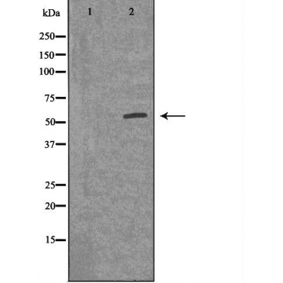
ACV1B Antibody - #DF3169
| Product: | ACV1B Antibody |
| Catalog: | DF3169 |
| Description: | Rabbit polyclonal antibody to ACV1B |
| Application: | WB IF/ICC |
| Reactivity: | Human, Mouse, Rat |
| Prediction: | Pig, Bovine, Horse, Sheep, Rabbit, Dog |
| Mol.Wt.: | 56 KD; 57kD(Calculated). |
| Uniprot: | P36896 |
| RRID: | AB_2835545 |
Product Info
*The optimal dilutions should be determined by the end user. For optimal experimental results, antibody reuse is not recommended.
*Tips:
WB: For western blot detection of denatured protein samples. IHC: For immunohistochemical detection of paraffin sections (IHC-p) or frozen sections (IHC-f) of tissue samples. IF/ICC: For immunofluorescence detection of cell samples. ELISA(peptide): For ELISA detection of antigenic peptide.
Cite Format: Affinity Biosciences Cat# DF3169, RRID:AB_2835545.
Fold/Unfold
Activin A receptor type 1B; Activin A receptor type II like kinase 4; Activin A type 1B receptor; Activin A type IB receptor; Activin receptor like kinase 4; Activin receptor type 1B; Activin receptor type IB; Activin receptor type-1B; Activin receptor-like kinase 4; ACTR IB; ACTR-IB; ACTRIB; ACV1B_HUMAN; ACVR 1B; Acvr1b; ACVRLK 4; ACVRLK4; ALK 4; ALK-4; ALK4; Serine(threonine) protein kinase receptor R2; Serine/threonine-protein kinase receptor R2; SKR 2; SKR2;
Immunogens
A synthesized peptide derived from human ACV1B, corresponding to a region within the internal amino acids.
Expressed in many tissues, most strongly in kidney, pancreas, brain, lung, and liver.
- P36896 ACV1B_HUMAN:
- Protein BLAST With
- NCBI/
- ExPASy/
- Uniprot
MAESAGASSFFPLVVLLLAGSGGSGPRGVQALLCACTSCLQANYTCETDGACMVSIFNLDGMEHHVRTCIPKVELVPAGKPFYCLSSEDLRNTHCCYTDYCNRIDLRVPSGHLKEPEHPSMWGPVELVGIIAGPVFLLFLIIIIVFLVINYHQRVYHNRQRLDMEDPSCEMCLSKDKTLQDLVYDLSTSGSGSGLPLFVQRTVARTIVLQEIIGKGRFGEVWRGRWRGGDVAVKIFSSREERSWFREAEIYQTVMLRHENILGFIAADNKDNGTWTQLWLVSDYHEHGSLFDYLNRYTVTIEGMIKLALSAASGLAHLHMEIVGTQGKPGIAHRDLKSKNILVKKNGMCAIADLGLAVRHDAVTDTIDIAPNQRVGTKRYMAPEVLDETINMKHFDSFKCADIYALGLVYWEIARRCNSGGVHEEYQLPYYDLVPSDPSIEEMRKVVCDQKLRPNIPNWWQSYEALRVMGKMMRECWYANGAARLTALRIKKTLSQLSVQEDVKI
Predictions
Score>80(red) has high confidence and is suggested to be used for WB detection. *The prediction model is mainly based on the alignment of immunogen sequences, the results are for reference only, not as the basis of quality assurance.
High(score>80) Medium(80>score>50) Low(score<50) No confidence
Research Backgrounds
Transmembrane serine/threonine kinase activin type-1 receptor forming an activin receptor complex with activin receptor type-2 (ACVR2A or ACVR2B). Transduces the activin signal from the cell surface to the cytoplasm and is thus regulating a many physiological and pathological processes including neuronal differentiation and neuronal survival, hair follicle development and cycling, FSH production by the pituitary gland, wound healing, extracellular matrix production, immunosuppression and carcinogenesis. Activin is also thought to have a paracrine or autocrine role in follicular development in the ovary. Within the receptor complex, type-2 receptors (ACVR2A and/or ACVR2B) act as a primary activin receptors whereas the type-1 receptors like ACVR1B act as downstream transducers of activin signals. Activin binds to type-2 receptor at the plasma membrane and activates its serine-threonine kinase. The activated receptor type-2 then phosphorylates and activates the type-1 receptor such as ACVR1B. Once activated, the type-1 receptor binds and phosphorylates the SMAD proteins SMAD2 and SMAD3, on serine residues of the C-terminal tail. Soon after their association with the activin receptor and subsequent phosphorylation, SMAD2 and SMAD3 are released into the cytoplasm where they interact with the common partner SMAD4. This SMAD complex translocates into the nucleus where it mediates activin-induced transcription. Inhibitory SMAD7, which is recruited to ACVR1B through FKBP1A, can prevent the association of SMAD2 and SMAD3 with the activin receptor complex, thereby blocking the activin signal. Activin signal transduction is also antagonized by the binding to the receptor of inhibin-B via the IGSF1 inhibin coreceptor. ACVR1B also phosphorylates TDP2.
Autophosphorylated. Phosphorylated by activin receptor type-2 (ACVR2A or ACVR2B) in response to activin-binding at serine and threonine residues in the GS domain. Phosphorylation of ACVR1B by activin receptor type-2 regulates association with SMAD7.
Ubiquitinated. Level of ubiquitination is regulated by the SMAD7-SMURF1 complex.
Ubiquitinated.
Cell membrane>Single-pass type I membrane protein.
Expressed in many tissues, most strongly in kidney, pancreas, brain, lung, and liver.
The GS domain is a 30-amino-acid sequence adjacent to the N-terminal boundary of the kinase domain and highly conserved in all other known type-1 receptors but not in type-2 receptors. The GS domain is the site of activation through phosphorylation by the II receptors.
Belongs to the protein kinase superfamily. TKL Ser/Thr protein kinase family. TGFB receptor subfamily.
Research Fields
· Cellular Processes > Cellular community - eukaryotes > Signaling pathways regulating pluripotency of stem cells. (View pathway)
· Environmental Information Processing > Signaling molecules and interaction > Cytokine-cytokine receptor interaction. (View pathway)
· Environmental Information Processing > Signal transduction > TGF-beta signaling pathway. (View pathway)
Restrictive clause
Affinity Biosciences tests all products strictly. Citations are provided as a resource for additional applications that have not been validated by Affinity Biosciences. Please choose the appropriate format for each application and consult Materials and Methods sections for additional details about the use of any product in these publications.
For Research Use Only.
Not for use in diagnostic or therapeutic procedures. Not for resale. Not for distribution without written consent. Affinity Biosciences will not be held responsible for patent infringement or other violations that may occur with the use of our products. Affinity Biosciences, Affinity Biosciences Logo and all other trademarks are the property of Affinity Biosciences LTD.
