


 Representative images of HE and TRAP staining, showing the reduced osteolytic legion and TRAP-positive OCs in the PF-treated groups. (D–G) Representative images of IHC staining of RANKL, OPG, OCN and TNF-α. (H) TRAPpositive OCs number. (H) Quantitative analysis of the expression of RANKL, OPG and RANKL/OPG ratio. The data are presented as the mean ± SD (*p < 0.05,**p < 0.01, ***p < 0.001).")
 Non-induced IF cells and MF cells showed
different gene expression patterns; Data are mean ± SD of n = 3 replicates, one-way ANOVA, *p<0.05, **p<0.01, ***p<0.001, ****p<0.0001. (B) Non-induced
and induced IF cells and MF cells showed different protein expression patterns. After induction with CLCCM and HERSCM, there were no significant changes of
the expression patterns of tooth eruption-related proteins in IF cells and MF cells. (C) The grey value ratios of Western blotting results; Data are mean ± SD of n = 3
replicates, one-way ANOVA followed by Tukey post hoc test, *p<0.05, **p<0.01, ***p<0.001, ****p<0.0001. (IF1, MF1 represent the cells cultured by α-MEM;
IF2, MF2 represent the cells cultured by α-MEM + CLCCM; IF3, MF3 represent the cells cultured by α-MEM+HERSCM).")
 Adipogenic differentiation was detected by oil red O staining; (B) osteogenic differentiation was detected by alizarin red staining; (C) the expressions of NF-κB, PPARγ, C/EBP-α, Runx2 and OPN in BMSCs were measured by WB. Actin was used as an internal control; (D) quantification of NF-κB/Actin, PPARγ/Actin, C/EBP-α/Actin, Runx2/Actin, and OPN/Actin protein expression. *, P<0.05, **, P<0.01, ***, P<0.001 vs. OIM or AIM group. TMAO, trimethylamine N-oxide; BMSCs, bone marrow mesenchymal stem cells. NF-κB, nuclear factor-κB; PPARγ, Peroxisome proliferator-activated receptor gamma; C/EBP-α, CCAAT/enhancer-binding protein alpha; Runx2, runt-related transcription factor 2; OPN, Osteopontin.")
 RT-PCR analysis was used to detect miRNA-19b-3p expression levels in BMSC-derived osteoblasts after miR-19b-3p-overexpressed or knockdown by corresponding lentivirus. (B) ALP activity in BMSC-derived osteoblasts after miRNA-19b-3p overexpression or knockdown by corresponding lentivirus. (C) RT-PCR was used to detect EBF2 mRNA expression levels in Journal Pre-proof 20 BMSC-derived osteoblasts after miRNA-19b-3p overexpression or knockdown by corresponding lentivirus. Western blot assay was used to detect (D and E) EBF2, (F-H) RANKL and OPG protein expression levels in BMSC-derived osteoblasts after miRNA-19b-3p overexpression or knockdown by corresponding lentivirus. (I) The ratio of OPG to RANKL in BMSC-derived osteoblasts after miRNA-19b-3p overexpression or knockdown by corresponding lentivirus (J) Alizarin red staining was used to detect cell mineralization at day 21 after differentiation after miRNA-19b-3p overexpression or knockdown at 100×. Data are analyzed using unpaired Student’s t-tests and presented as the mean ± SD from three independent experiments. * p < 0.05; ** p < 0.01 vs. the indicated group.")
 Non-induced IF cells and MF cells showed
different gene expression patterns; Data are mean ± SD of n = 3 replicates, one-way ANOVA, *p<0.05, **p<0.01, ***p<0.001, ****p<0.0001. (B) Non-induced
and induced IF cells and MF cells showed different protein expression patterns. After induction with CLCCM and HERSCM, there were no significant changes of
the expression patterns of tooth eruption-related proteins in IF cells and MF cells. (C) The grey value ratios of Western blotting results; Data are mean ± SD of n = 3
replicates, one-way ANOVA followed by Tukey post hoc test, *p<0.05, **p<0.01, ***p<0.001, ****p<0.0001. (IF1, MF1 represent the cells cultured by α-MEM;
IF2, MF2 represent the cells cultured by α-MEM + CLCCM; IF3, MF3 represent the cells cultured by α-MEM+HERSCM).")
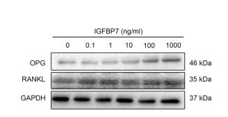
 promoted the differentiation and mineralization of osteoblasts. A) Osteocalcin-positive rate of SaOS-2 cells was detected by flow cytometry. B) The expression of osteocalcin in SaOS-2 cells was observed by immunofluorescence staining, and the fluorescence intensity was exhibited on the right. C) Alizarin Red S staining was performed on SaOS-2 cells to evaluate the mineralization. D) The protein expression of RANKL, M-CSF and OPG was detected by Western blot.")
 in the Control group, marked increase in expression (black arrows) in L-MTZ, increased expression (black arrows) in S-MTZ. BMP-2: expression findings between the groups, moderate expression (black arrow) in the Control group, increase in expression (black arrows) in L-MTZ, marked expression (black arrow) in S-MTZ. Runx2: moderate expression (black arrows) in the Control group, increase in expression (black arrows) in L-MTZ, marked expression (black arrows) in S-MTZ. ALP: moderate expression (black arrow) in the Control group, marked increase in expression (black arrows) in L-MTZ, moderate expression (black arrow) in S-MTZ. OCN: moderate expression (black arrow) in the control group, markedly increased expression (black arrow) in L-MTZ, and increased expression (black arrow) in S-MTZ. RANKL: Similar RANKL expressions (black arrows) in (A) Control group, (B) L-MTZ, and (C) S-MTZ, Streptavidin biotin peroxidase method, Scale bars = 50 μm Representative histopathological images among the groups. (A) Marked fibrous tissue, moderate new bone formation (white arrow with a black border), and residual graft materials (RG) in the Control group. (B) Decreased fibrous tissue, moderate residual graft material (RG), and marked new bone formation (white arrow with a black border) in L-MTZ. (C) Decreased fibrous tissue, moderately increased new bone formation (white arrow with a black border), and residual graft materials (RG) in S-MTZ. HE, Scale bars = 200 μm")
| Product: | RANKL Antibody |
| Catalog: | AF0313 |
| Description: | Rabbit polyclonal antibody to RANKL |
| Application: | WB IHC IF/ICC |
| Cited expt.: | WB, IHC, IF/ICC |
| Reactivity: | Human, Mouse, Rat |
| Prediction: | Pig, Bovine, Horse, Sheep, Rabbit, Dog |
| Mol.Wt.: | 40kDa(Observed); 35kD(Calculated). |
| Uniprot: | O14788 |
| RRID: | AB_2833477 |
Control Products

Related Downloads
Protocols
Product Info
*The optimal dilutions should be determined by the end user. For optimal experimental results, antibody reuse is not recommended.
*Tips:
WB: For western blot detection of denatured protein samples. IHC: For immunohistochemical detection of paraffin sections (IHC-p) or frozen sections (IHC-f) of tissue samples. IF/ICC: For immunofluorescence detection of cell samples. ELISA(peptide): For ELISA detection of antigenic peptide.
Cite Format: Affinity Biosciences Cat# AF0313, RRID:AB_2833477.
Fold/Unfold
CD254; hRANKL2; ODF; OPGL; OPTB2; Osteoclast differentiation factor; Osteoprotegerin ligand; RANKL; Receptor activator of nuclear factor kappa B ligand; Receptor activator of nuclear factor kappa-B ligand; sOdf; TNF related activation induced cytokine; TNF-related activation-induced cytokine; TNF11_HUMAN; TNFSF 11; Tnfsf11; TRANCE; Tumor necrosis factor (ligand) superfamily member 11; Tumor necrosis factor ligand superfamily member 11; Tumor necrosis factor ligand superfamily member 11, soluble form;
Immunogens
A synthesized peptide derived from human RANKL, corresponding to a region within the internal amino acids.
Highest in the peripheral lymph nodes, weak in spleen, peripheral blood Leukocytes, bone marrow, heart, placenta, skeletal muscle, stomach and thyroid.
- O14788 TNF11_HUMAN:
- Protein BLAST With
- NCBI/
- ExPASy/
- Uniprot
MRRASRDYTKYLRGSEEMGGGPGAPHEGPLHAPPPPAPHQPPAASRSMFVALLGLGLGQVVCSVALFFYFRAQMDPNRISEDGTHCIYRILRLHENADFQDTTLESQDTKLIPDSCRRIKQAFQGAVQKELQHIVGSQHIRAEKAMVDGSWLDLAKRSKLEAQPFAHLTINATDIPSGSHKVSLSSWYHDRGWAKISNMTFSNGKLIVNQDGFYYLYANICFRHHETSGDLATEYLQLMVYVTKTSIKIPSSHTLMKGGSTKYWSGNSEFHFYSINVGGFFKLRSGEEISIEVSNPSLLDPDQDATYFGAFKVRDID
Predictions
Score>80(red) has high confidence and is suggested to be used for WB detection. *The prediction model is mainly based on the alignment of immunogen sequences, the results are for reference only, not as the basis of quality assurance.
High(score>80) Medium(80>score>50) Low(score<50) No confidence
Research Backgrounds
Cytokine that binds to TNFRSF11B/OPG and to TNFRSF11A/RANK. Osteoclast differentiation and activation factor. Augments the ability of dendritic cells to stimulate naive T-cell proliferation. May be an important regulator of interactions between T-cells and dendritic cells and may play a role in the regulation of the T-cell-dependent immune response. May also play an important role in enhanced bone-resorption in humoral hypercalcemia of malignancy. Induces osteoclastogenesis by activating multiple signaling pathways in osteoclast precursor cells, chief among which is induction of long lasting oscillations in the intracellular concentration of Ca (2+) resulting in the activation of NFATC1, which translocates to the nucleus and induces osteoclast-specific gene transcription to allow differentiation of osteoclasts. During osteoclast differentiation, in a TMEM64 and ATP2A2-dependent manner induces activation of CREB1 and mitochondrial ROS generation necessary for proper osteoclast generation (By similarity).
The soluble form of isoform 1 derives from the membrane form by proteolytic processing (By similarity). The cleavage may be catalyzed by ADAM17.
Cell membrane>Single-pass type II membrane protein.
Cell membrane>Single-pass type II membrane protein.
Cytoplasm.
Secreted.
Highest in the peripheral lymph nodes, weak in spleen, peripheral blood Leukocytes, bone marrow, heart, placenta, skeletal muscle, stomach and thyroid.
Belongs to the tumor necrosis factor family.
Research Fields
· Environmental Information Processing > Signaling molecules and interaction > Cytokine-cytokine receptor interaction. (View pathway)
· Environmental Information Processing > Signal transduction > NF-kappa B signaling pathway. (View pathway)
· Human Diseases > Cancers: Specific types > Breast cancer. (View pathway)
· Human Diseases > Immune diseases > Rheumatoid arthritis.
· Organismal Systems > Development > Osteoclast differentiation. (View pathway)
· Organismal Systems > Endocrine system > Prolactin signaling pathway. (View pathway)
References

Application: WB Species: Mouse Sample:
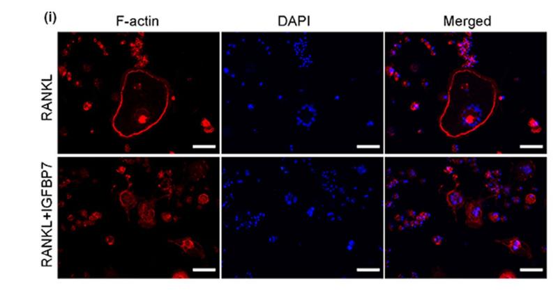
Application: IF/ICC Species: mice Sample: RAW264.7 cells

Application: IF/ICC Species: mouse Sample: osteoblastic cel
Application: WB Species: mouse Sample: MC3T3-E1

Application: WB Species: Mouse Sample:
Restrictive clause
Affinity Biosciences tests all products strictly. Citations are provided as a resource for additional applications that have not been validated by Affinity Biosciences. Please choose the appropriate format for each application and consult Materials and Methods sections for additional details about the use of any product in these publications.
For Research Use Only.
Not for use in diagnostic or therapeutic procedures. Not for resale. Not for distribution without written consent. Affinity Biosciences will not be held responsible for patent infringement or other violations that may occur with the use of our products. Affinity Biosciences, Affinity Biosciences Logo and all other trademarks are the property of Affinity Biosciences LTD.