Product: SNAI2 Antibody
Catalog: AF4002
Description: Rabbit polyclonal antibody to SNAI2
Application: WB
Reactivity: Human, Mouse, Rat
Prediction: Pig, Zebrafish, Bovine, Horse, Sheep, Rabbit, Dog, Chicken, Xenopus
Mol.Wt.: 30 kDa; 30kD(Calculated).
Uniprot: O43623
RRID: AB_2835331

View similar products>>

   Size Price Inventory
 50ul $250 In stock
 100ul $350 In stock
 200ul $450 In stock

Lead Time: Same day delivery

For pricing and ordering contact:
Local distributors

Product Info

Source:
Rabbit
Application:
WB 1:500-1:2000
*The optimal dilutions should be determined by the end user.
*Tips:

WB: For western blot detection of denatured protein samples. IHC: For immunohistochemical detection of paraffin sections (IHC-p) or frozen sections (IHC-f) of tissue samples. IF/ICC: For immunofluorescence detection of cell samples. ELISA(peptide): For ELISA detection of antigenic peptide.

Reactivity:
Human,Mouse,Rat
Prediction:
Pig(100%), Zebrafish(100%), Bovine(100%), Horse(100%), Sheep(100%), Rabbit(100%), Dog(100%), Chicken(100%), Xenopus(100%)
Clonality:
Polyclonal
Specificity:
SNAI2 Antibody detects endogenous levels of total SNAI2.
RRID:
AB_2835331
Cite Format: Affinity Biosciences Cat# AF4002, RRID:AB_2835331.
Conjugate:
Unconjugated.
Purification:
The antiserum was purified by peptide affinity chromatography using SulfoLink™ Coupling Resin (Thermo Fisher Scientific).
Storage:
Rabbit IgG in phosphate buffered saline, pH 7.4, 150mM NaCl, 0.02% sodium azide and 50% glycerol. Store at -20 °C. Stable for 12 months from date of receipt.
Alias:

Fold/Unfold

dJ710H13.1; MGC10182; Neural crest transcription factor Slug; Protein sna; Protein snail homolog 1; Protein snail homolog 2; Protein snail homolog; Slug homolog zinc finger protein; Slug zinc finger protein; SLUGH; SLUGH 1; SLUGH1; SLUGH2; SNA; Sna protein; SNAH; SNAI 2; snai1; SNAI1_HUMAN; Snai2; SNAI2_HUMAN; Snail 2; Snail homolog 1 (Drosophila); Snail homolog 2; Snail2; WS 2D; WS2D; Zinc finger protein SLUG; Zinc finger protein SNAI1; Zinc finger protein SNAI2;

Immunogens

Immunogen:
Uniprot:
Gene(ID):
Expression:
O43623 SNAI2_HUMAN:

Expressed in most adult human tissues, including spleen, thymus, prostate, testis, ovary, small intestine, colon, heart, brain, placenta, lung, liver, skeletal muscle, kidney and pancreas. Not detected in peripheral blood leukocyte. Expressed in the dermis and in all layers of the epidermis, with high levels of expression in the basal layers (at protein level). Expressed in osteoblasts (at protein level). Expressed in mesenchymal stem cells (at protein level). Expressed in breast tumor cells (at protein level).

Description:
This gene encodes a member of the Snail family of C2H2-type zinc finger transcription factors. The encoded protein acts as a transcriptional repressor that binds to E-box motifs and is also likely to repress E-cadherin transcription in breast carcinoma. This protein is involved in epithelial-mesenchymal transitions and has antiapoptotic activity.The tumor suppressor protein p53 induces Slug expression in gamma-irradiated cells; Slug protects damaged cells from apoptosis by repressing p53-induced transcription of the proapoptotic Bcl-2 family protein Puma. Mutations in this gene may be associated with sporatic cases of neural tube defects.
Sequence:
MPRSFLVKKHFNASKKPNYSELDTHTVIISPYLYESYSMPVIPQPEILSSGAYSPITVWTTAAPFHAQLPNGLSPLSGYSSSLGRVSPPPPSDTSSKDHSGSESPISDEEERLQSKLSDPHAIEAEKFQCNLCNKTYSTFSGLAKHKQLHCDAQSRKSFSCKYCDKEYVSLGALKMHIRTHTLPCVCKICGKAFSRPWLLQGHIRTHTGEKPFSCPHCNRAFADRSNLRAHLQTHSDVKKYQCKNCSKTFSRMSLLHKHEESGCCVAH

Predictions

Predictions:

Score>80(red) has high confidence and is suggested to be used for WB detection. *The prediction model is mainly based on the alignment of immunogen sequences, the results are for reference only, not as the basis of quality assurance.

Species
Results
Score
Pig
100
Horse
100
Bovine
100
Sheep
100
Dog
100
Xenopus
100
Zebrafish
100
Chicken
100
Rabbit
100
Model Confidence:
High(score>80) Medium(80>score>50) Low(score<50) No confidence

PTMs - O43623 As Substrate

Site PTM Type Enzyme
S4 Phosphorylation
K8 Acetylation
S54 Phosphorylation
S87 Phosphorylation P28482 (MAPK1)
S92 Phosphorylation P49841 (GSK3B)
S96 Phosphorylation P49841 (GSK3B)
S100 Phosphorylation P49841 (GSK3B)
S104 Phosphorylation P28482 (MAPK1) , P49841 (GSK3B)
K116 Acetylation
T136 Phosphorylation
S158 Phosphorylation
K166 Acetylation
K175 Ubiquitination
T206 Phosphorylation
T208 Phosphorylation
S247 Phosphorylation
S251 Phosphorylation
S254 Phosphorylation

Research Backgrounds

Function:

Transcriptional repressor that modulates both activator-dependent and basal transcription. Involved in the generation and migration of neural crest cells. Plays a role in mediating RAF1-induced transcriptional repression of the TJ protein, occludin (OCLN) and subsequent oncogenic transformation of epithelial cells (By similarity). Represses BRCA2 expression by binding to its E2-box-containing silencer and recruiting CTBP1 and HDAC1 in breast cells. In epidermal keratinocytes, binds to the E-box in ITGA3 promoter and represses its transcription. Involved in the regulation of ITGB1 and ITGB4 expression and cell adhesion and proliferation in epidermal keratinocytes. Binds to E-box2 domain of BSG and activates its expression during TGFB1-induced epithelial-mesenchymal transition (EMT) in hepatocytes. Represses E-Cadherin/CDH1 transcription via E-box elements. Involved in osteoblast maturation. Binds to RUNX2 and SOC9 promoters and may act as a positive and negative transcription regulator, respectively, in osteoblasts. Binds to CXCL12 promoter via E-box regions in mesenchymal stem cells and osteoblasts. Plays an essential role in TWIST1-induced EMT and its ability to promote invasion and metastasis.

PTMs:

GSK3B-mediated phosphorylation results in cytoplasmic localization and degradation.

Subcellular Location:

Nucleus. Cytoplasm.
Note: Observed in discrete foci in interphase nuclei. These nuclear foci do not overlap with the nucleoli, the SP100 and the HP1 heterochromatin or the coiled body, suggesting SNAI2 is associated with active transcription or active splicing regions.

Extracellular region or secreted Cytosol Plasma membrane Cytoskeleton Lysosome Endosome Peroxisome ER Golgi apparatus Nucleus Mitochondrion Manual annotation Automatic computational assertionSubcellular location
Tissue Specificity:

Expressed in most adult human tissues, including spleen, thymus, prostate, testis, ovary, small intestine, colon, heart, brain, placenta, lung, liver, skeletal muscle, kidney and pancreas. Not detected in peripheral blood leukocyte. Expressed in the dermis and in all layers of the epidermis, with high levels of expression in the basal layers (at protein level). Expressed in osteoblasts (at protein level). Expressed in mesenchymal stem cells (at protein level). Expressed in breast tumor cells (at protein level).

Subunit Structure:

Interacts (via SNAG domain) with LIMD1 (via LIM domains), WTIP (via LIM domains) and AJUBA (via LIM domains) (By similarity). Interacts (via zinc fingers) with KPNA2, KPNB1, and TNPO1. May interact (via zinc fingers) with IPO7.

Family&Domains:

Repression activity depends on the C-terminal DNA-binding zinc fingers and on the N-terminal repression domain.

Belongs to the snail C2H2-type zinc-finger protein family.

Research Fields

· Cellular Processes > Cellular community - eukaryotes > Adherens junction.   (View pathway)

· Environmental Information Processing > Signal transduction > Hippo signaling pathway.   (View pathway)

References

1). Non-canonical signaling pathway of SNAI2 induces EMT in ovarian cancer cells by suppressing miR-222-3p transcription and upregulating PDCD10. Theranostics, 2020 (PubMed: 32483426) [IF=12.4]

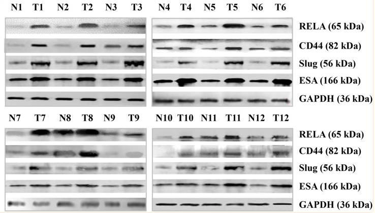
2). MiR-7 reduces the BCSC subset by inhibiting XIST to modulate the miR-92b/Slug/ESA axis and inhibit tumor growth. BREAST CANCER RESEARCH, 2020 (PubMed: 32143670) [IF=7.4]

Application: WB    Species: Human    Sample: breast cancer cells

Fig. 1 Detection of miR-7 and BCSC-related molecular expression. a Expression of miR-7, XIST, miR-92b, RELA, CD44, Slug, and ESA in breast cancer postsurgery samples analyzed by RT-qPCR. b–h Relative expression levels of miR-7 and XIST, miR-7 and RELA, miR-7 and Slug, miR-92b and XIST, miR-92b and Slug, RELA and CD44, and ESA and Slug in that order. i Relative expression levels of RELA, CD44, Slug, and ESA analyzed by Western blotting. All the data represent the mean ± S.D. (n = 12). Blue points represent adjacent noncancerous tissues; red points represent tumor tissues. N noncancerous tissues, T tumor tissues

3). MicroRNA-181a regulates epithelial-mesenchymal transition by targeting PTEN in drug-resistant lung adenocarcinoma cells. INTERNATIONAL JOURNAL OF ONCOLOGY, 2015 (PubMed: 26323677) [IF=5.2]

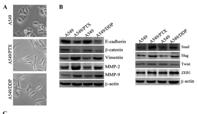
Application: WB    Species: human    Sample: A549 cells

Figure 3. A549/DDP and A549/PTX cells showed molecular and morphological changes that were consistent with EMT. (A) microscopy at x200 magnification was used to assess cell morphology. The A549 cells (parental cells) had an epithelioid, rounded cobblestone appearance and there was limited formation of pseudopodia. A549/PTX and A549/DDP cells exhibited a spindle-shaped morphology and an increased formation of pseudopodia, indicating a loss of cell polarity. (B) E-cadherin, β-catenin, vimentin, MMP-2 and MMP-9 which are EMT-related proteins, were assessed in terms of expression levels. EMT-related transcription factors (Snail, Slug, Twist and ZEB1) were measured in A549/PTX and A549/DDP cells using western blot analysis. (C) The expression changes were confirmed at the mRNA level by qRT-PCR. Expression was standardized to the expression of GAPDH and normalized to 1.0 in the parental cells (compared with the parental A549 cells, means ± SEM, n=3, * P<0.05)

Application: WB    Species: human    Sample: A549/PTX cells

Figure 7.| miR-181a downregulation in A549/PTX cells reverses both drug resistance and morphological and molecular changes. (C) Western blot analysis was used to detect the expression of E-cadherin, β-catenin, vimentin, MMP-9, MMP-2, Snail, Slug,Twist and ZEB1 after transfection.

4). The regulatory effects of metformin on the [SNAIL/miR-34]:[ZEB/miR-200] system in the epithelial-mesenchymal transition(EMT) for colorectal cancer(CRC). European Journal of Pharmacology, 2018 (PubMed: 30017802) [IF=5.0]

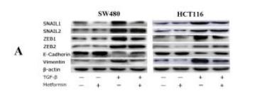
Application: WB    Species: human    Sample: SW480 and HCT116 cells

Fig. 3. |Western-blot for SW480 and HCT116 cells (A)(B) Protein strips and quantitative analysis: significant decreases in the SNAIL1, ZEB1 and vimentin and a significant increase in the E-cadherin of metformin and TGF-β-treated samples compared with only TGF-β-treated samples for both SW480 and HCT116 cells; a significant increase in the ZEB2 of metformin and TGF-β-treated samples compared with only TGF-β-treated samples for SW480 cells; significant decreases in the SNAIL2, ZEB1 and ZEB2 of metformin-treated samples compared with negative control samples for HCT116 cells. *P<0.05

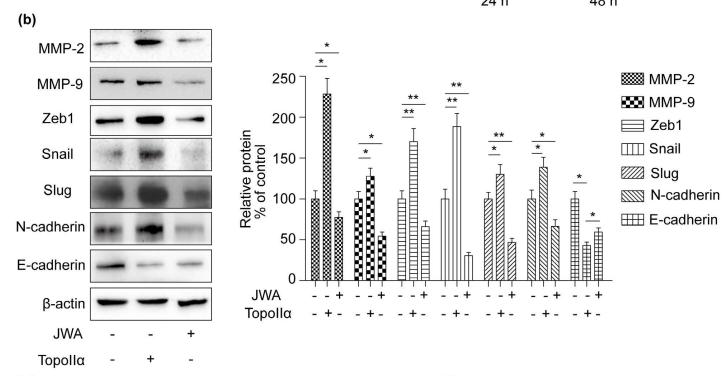
5). EGCG regulates the cross-talk between JWA and topoisomerase IIα in non-small-cell lung cancer (NSCLC) cells. Scientific Reports, 2015 (PubMed: 26046674) [IF=4.6]

Application: WB    Species: human    Sample: NCI-H460 cells


6). MiR-524-5p inhibits angiogenesis through targeting WNK1 in colon cancer cells. AMERICAN JOURNAL OF PHYSIOLOGY-GASTROINTESTINAL AND LIVER PHYSIOLOGY, 2020 (PubMed: 32174132) [IF=4.5]

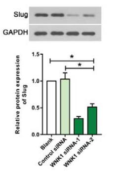
Application: WB    Species: human    Sample: HT29 cells

Fig. 6.| WNK1 silence inhibited angiogenesis in vitro. (A) The relative mRNA and protein levels of WNK1 in HT29 cells.(B) Cell viability was measured by CCK8 assay in HT29 cells after WNK1 silence. (C) Angiogenesis in HUVECs cultured with TCM from WNK1 silenced HT29 cells. Representative western blot for (D) MMP2,(E) MMP9 and (F) Slug in HT29 cells after WNK1 silence.

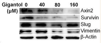
7). Gigantol Attenuates the Metastasis of Human Bladder Cancer Cells, Possibly Through Wnt/EMT Signaling. OncoTargets and Therapy, 2020 (PubMed: 33177841) [IF=4.0]

Application: WB    Species: Human    Sample: bladder cancer cells

Figure 3 Cell invasion assays and expression analysis of Wnt/EMT related genes in cells treated with gigantol. (A) Cell invasion was measured by transwell assay in bladder cancer cells treated with increasing concentrations of gigantol (magnification ×100). (B) qRT-PCR analysis of the Wnt target genes and EMT markers in bladder cancer cells treated with gigantol. (C) Western blot analysis of Wnt/EMT markers in bladder cancer cells treated with indicated concentrations of gigantol. Data in (B) were shown as means ± SD (n = 3), the statistically significant differences were considered at *p<0.05, **p<0.01, ***p<0.001.

8). miR-205-5p regulates epithelial-mesenchymal transition by targeting PTEN via PI3K/AKT signaling pathway in cisplatin-resistant nasopharyngeal carcinoma cells. GENE, 2019 (PubMed: 31158447) [IF=3.5]

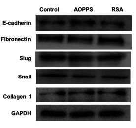
9). Advanced oxidation protein products trigger apoptosis and block epithelial‑to‑mesenchymal transition in crypt epithelial cells. Experimental and Therapeutic Medicine, 2021 (PubMed: 34194563) [IF=2.7]

Application: WB    Species: Rat    Sample: crypt epithelial cells

Figure 2 AOPPs inhibited the EMT of rat crypt epithelial cells. (A) EMT-related protein expression; representative blots of the related proteins. (B) Quantitative data of the related protein expression. (C) EMT-related gene expression at mRNA level. *P<0.05 vs. control; #P<0.05 vs. AOPPs. AOPPs; EMT, epithelial-mesenchymal transition.

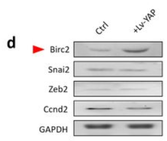
10). Wnt/PCP-YAP-BIRC2 axis maintains cartilage stem/progenitor cell homeostasis in osteoarthritis. Research Square, 2022

Application: WB    Species: Human    Sample: CSPCs

Figure 7. Transcriptomic analysis. a KEGG and GO enrichment analysis of DEGs. b The detailed DEGs that enriched in cell cycle, cellular senescence, autophagy, apoptosis, Hippo signaling pathway, and TNF signaling pathways. c The direct downstream target genes of YAP. d The protein level of Birc2, Snai2, Zenb2, and Ccnd2 in CSPCs.

Restrictive clause


Affinity Biosciences tests all products strictly. Citations are provided as a resource for additional applications that have not been validated by Affinity Biosciences. Please choose the appropriate format for each application and consult Materials and Methods sections for additional details about the use of any product in these publications.

For Research Use Only.
Not for use in diagnostic or therapeutic procedures. Not for resale. Not for distribution without written consent. Affinity Biosciences will not be held responsible for patent infringement or other violations that may occur with the use of our products. Affinity Biosciences, Affinity Biosciences Logo and all other trademarks are the property of Affinity Biosciences LTD.