Product: DFNA5/GSDME Antibody - N-terminal
Catalog: AF4016
Description: Rabbit polyclonal antibody to DFNA5/GSDME - N-terminal
Application: WB
Reactivity: Human, Mouse
Mol.Wt.: 30kDa; 55kD(Calculated).
Uniprot: O60443

View similar products>>

   Size Price Inventory
 100ul $280 In stock
 200ul $350 In stock

Lead Time: Same day delivery

For pricing and ordering contact:
Local distributors

Product Info

Source:
Rabbit IgG
Application:
WB 1:500-1:2000
*The optimal dilutions should be determined by the end user.
*Tips:

WB: For western blot detection of denatured protein samples. IHC: For immunohistochemical detection of paraffin sections (IHC-p) or frozen sections (IHC-f) of tissue samples. IF/ICC: For immunofluorescence detection of cell samples. ELISA(peptide): For ELISA detection of antigenic peptide.

Reactivity:
Human,Mouse
Clonality:
Polyclonal
Specificity:
DFNA5/GSDME Antibody detects N-terminal fragment of DFNA5/GSDME.
Conjugate:
Unconjugated.
Purification:
The antiserum was purified by peptide affinity chromatography using SulfoLink™ Coupling Resin (Thermo Fisher Scientific).
Storage:
Rabbit IgG in phosphate buffered saline , pH 7.4, 150mM NaCl, 0.02% sodium azide and 50% glycerol. Store at -20 °C. Stable for 12 months from date of receipt.
Alias:

Fold/Unfold

2310037D07Rik; 4932441K13Rik; Deafness, autosomal dominant 5; Deafness, autosomal dominant 5 protein; DFNA5; DFNA5 gene; DFNA5_HUMAN; Dfna5h; EG14210; Fin15; ICERE 1; ICERE-1; Inversely correlated with estrogen receptor expression 1; Non-syndromic hearing impairment protein 5; Nonsyndromic hearing impairment protein; Gasdermin-E;GSDME_HUMAN;ICERE1;

Immunogens

Immunogen:

A synthesized peptide derived from human DFNA5(Accession O60443), corresponding to N-terminal amino acid.

Uniprot:
Gene(ID):
Expression:
O60443 GSDME_HUMAN:

Expressed in cochlea. Low level of expression in heart, brain, placenta, lung, liver, skeletal muscle, kidney and pancreas, with highest expression in placenta.

Sequence:
MFAKATRNFLREVDADGDLIAVSNLNDSDKLQLLSLVTKKKRFWCWQRPKYQFLSLTLGDVLIEDQFPSPVVVESDFVKYEGKFANHVSGTLETALGKVKLNLGGSSRVESQSSFGTLRKQEVDLQQLIRDSAERTINLRNPVLQQVLEGRNEVLCVLTQKITTMQKCVISEHMQVEEKCGGIVGIQTKTVQVSATEDGNVTKDSNVVLEIPAATTIAYGVIELYVKLDGQFEFCLLRGKQGGFENKKRIDSVYLDPLVFREFAFIDMPDAAHGISSQDGPLSVLKQATLLLERNFHPFAELPEPQQTALSDIFQAVLFDDELLMVLEPVCDDLVSGLSPTVAVLGELKPRQQQDLVAFLQLVGCSLQGGCPGPEDAGSKQLFMTAYFLVSALAEMPDSAAALLGTCCKLQIIPTLCHLLRALSDDGVSDLEDPTLTPLKDTERFGIVQRLFASADISLERLKSSVKAVILKDSKVFPLLLCITLNGLCALGREHS

PTMs - O60443 As Substrate

Site PTM Type Enzyme
K4 Ubiquitination
T6 Phosphorylation
Y80 Phosphorylation
K83 Ubiquitination
K98 Ubiquitination
K100 Ubiquitination
S106 Phosphorylation
T117 Phosphorylation
S132 Phosphorylation
C156 S-Nitrosylation
K248 Ubiquitination
S252 Phosphorylation
Y254 Phosphorylation
K286 Ubiquitination
K440 Ubiquitination
K467 Ubiquitination

Research Backgrounds

Function:

Plays a role in the TP53-regulated cellular response to DNA damage probably by cooperating with TP53.

Switches CASP3-mediated apoptosis induced by TNF or danger signals, such as chemotherapy drugs, to pyroptosis. Produced by the cleavage of GSDME by CASP3, perforates cell membrane and thereby induces pyroptosis. After cleavage, moves to the plasma membrane where it strongly binds to inner leaflet lipids, bisphosphorylated phosphatidylinositols, such as phosphatidylinositol (4,5)-bisphosphate. Mediates secondary necrosis downstream of the mitochondrial apoptotic pathway and CASP3 activation as well as in response to viral agents. Exhibits bactericidal activity.

PTMs:

Cleavage at Asp-270 by CASP3 (mature and uncleaved precursor forms) relieves autoinhibition and is sufficient to initiate pyroptosis.

Subcellular Location:

Cell membrane.

Cytoplasm>Cytosol.

Extracellular region or secreted Cytosol Plasma membrane Cytoskeleton Lysosome Endosome Peroxisome ER Golgi apparatus Nucleus Mitochondrion Manual annotation Automatic computational assertionSubcellular location
Tissue Specificity:

Expressed in cochlea. Low level of expression in heart, brain, placenta, lung, liver, skeletal muscle, kidney and pancreas, with highest expression in placenta.

Subunit Structure:

The N-terminal moiety forms homooligomer; disulfide-linked. May form an 16-mer complex. Oligomerization occurs in the presence of membranes.

Family&Domains:

Intramolecular interactions between N- and C-terminal domains may be important for autoinhibition in the absence of activation signal. The intrinsic pyroptosis-inducing activity is carried by the N-terminal domain, that is released upon cleavage by CASP3.

Belongs to the gasdermin family.

References

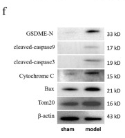
1). Iron induces B cell pyroptosis through Tom20–Bax–caspase–gasdermin E signaling to promote inflammation post-spinal cord injury. Journal of Neuroinflammation (PubMed: 37480037) [IF=9.3]

Application: WB    Species: Mouse    Sample: B cells

Fig. 3 Pyroptosis induced by iron accumulation may occur in splenic B cells after SCI. a Three days after SCI, serum samples were collected for quantification of the protein expression levels of MCP-1, IL-1β, IL-6, and TNF-α by ELISA. b Three days after SCI, injured spinal cord homogenates were collected for quantification of the protein expression levels of MCP-1, IL-1β, IL-6, and TNF-α by ELISA. c Three days after SCI, the spleen was stained with Prussian blue to detect the level of iron ion. d Three days after SCI, serum samples were collected to quantify the concentration of iron ions. e Three days after SCI, B cell lysates were collected to quantify the concentration of iron ions. f Detection of Tom20-Bax-caspase-GSDME pathway-related protein expression in B cells by western blot. g The knock-down efficiency of AAV on Tom20 in B cells was verified. h, l, m The expression levels of MCP-1, IL-1 β, IL-6, TNF- α, IgG and IgM in serum of different groups were detected by ELISA. i–k Three days after SCI, the spleens of mice were observed, and the spleen length and organ index of different groups were compared. n, o, q The spleen was taken for HE staining, Prussian blue staining, and immunofluorescence. CD19+ was red, dapi was blue, and merge was combined. p Detection of nerve evoked potentials in different groups of mice. r Detection of the expression of Tom20-Bax-caspsae-GSDME pathway-related proteins in different groups of B cells. All data are expressed as the mean ± SD (n ≥ 3 replicates per group). ns P > 0.05, * P 

2). Liproxstatin‑1 induces cell cycle arrest, apoptosis, and caspase‑3/GSDME‑dependent secondary pyroptosis in K562 cells. International Journal of Oncology (PubMed: 36004469) [IF=5.2]

Application: WB    Species: Human    Sample: K562 cells

Figure 6 - Lip-1 induces pyroptosis in K562 leukemia cells via caspase-3/GSDME activation. K562 cells were treated with 10 and 20 µM Lip-1 for 24 h in most experiments; part F shows an experiment in which K562 cells were pretreated with z-DEVD-FMK for 3 h and further treated with 20 µM Lip-1 for 12 h. (A) K562 cells double-stained with Annexin V-FITC/PI for flow cytometry. (B) Expression levels of TNF-α assessed by real-time polymerase chain reaction and normalized to RPLP0. (C) Protein levels of TNF-α in cell culture supernatants and cell lysates detected by enzyme-linked immunosorbent assay (minimum detectable concentration, 6.9 pg/ml). (D) BAX, cleaved caspase-3, and cleaved PARP protein levels quantified by western blotting. (E) Lip-1 induced caspase-3, but not caspase-1, activation in a concentrationand time-dependent manner. Caspase-1/-3 activities were measured by colorimetric assay. (F) Comparison of cell viability and the protein level of N-terminal GSDME in Lip-1-treated K562 cells and the effects of z-DEVD-FMK inhibition. SH-SY5Y whole cell lysate was used as a positive control. (G) Expression levels of GSDME assessed by real-time polymerase chain reaction and normalized to RPLP0. (H) GSDME and cleaved N-terminal GSDME protein levels quantified by western blotting. SH-SY5Y whole cell lysate was used as a positive control. (I) TUNEL assay used to determine DNA fragmentation. Specimens treated with DNase Ⅰ were used as positive controls. Data represent the mean ± SD from at least three independent experiments. *P<0.05, **P<0.01 and ***P<0.001 vs. Control. Lip-1, liproxstatin-1; ns, not significant.

Restrictive clause


Affinity Biosciences tests all products strictly. Citations are provided as a resource for additional applications that have not been validated by Affinity Biosciences. Please choose the appropriate format for each application and consult Materials and Methods sections for additional details about the use of any product in these publications.

For Research Use Only.
Not for use in diagnostic or therapeutic procedures. Not for resale. Not for distribution without written consent. Affinity Biosciences will not be held responsible for patent infringement or other violations that may occur with the use of our products. Affinity Biosciences, Affinity Biosciences Logo and all other trademarks are the property of Affinity Biosciences LTD.