- WB
- IHC
- IF/ICC
- Cites


 and mouse anti-beta tubulin Ab(T0023 1:200) for 1 hour at 37°C. An AlexaFluor594 conjugated goat anti-rabbit IgG(H+L) Ab(Red) and an AlexaFluor488 conjugated goat anti-mouse IgG(H+L) Ab(Green) were used as the secondary antibody.
The nuclear counter stain is DAPI(blue).")
 and (B) NIH-3T3 and HaCat Cells were treated with PnAg at different concentrations and cell viability was tested using MTT analysis. (C) Wound healing assay reflected the effect of PnAg on cell migration. (D) Binding mode of PnAg in the active pocket of gp130. (E) and (F) MMPs activity and expression levels of Stat3, VEGF, TGFB-1, and TGFB1 detected using zymographic and Western blot assays. (G) Diagram of the proposed function of PnAg in wound inflammation and re-epithelialization controls.")
 Photographs of rat skin full-thickness excision wounds on different post-excision days. (B) Change in wound areas of SD rats after treatment; (C) and (D) Expression levels of collagen I, NF-κB, TGF-ß, MMP-2, and MMP-9 in tissues on day 7 and 17 detected by immunohistochemistry. (E) Histogram of protein expression levels in these tissues. (F) and (G) Histomorphological changes in wound tissues stained by Masson trichrome and HE on day 17.")
. BMFBs were pretreated with PD98059 2 h before the indicated treatments with 5 ng/ ml TGF-b1. b-actin was used as a loading control for each condition (B). The data shown represent one of three independent experiments.")

 Lung fibroblasts were treated with/without TGF-β1 (5 ng/mL) and compounds 42, 44 and 45 (10 μΜ) for 24 h. The expression of α-SMA in cell extracts, Col1 in the medium (supernatant) were examined by Western blot. Lung fibroblasts were treated with/without TGF-β1 (5 ng/mL) and compound 42, 44 and 45 (10 μΜ) for 30min, the expression of p-Smad3 was examined by Western blot. β-Actin was used as a loading control.")
 Immunohistochemical analysis of α-SMA, Col1 and p-Smad3 in lung sections. Representative images of the staining are shown (n = 10 per group). Error bars indicate mean ± SEM.")
 were subjected to sham operation or 30 min of unilateral ischemia of left kidney followed by reperfusion. For biochemical and histological analyses, kidney tissues were collected at 2, 7 or 14 days after renal ischemia. To determine the function of postischemic left kidney, right nephrectomy was performed one day before animal sacrifice and blood samples were then collected at animal sacrifice to measure serum creatinine.(A) Post-ischemic kidney weight/body weight (mg/g). (B) Contralateral kidney weight/body weight (mg/g). (C) Serum creatinine(mg/dL). (D) Representative histology images of hematoxylin-eosin staining. Bar = 200 μm. (E) Pathological score of tubular atrophy. (F) Representative immunoblots of collagen 1, α-SMA, vimentin, and GAPDH. GAPDH was used as a loading control.")
 measured by PicoGreen staining in LX2 cells; Scale bar, 10 μm. b Representative western blots of TOM20 with GAPDH serving as the internal reference. Bar graph represents the mean ± SEM. *P < 0.05 and **P < 0.01 vs. the indicated groups. c TUNEL staining of LX2 cells from the indicated groups; scale bar, 25 μm. Bar graph represents the mean ±SEM. **P < 0.01 and ***P < 0.001 vs. the indicated groups. d Representative western blots of cleaved caspase3, cleaved caspase9, collagen I and α-SMA. Bar graph represents the mean ± SEM of three different experiments. *P < 0.05 and **P < 0.01 vs. the indicated groups")
 H&E staining of liver sections from mice, n=6 per group, scale bar, 50 μm. (B) Masson’s trichrome staining for collagen in the mouse liver,n=6 per group, scale bar, 50 μm. Immunohistochemistry staining for collagen I (C) and α-SMA (D)in mouse liver sections. n=6 per group, bar graph represents the mean ± SEM, scale bar, 25 μm.")
 Protein extracts from NRK-49F and NRK-52E cells were analyzed by western blot for E-cadherin, collagen I, α-SMA, FN, PAI1 and collagen IV. GAPDH was used as a control. Data are representative images or expressed as the mean ± SD of each group (n = 3 per group). Densitometry analysis was performed with Image J software for quantification. ##P < 0.01; ###P < 0.001; ####P < 0.0001 vs control group. *P < 0.05; **P < 0.01; ***P < 0.001 vs TGF-β1 group. All statistical tests were two-sided.")
 CCK-8 assay of hBMSCs on the indicated surfaces after 1, 3 and 7 days of culture.
* denotes p < 0.05 and ** denotes p < 0.01 compared to Ti-NTs, # denotes p < 0.05 compared to the corresponding control group without peptides. Error bars denote the
standard deviations over quadruplicate measurements with separately implants. (B) Confocal fluorescence microscopy images of hBMSCs stained with vinculin, F-actin and DAPI
after being cultured for 24 h. Scale bar, 50 μm. (C-F) qRT-PCR assay of osteogenic gene expression of (C) ALP, (D) RUNX-2, (E) COL1 and (F) OPN of hBMSCs after 7 and 14
days of culture. * denotes p < 0.05, ** denotes p < 0.01 and *** denotes p < 0.001 compared to Ti-NTs-P-A; # denotes p < 0.05, & denotes p < 0.01 and $ denotes p < 0.001
compared to the corresponding control group without peptides. All error bars denote the standard deviations over quadruplicate measurements with separately implants. (G)
Immunofluorescence staining of hBMSCs cultured on Ti-NTs-P-A for 7 days (ALP and RUNX-2) and 14 days (OPN). The images were obtained by confocal fluorescence
microscopy. Scale bar, 50 μm. (H) Western blotting of hBMSCs cultured on the substrates for 7 and 14 days. At each time point, left lane was Ti-NTs, middle lane was Ti-NTs-A
and right lane was Ti-NTs-P-A. * denotes p < 0.05, ** denotes p < 0.01 and *** denotes p < 0.001 compared to Ti-NTs-P-A. Error bars denote the standard deviations over
triplicate measurements with separately Western blotting results.")
 Representative Western blot and summarized data showing the effects of
Ang II on the expression of alpha-SMA and GAPDH in 24, 48, and 72 h (N = 4). *p < 0.05 vs. Ang II 0 nM. (D–F) NRK-52E were stimulated with different
concentrations of TGF-beta (0, 5, 10 ng/ml) for 24, 48, and 72 h and total proteins that analyzed by Western blot using antibodies against alpha-SMA (N = 4).
*p < 0.05 vs. TGF-beta 0 ng/ml. (G–I) Representative Western blot and summarized data of CollagenI and GAPDH in 24, 48, and 72 h (N = 4). *p < 0.05, vs.
TGF-beta 0 ng/ml.")
. (A) Immunohistochemical validation of the essential extracellular
matrix proteins of the native testis (laminin, collagen type I, collagen type IV, and fibronectin) in the DTM. Scale bar, 200 µm. (B–E) Relative quantity of collagen I,
collagen IV, fibronectin, and laminin. ImageJ software was used to determine the average optical density (AOD). Data were shown as mean ± SD, n = 8. Differences
were considered statistically significant at *p < 0.05, **p < 0.01.")

 CRLF1 expression induced by TGF-β1 in LF cells transfected with siRNAs. (B,C) ERK signaling pathway
activation by CRLF1 and TGF-β1. (D) siCRLF1 attenuated the ERK phosphorylation induced by TGF-β1. (E) siCRLF1 reduced the fibrosis markers expression
induced by TGF-β1. (F,G) Inhibition of the ERK pathway attenuated the CRLF1- and TGF-β1-induced expressions of fibrosis markers in LF cells. (H) siCRLF1
significantly reduced the pro-fibrotic effect of IL-1β (20 ng/ml). (I) siCRLF1 significantly reduced the pro-fibrotic effect of mechanical stretching forces.")
. (A) Representative images of contrast microscopy to show cellular morphologic changes. Bar:100μm. (B) Representative immunoblot of Fibronectin (FN), Collagen 1, PARK7 and GAPDH (protein loading control) in whole cell lysates. (C–E) Densitometry of FN, Collagen 1 and PARK7(n=3). The signal of FN, Collagen 1, or PARK7 was normalized to the GAPDH signal of the same samples to determine the ratios. The values are expressed as fold change over control cells. Each symbol (circle) represents an independent experiment. (F) Quantification of Park7 mRNA levels. Error bars: SEM. *p < 0.05, **p < 0.01, ***p < 0.001. ns, not significant.")
 of animals after PTL treatment. b Hydroxyproline content in lung tissues. c Western blot analysis of type I collagen (Col1) and α-SMA in lung tissues was performed. GAPDH was used as a loading control.")

 from day 7–13 after administrating BLM (A) Immunohistochemistry was used to analyze the expression levels of α-SMA, Col1 and Fn (n = 3). Quantitative analysis was shown beside. Scale bars: 50 μm (B–C) The expression levels of α-SMA and Col1 were detected by immunofluorescence in lung sections. The analyses of mean gray value were shown beside. Scale bars: 50 μm (D) Lung homogenization was used to analysis the α-SMA, LC3-II/I and p-mTOR (S2248), mTOR expression levels by Western blot (n = 6). Densitometric analyses were shown beside. Data in (A,D) are means ± Standard Error of Mean, *p < 0.05, **p < 0.01, ***p < 0.001, and NS: nonsignificant (one-way ANOVA). β-tubulin was used as a loading control.")
 and Fibronectin (B). RDP treatment decreases the protein expressions of collagen I (C), fibronectin (D), vimentin (E), α-SMA(F) and increases the protein expression of E-cadherin (G) detected by western blot. Data are presented as mean ±SEM (n = 3). #P < 0.05, ##P < 0.01, UUO group vs. the sham group; *P < 0.05, **P <0.01, UUO + RDP group vs. UUO group")
 and high (bottom) power images of control and silica-exposed lung after van Gieson (VG) staining. Right). Quantitative assessment of area and number silicotic nodules in the lungs of control and silica-exposed rats. Western blot analysis for collagen I (COLI) and α-smooth muscle actin (-SMA) in whole lung tissues and isolated fibroblasts from control and silica-exposed rats Abbreviations: C, control; S,silicosis. Statistical significance was assessed using the Student's t-test")
 or by measuring the expression of FN, Col-I,CTGF, and a-SMA by Western blotting and further quantified (B). One representative of 3 independent experiments is shown.N.S., not significant. Data represent means 6 SD. *P , 0.05 vs. vehicle-sham, **P , 0.01 vs. vehicle-sham, ***P , 0.001 vs.vehicle-sham, #P , 0.05 vs. vehicle-UUO, ###P , 0.001 vs. vehicle-UUO")
-β1-induced activation, extracellular matrix (ECM)
accumulation, and the migration of pulmonary fibroblasts. (A,B) Mouse lung fibroblast (Mlg) or NaCl-primary pulmonary
fibroblast (PPF) cells were exposed to TGF-β1(5 ng/mL) and/or regorafenib (RG) (2 µM, 4 µM) for 24 h to detect the
expression levels of α-SMA and Col 1 by Western blotting. Densitometric analyses are shown; (C) Mlg cells were exposed to
TGF-β1 (5 ng/mL) and/or RG (2 µM, 4 µM) for 24 h, and quantitative real-time PCR was used to detect the mRNA levels
of α-SMA, Col 1a, and Fn; (D) RG (2 µM, 4 µM) was exposed to Mlg cells for 24 h, and the α-SMA expression level was
detected by immunofluorescence. Relative pixels intensity is shown. Scale bars: 50 µm; (E) Mlg cells were treated with TGF-
β1 and/or RG (2 µM, 4 µM) for 0, 12, 24, or 36 h. Distance analyses are shown below. Scale bars: 100 µm; (F) The PPF cells
isolated from NaCl-treated and BLM-treated mice were incubated RG (2 µM, 4 µM) for a series of time points (0, 12, 24,
36 h). Distance analyses are shown below. Scale bars: 100 µm; Data in (A–F) are means ± standard error of mean (SEM),
and β-tubulin was used as a loading control. ### p < 0.001, ** p < 0.01, *** p < 0.001 (one-way ANOVA), NS: not significant.")
-induced pulmonary fibrosis in mice. (A) Regorafenib (15 mg/kg,
30 mg/kg) and nintedanib (100 mg/kg) were given orally once a day from days 7–13 after BLM treatment, lungs were
harvested at day 14, and body weight loss was measured every day; (B) Percentages of surviving mice were plotted from
days 7–14 after bleomycin treatment; (C) Representative images of hematoxylin–eosin (H&E) and Masson staining of lung
tissue sections. Scale bars: 50 µM; (D) Percentages of fibrotic area in lung tissues; (E) Hydroxyproline contents in right lung
tissues; (F) Immunohistochemistry of lung sections was used to analyze the expression levels of α-smooth muscle actin
(α-SMA), collagen 1 (Col 1), and fibronectin (Fn). Quantitative analysis is shown below. Scale bars: 50 µm; (G–L) Pulmonary
function parameters, such as forced vital capacity (FVC), forced expiratory volume in 0.1 s (FEV0.1), forced expiratory
volume in 0.3 s (FEV0.3), expiratory resistance (Re), inspiratory resistance (Ri), and dynamic compliance (Cydn). Data in
(A,B) and (D–L) are means ± standard error of mean (SEM), n = 6; # p < 0.05, ## p < 0.01, ### p < 0.001, * p < 0.05, ** p < 0.01,
*** p < 0.001 (one-way ANOVA), NS: not significant.")

 The Sirius Red-positive area in the CCl4/Tg group was markedly larger than that in the CCl4/WT group(200x). (b) The hydroxyproline content in the CCl4/Tg group was higher than that in the CCl4/WT group. (c–e) The Collagen1α1 expressions in the CCl4/Tg group were remarkably higher than those in the CCl4/WT group detected by RT-PCR, immunohistochemical staining (200x), and Western blot. Data are expressed as mean ± SD, ∗p < 0 05 and ∗∗p < 0 01.")
 Representative western blot showing α-SMA, E-Cadherin and collagen I. (B) Representative western blot showing TGF-β1, p-Smad3 and p-p38 MAPK. (C)-(H) Quantitative analyses of the results from western blot. The each protein level was normalized to β-actin. *P < 0.05, n=3.")
 After osteogenic induction of hPDLSCs with various concentrations of TGF-β3 for 7 and 14 d, expression of osteogenic proteins (COL I, COL II, and ALP) was detected by (A) western blotting and analyzed by grayscale scanning for (B) COL I, (C) COL II, and (D) ALP (n = 3). ns means no significant differences,p >0.05 vs. control; ** p < 0.01 vs. control; ***p < 0.001 vs. control. Abbreviations: Col I, type I collagen;Col II, type II collagen; ALP, alkaline phosphatase")
Immunohistochemistry for a-SMA and collagenI(Brownarea) in bleomycin-induced pulmonary fibrosis in mice on day 14. Representative images of lung sections from each group are shown.Dashed boxes on top panels indicate region shown at higher magnification in bottom panels.Magnification=9200(toppanels),9400(bottompanels).")
 Immunohistochemical staining for collagens I, III and α-SMA,scale bars=100 µm. Bar graphs show the percentage of (B) collagens I, (C) III and (D) α-SMA. (E) Western blotting detection of collagens I, III and (F) α-SMA.")
 Immunohistochemical staining for collagens I, III and α-SMA,scale bars=100 µm. Bar graphs show the percentage of (B) collagens I, (C) III and (D) α-SMA. (E) Western blotting detection of collagens I, III and (F) α-SMA.")
 Protein levels of FGFR4, TGF-β1, α-SMA, and COL1A1 measured by western blot. LPS:lipopolysaccharides; NC: scramble-miR control. ∗P < 0:05, ∗∗P < 0:01, and ∗∗P < 0:001 versus control; #P < 0:05, ##P < 0:01,and ###P < 0:001 versus NC+LPS.")

. Scale bar,100 μm. B Representative immunoblots of Col IA/Col IIIA/α-SMA and summarized intensities of blots in the cortex and medulla (n = 6–8 per group).")
 Cell lysates were obtained for Western blot analysis.")
 mRNA and (E) protein expression levels of α-SMA, collagen I and collagen III. *P<0.05, **P<0.01 and ***P<0.001. ALK1, activin receptor-like kinase 1;Dor, dorsomorphin-1; BMP9, bone morphogenetic protein 9; α-SMA, α-smooth muscle actin; ctrl, control.")
 Western blot analysis of the protein levels of Fn, Col1 and α-SMA in lung tissues.")
 Immunohistochemical staining of α-SMA, Col1 and Fn in lung tissues. Scale bar = 50 µm.")
, Scale bar = 50 μm; the mean IOD was calculated as total IOD value/total
region in similar scale lung area from each group. Data are presented as the mean ± SD of at least three repeat experiments, **P < 0.01, ***P < 0.001 vs. the
control group; #P < 0.05, ##P < 0.01 vs. the silicosis group.")
 Immunochemistry staining of α-SMA expression in the lungs of wild-type mice, n = 6 in each group. Magnification, ×40 (top panel) and ×200 (bottom panel). Silicotic mice underwent various treatment combinations with DIZE (ACE2 activator), A779 (Mas receptor blocker), and MLN-4760 (ACE2 inhibitor). (B) The proportion of silicotic areas in the lung samples in (A). (C) Western blot showing the protein expression of α-SMA (D), Vim (E), pro-Col I (F), pro-Col III (G), and E-cad (H) in the lungs of mice from the various treatment groups. Values represent the mean ± SD, n = 3 independent experiments, fold change is expressed relative to the control (no treatments), *P < 0.05 vs corresponding group, **P < 0.01 vs corresponding group.")
 Kidney tissue lysates were subjected to immunoblot analysis with specific antibodies against fibronectin, collagen I, or glyceraldehyde 3-phosphate dehydrogenase (GAPDH). (B) Expression levels of fibronectin were quantified by densitometry and normalized to GAPDH. (C) Expression levels of collagen I were quantified by densitometry and normalized to GAPDH. (D) Photomicrographs (original magnification, ×400) illustrate immunohistochemical fibronectin staining of the kidney tissues. RGTZ, rosiglitazone. Data are represented as the mean ± SEM. *p < 0.05 vs sham group; #p < 0.05 vs sham + RGTZ group. **p < 0.05 vs HN group.")
 Predicted binding sites of miR-29a-3p in circKRT7 and COL1A1 3ʹUTR. (B) Localization of circKRT7 and miR-29a-3p in ES-2 cells. (C) Luciferase activity assay analyzes the binding ability of miR-29a-3p and circKRT7. (D) Luciferase activity assay analyzes the binding ability of miR-29a-3p and COL1A1 3ʹUTR. (E) miR-29a-3p and circKRT7 expression after over-expression of wild type or mutated circKRT7. (F) Western blot analysis COL1A1 protein level after treated with circKRT7 shRNA or along with miR-29a-3p ASO. Experiments were performed in triplicate. Statistical significance was considered at P < 0.05 and labeled with *.")
 Representative Western blotting and quantitative densitometry analysis images for caspase 3. (B)
Representative Western blotting and quantitative densitometry analysis images for TGF-b1. (C) Representative Western blotting and quantitative densitometry analysis images for MMP9. (D)
Representative Western blotting and quantitative densitometry analysis images for collagen I. (E) Representative Western blotting and quantitative densitometry analysis images of collagen
III. (n = 4). ∗
P < 0.01 vs. sham; +
P < 0.01 vs. the AMI group. All protein levels are normalized to GAPDH levels. AMI: Acute myocardial infarction; ASX: Astaxanthin; GAPDH: Glyceraldehyde3-phosphate dehydrogenase; MMP9: Metalloproteinase 9; TGF-b1: Transforming growth factor b1.")

 Western blotting detection of collagen Ⅰ, collagen Ⅲ, and α-smooth muscle actin (α-SMA). Bar graphs show fold changes for the collagen Ⅰ,collagen Ⅲ, and α-SMA expression as analyzed by western blotting. Glyceraldehyde 3-phosphate dehydrogenase (GAPDH) was used as a loading control (n 3). Datawere shown as mean ± SEM. **p < 0.01 vs. control, #p < 0.05 vs. ISO, ##p < 0.01 vs. ISO.")
 The effects of different doses of BEL (15, 30, 60 μM)on α-SMA, collagen Ⅰ, and collagen Ⅲ expression induced by TGF-β1 using immunofluorescence staining.")
 Effects of emodin at various concentration on protein
levels of FN, Col-I, a-SMA. (B) Effects of emodin at various concentration on protein levels of pSmad3, Smad3, CTGF. One representative of 3 independent experiments is shown. Data
represent means ± SD. **P < 0.01 vs. sham, #P < 0.05 vs. UUO, ##P < 0.01 vs. UUO.")
 mRNA expression of Collagen I, fibronectin and α-SMA. (B) Immunohistochemical staining of lung sections in different groups. Magnification, ×400. Scale bar, 25 µm. (C) Average IOD of Collagen I, fibronectin and α-SMA in different groups. ***P<0.001 vs. Control; ###P<0.001 vs. Silicosis. α-SMA, α-smooth muscle actin; ASV, astragaloside IV; IOD, integral light density.")
 Detection of overexpression efficiency of CCL19 plasmid was measured by qRT-PCR. The inflammatory cytokine IL1-α (B), IL-1β (C), TNF-α (D), IL-6 (E) and the deposition of ECM such as collagen1 (F), collagen3 (G) and OPN (H) were detected by qRT-PCR.n=5 (I) Western blot showed the expression of proteins collagen1 and IL-6. n=4. Migration (J) and proliferation (K) of VSMCS after overexpressing CCL19. n=6. EV: Empty vector. *P<0.05; **P<0.01.")
 MC3T3-E1 cells were transfected with adenoviruses. GFP marker genes were detected 48 h after transfection under both bright field and fluorescence microscopy (100×). (B) Real-time PCR analysis showing Nampt gene expression normalized to β-actin and (C) Western blot results showing the protein expression of Nampt 3 days after the transfection. On the 7th d of culture in OBM, (D) ALP staining and (E) Measurement of ALP activity showing that ALP was decreased in sh-Nampt cells and increased in ad-Nampt cells. (F) Western blot results showing the protein expression of Sirt1, Runx2, Col1a1 and OCN. Con (control) cells were not transfected; sh-con and ad-con (negative control) cells were transfected with nontargeting control viruses; sh-Nampt cells were transfected with Nampt knockdown shRNA; ad-Nampt cells were transfected with vector over expressing Nampt. The data are expressed as the mean ± SD. *, p<0.05; **, p<0.01.")
. mRNA expression level of RUNX2 (B), osterix (D), and COL1A1 (F) of the callus ares. Immunohistochemistry of RUNX2 (C), osterix (E), and type I collagen (G) within the callus area. Magnification, ×200, scale bar = 100 µm. The typical positive stained cells or regions were indicated by arrows. n=6, Mean ± SD, **p < 0.01.")
. mRNA expression level of RUNX2 (B), osterix (D), and COL1A1 (F) of the callus ares. Immunohistochemistry of RUNX2 (C), osterix (E), and type I collagen (G) within the callus area. Magnification, ×200, scale bar = 100 µm. The typical positive stained cells or regions were indicated by arrows. n=6, Mean ± SD, **p < 0.01.")

 Western blot analysis for α-smooth muscle actin (α-SMA), collagen I, transforming growth factor (TGF)-β1 and connective tissue growth factor (CTGF). The relative expression of (B) α-SMA, (C) collagen I, (D) TGF-β1 and (E) CTGF. Data were shown as mean value ± SD (n=6). ***P<0.001, versus Control group. &&P<0.01, &&&P<0.001, versus NC agomir group.")
.")
 collagen Ⅰ and (B) collagen III in atrial tissues of heart were determined via immunohistochemistry. At the same time, quantitative analysis was carried out.")
 RT-PCR showing relative mRNA expression of collagen type I alpha 1 chain (COL1A1), collagen type I alpha 2 chain (COL1A2), collagen type III alpha 1 chain (COL3A1), matrix metalloproteinases MMP1, MMP2, and MMP3 in fibroblasts treated with different media (versus GAPDH mRNA). (B) Western blotting showing protein levels of collagen I, collagen III, MMP1, and MMP2 in the two groups of fibroblasts. (C) Relative collagen I, collagen III, MMP1 and MMP2 protein levels (versus GAPDH). Data are shown as means ± standard deviation (SD). *P < 0.05; **P < 0.01; ***P < 0.001. BMSC-CM, bone marrow stem cell-conditioned medium; con, control medium treatment group; CM, BMSC-CM treatment group.")
 Immunohistochemical staining of osteogenesis-associated proteins Runx2, BMP-2, and COL I. Red arrows indicate positive cells (B) Runx2 positive statistics (C) BMP-2 positive statistics (D) COL I positive statistics (n = 3, *p < 0.05, **p < 0.01, ***p < 0.001 vs model).")
 HE staining to detect morphological changes; Masson trichrome staining to detect cardiac collagen deposition (B–D) Western blotting detection of collagen Ⅰ, collagen Ⅲ, and α-smooth muscle actin (α-SMA). Bar graphs show fold changes for the collagen Ⅰ, collagen Ⅲ, and α-SMA expression as analyzed by western blotting. Glyceraldehyde 3-phosphate dehydrogenase (GAPDH) was used as a loading control (n = 3). Data were shown as mean ± SEM. **p < 0.01 vs. control, #p < 0.05 vs. ISO, ##p < 0.01 vs. ISO.")
 EMT-related protein expression; representative blots of the related proteins. (B) Quantitative data of the related protein expression. (C) EMT-related gene expression at mRNA level. *P<0.05 vs. control; #P<0.05 vs. AOPPs. AOPPs; EMT, epithelial-mesenchymal transition.")
 Expression of fibrosis-related proteins in serum by western blot analysis.")
 Mlg and NaCl-PPF cells were exposed to Wnt3a(100 ng/ml) and/or Ellagic acid (5 µM, 10 µM) 24 h to detect expression level of Col1 by using Western blot.")
 on diabetic-induced cardiac remodeling.(F) The percentage of fibrosis was calculated. Immunohistochemical staining of panel (G) collagen I, (H) collagen III. Scale bar,50 µm. Data are presented as mean ± SD. **P < 0.01 CON vs. SHAM; ##P < 0.01 SG vs. SHAM. n = 3 in each group.")
 Representative photomicrographs of HE, Masson’s staining, and IHC (Col I and Col III) of the myocardium of rats. (B) The area of fibrosis in the myocardium was calculated using the Image J software. (C) The area of Col I in the myocardium was calculated using the Image J software. (D) The area of Col III in the myocardium was calculated using the Image J software. *p < 0.05 vs. WKY group; #p < 0.05 vs. SHR group; ▲p < 0.05 vs. OBH group; ⋄p < 0.05 vs. OBH + HQQR(L) group; Δp < 0.05 vs. OBH + HQQR(H) group.")
 The protein expression of Col I and Col III was analyzed by western blotting. (B) The expression of IL-1β in the supernatant of the different groups of CFs. (C) The mRNA expression of NLRP3, caspase-1, and IL-1β was analyzed by western blotting. (D) The relative protein expression levels of NLRP3, caspase-1, and IL-1β among the different groups were quantified using the Image J software. *p < 0.05 vs. control group; #p < 0.05 vs. Ang II group; ▲p < 0.05 vs. HQQR(L) group; ⋄p < 0.05 vs. HQQR(H) group; ♦p < 0.05 vs. Ang II + Opti-men group; ▼p < 0.05 vs. si-NLRP3 group; ‡p < 0.05 vs. vector + HQQR group.")

and collagen deposition was detected by masson staining (B). Scale bar = 100 μm. The level of hydroxyproline in the lungs was examined with a hydroxyproline
detection kit (C). The levels of TGF-β1, collagen I, and collagen III in the lungs were detected by western blot (D-E). The results are presented as mean ± SD. N = 6 for
each group.")
 collagen I and (B) collagen III in atrial tissues of heart were determined via immunohistochemistry. At the same time, quantitative analysis was carried out. (C) Protein expression levels of TGF-β1, CTGF, fibronectin and α-SMA in atrial tissues of heart were assessed via western blotting. The results are presented as the mean ± standard deviation (n=6). ###P<0.001 vs. sham group; **P<0.01 and ***P<0.001 vs. AF + LV-NC group. CTGF, connective tissue growth factor; α-SMA, α-smooth muscle actin; AF, atrial fibrillation; LV-NC, lentivirus negative control; miR, microRNA.")
 and COL-1(red fluorescence) in
LX-2 cells.*p < 0.05; **p < 0.01; ***p < 0.001. All data are expressed as the means ± SD from three independent experiments, each performed in triplicate. (For interpretation of the
references to colour in this figure legend, the reader is referred to the Web version of this article.)")
 Proteins of lung tissues were extracted and western blotting was used to detect proteins with antibodies as indicated (n = 3). Relative intensity of each band
was normalized to GAPDH protein. (B) Immunofluorescence was performed on cryosection of lung tissues with indicated antibodies (n = 3). Scale bar: 100 μm.
Relative fluorescence intensity was analyzed. Data are mean ± SD, compared using one-way ANOVA test.*, p < 0.05.")
 HE staining of the kidneys of UUO mice and the IHC analysis of CoL-1, Fn, and Sp1 in renal tissues (400×). (B-D) Areas showing positive immunostaining for CoL-1, Fn, and Sp1 in mice kidneys. Data are presented as mean ±SEM (n=6). (E-F) Correlation analysis of CoL-I and Fn expression levels with Sp1, as determined by the IHC analysis of mice kidneys (n=6). (G-H) Western blotting and densitometric analyses of Sp1 expression in the kidneys of UUO mice; *P<0.05, data are presented as the mean ±SEM (n=4).")
 qRT-PCR, western blotting and densitometric analyses of Sp1 expression in BUMPT cells following the transfection of Sp1 siRNA. (D-E) qRT-PCR analysis of the mRNA expression levels of CoL-I and Fn in TGF-β1-stimulated BUMPT cells following transfection with Sp1 siRNA. (F-G) Western blotting and densitometric analyses of CoL-I and Fn expression level in TGF-β1-stimulated BUMPT cells following Sp1 siRNA transfection. *P<0.05, #P<0.05; data are presented as the mean ±SEM (n=3).")
 RT-PCR was performed to detect mRNA levels of α-SMA, Fibronectin and Collagen I in lung tissues. (B) Protein levels of α-SMA and Collagen I were verified by Western blot in lung tissues. GAPDH was used as an internal reference in densitometric analysis (C) Immunohistochemical staining analysis of α-SMA and Fibronectin in lung tissues. (D) Protein expression levels of E-cadherin and N-cadherin were assessed by Western blot in lung tissues. GAPDH was used as a loading control. Data was presented as the means ± SD, n = 3. * p < 0.05, ** p < 0.01, *** p < 0.001.")
 Expression levels of COL-I and α-SMA were determined in LX2 cells exposed to different concentrations of TGF-β1 by western blotting. *P<0.05 vs. CTL group. (B) Expression levels of LC3 and P62 were determined in TGF-β1-treated LX-2 cells. *P<0.05 vs. CTL group. (C) The expression of P62 was detected in TGF-β1-treated LX2 cells by immunofluorescence. The cells were immunostained against antibodies P62 (green), DAPI was used to stain nuclei (blue). (D) Expression levels of COL-I and α-SMA were determined in TGF-β1-treated LX-2 cells after RAPA treatment by western blotting. *P<0.05 vs. TGF-β1-treated group. TGF-β1, transforming growth factor-β; COL-I, collagen-I; α-SMA, α-smooth muscle actin; RAPA, rapamycin; CTL, control.")

 by inhalation from day 21 to day 42. Mice were intraperitoneally injected with 20 mg/kg Rhy-SLNs or 20 mg/kg Rhy at one hour before the airway challenge with OVA. a, the representative images of Masson-stained airway in lung tissues. b and c, the representative images of periodic acid-Schiff-stained goblet cells of airway in lung tissues, which were quantified with the mucus score. Black arrows represented the goblet cells. d, the protein levels of α-SMA and collagen I. n = 6 in each group. *p < 0.05, **p < 0.01. OVA, ovalbumin; Rhy, rhynchophylline; Rhy-SLNs, rhynchophylline-solid lipid nanoparticles; α-SMA, alpha-smooth muscle actin")
. To fit into the manuscript properly, the gel was reasonably trimmed. aa, P < 0.01 vs the control group; b, P < 0.05, bb, P < 0.01 vs the Wnt-1 group. ##, P < 0.01 vs the normal group; *, P < 0.05 and **, P < 0.01 vs the model group")

 Representative immunoblots of SIRT3, p-P38, p-JNK, c-Jun, MMP-1, MMP-3, TGF-β, Smad3 and Collagen I protein expressions in skin tissue. (B) Optical density histograms of SIRT3/Tubulin, p-P38/Tubulin, p-JNK/Tubulin, c-Jun/Tubulin, MMP-1/Tubulin, MMP-1/Tubulin, MMP-3/Tubulin, TGF-β/Tubulin, Smad3/Tubulin and Collagen I/Tubulin. # indicates significant difference between the model group and the blank group (#p < 0.05; ##p < 0.01); * indicates significant difference between the administered group and the model group (*p < 0.05; **p < 0.01).")
 Dose-dependent cytotoxicity of anlotinib in primary mouse lung fibroblast isolated from healthy mice by CCK-8. (B) Western blots analysis of Fibronectin, Collagen I, α-SMA and β-actin in mouse lung fibroblasts treated with anlotinib (1 µM) for 3 h and then exposed to TGF-β1 (10 ng/ml) for 3 h, followed by TGF-β1 for an additional 24 h. (C) Quantification for the indicated protein (mean ± SD, n = 3). Immunofluorescence for α-SMA (green) (D) and Fibronectin (green) (E). DAPI-stained nuclei (blue). Scale bar, 25 µm. (F) An EdU assay was used to observe the proliferative cells. Scale bar, 100 µm. (G) The number of EdU-positive cells was recorded (mean ± SD, n = 3). (H) A cck8 assay was used to observe the proliferative cells. (I) Representative images to show scratch-wound assay. Scale bars, 100 µm. Experiments were performed as in B. Images were taken 0, and 24 h after assay (white lines indicate wound edge). (J) Quantitative analysis of migration distance (mean ± SD, n = 3). (K) Western blots analysis of Fibronectin, Collagen I, α-SMA and β-actin in IMR90 cells treated with anlotinib (1 µM) for 3 h and then exposed to TGF-β1 (10 ng/ml) for an additional 24 h. (L) Quantification for the indicated proteins in IMR90 cells (mean ± SD, n = 3). *p < 0.05, **p < 0.01, ***p < 0.001 VS TGF-β1-treated group (ANOVA).")

 Serum levels of ALT and AST in Oil+vehicle, Oil+sTREM‐1, CCl4+vehicle and CCl4+sTREM‐1 groups. (B) Liver histology (HE staining, 100X). (C) Liver fibrosis (Sirius Red staining; 100X). (D) Collagen I and α‐SMA expressions (IHC staining, 200X). (E, F) Quantification of collagen I and α‐SMA protein expressions (Western blot). ns: not significant, * p < 0.05, ** p < 0.01, *** p < 0.001")
 Serum levels of ALT and AST in Oil+vehicle, Oil+sTREM‐1, CCl4+vehicle and CCl4+sTREM‐1 groups. (B) Liver histology (HE staining, 100X). (C) Liver fibrosis (Sirius Red staining; 100X). (D) Collagen I and α‐SMA expressions (IHC staining, 200X). (E, F) Quantification of collagen I and α‐SMA protein expressions (Western blot). ns: not significant, * p < 0.05, ** p < 0.01, *** p < 0.001")
 Western blot of the effect of recombinant LAP on α-SMA, collagen I and FN expression in H9C2 cells induced by TGF-β1; (B–D) Semi-quantitative analysis of α-SMA, collagen I and FN expression presented as the relative ratio to β-actin. The expression levels of α-SMA, collagen I and FN in H9C2 cells treated with TGF-β1 were significantly higher than the control group (α-SMA, NC vs TGF-β1, P = 0.0013; collagen I, NC vs TGF-β1, P = 0.0004; FN, NC vs TGF-β1, P = 0.0005). Compared with the TGF-β1 group, the expressions of α-SMA, collagen I and FN in the LAP group were significantly reduced (α-SMA, TGF-β1 vs TGF-β1+LAP, P = 0.0311; collagen I, TGF-β1 vs TGF-β1+LAP, P = 0.0096; FN, TGF-β1 vs TGF-β1+LAP, P = 0.0276). NC: control group; TGF-β1: 10 ng/mL TGF-β1 group; TGF-β1+LAP: 10 ng/mL TGF-β1+60 μg/mL LAP group. n = 3, *P < 0.05 vs NC group; **P < 0.01 vs NC group; ***P < 0.001 vs NC group; #P < 0.05 vs TGF-β1 group; ##P < 0.01 vs TGF-β1 group.")
 Effect of TGF-β1 on cell proliferation was measured by MTT. (B-D) The protein expression of COL I and α-SMA was analyzed by western blot assay. Data from the three independent trials were represented by mean ± S.D. ∗∗∗p <0.001 TGF-β1 vs control; ∗∗p <0.01 TGF-β1 vs control; ∗p <0.05 TGF-β1 vs control.")
 Representative images of IHC staining for collagen I in the lungs, with original magnification ×100; (b) representative images of IHC staining for collagen I in the lungs, with original magnification ×400; (c) quantitative analysis of the lung inflammation, n = 6. Note: compared with the CON group, # represents p < 0.05; compared with MOD group, * represents p < 0.05.")

 (n = 4); E-G: Col 1 and α-smooth muscle actin. Data are presented as mean ± SD. aP < 0.05, bP < 0.01, cP < 0.001, dP < 0.0001. α-SMA: α-smooth muscle actin.")
 Expression levels of WTAP in low-fibrosis and high-fibrosis samples derived from the GEO database. (B) Small interfering RNA of WTAP was screened by RT-qPCR assay. (C) Optimal stimulation time of WTAP small interfering RNA was screened by RT-qPCR assay. (D) Optimal stimulation concentration of WTAP small interference RNA was screened by RT-qPCR assay. (E) Cell proliferation was detected by CCK8 assay. (F) The phase of the cell cycle was detected by flow cytometry. a, control group. b, model group. c, si-WTAP group. d, si-NC group. (G) Quantification of the cell cycle results. (H) mRNA expression level of α-SMA. (I) The mRNA expression level of collagen Ⅰ. (J) Protein expression levels of α-SMA. (K) Protein expression levels of collagen Ⅰ. (L) Semiquantitative analysis of α-SMA protein. (M) Semiquantitative analysis of collagen Ⅰ protein.")
 qRT-PCR analyses showed mRNA expression level of COL1A1, COL3A1 and ELASTIN in vagina tissue (N: non-prolapsed tissue, P: prolapsed tissue, n = 4 in each group). (B) Western blots analyses of COL1A1, COL3A1 and ELASTIN in vagina tissue. Relative content of each protein were quantified on the right, the levels of protein expressed relative to GAPDH, (n = 4 in each group). (C) Immunofluorescence staining for visualization (red) of COL1A1, COL3A1 and ELASTIN in vagina tissue (White dotted line indicated the interface between mucus layer and submucosa layer). The proportion of positive signal area (red) in each High Power Field of vision (HPF) of each protein were quantified on the right, (n = 6 in each group).")
 qRT-PCR analyses showed mRNA expression level of COL1A1, COL3A1 and ELASTIN in vagina tissue (N: non-prolapsed tissue, P: prolapsed tissue, n = 4 in each group). (B) Western blots analyses of COL1A1, COL3A1 and ELASTIN in vagina tissue. Relative content of each protein were quantified on the right, the levels of protein expressed relative to GAPDH, (n = 4 in each group). (C) Immunofluorescence staining for visualization (red) of COL1A1, COL3A1 and ELASTIN in vagina tissue (White dotted line indicated the interface between mucus layer and submucosa layer). The proportion of positive signal area (red) in each High Power Field of vision (HPF) of each protein were quantified on the right, (n = 6 in each group).")
 collagen-1, (b) fibronectin (Fn), and (c) α-SMA. (d) Statistical expressions of fibrosis biomarkers in the immunohistochemical assay (∗∗p < 0.01 vs. the Sham group; ##p < 0.01 vs. the DKD group, n = 6). (e) Detection of fibrosis biomarker protein expressions by western blot analysis. (f) Statistical expressions of fibrosis biomarkers in the western blot assay (∗p < 0.05, ∗∗p < 0.01 vs. the Sham group; ##p < 0.01 vs. the DKD group, n = 3). The statistical results were compared with the Sham operation group and other groups.")
 collagen-1, (b) fibronectin (Fn), and (c) α-SMA. (d) Statistical expressions of fibrosis biomarkers in the immunohistochemical assay (∗∗p < 0.01 vs. the Sham group; ##p < 0.01 vs. the DKD group, n = 6). (e) Detection of fibrosis biomarker protein expressions by western blot analysis. (f) Statistical expressions of fibrosis biomarkers in the western blot assay (∗p < 0.05, ∗∗p < 0.01 vs. the Sham group; ##p < 0.01 vs. the DKD group, n = 3). The statistical results were compared with the Sham operation group and other groups.")
 were subjected to two weekly injections of 0.25 mg/kg TM (or DMSO for control) to collect kidney tissues 7 days or 14 days later. A, B Immunoblot analysis of proteins in kidney cortical tissues to indicate ER stress and autophagy. For quantification, the proteins bands were analyzed by densitometry and their signals were normalized with that of GAPDH and values expressed relative to mean of control counterparts. C Representative images and quantification of the staining of LC3. Bar = 20 μm. D, E Immunoblot analysis of fibrosis marker proteins and cleaved caspase-3 in kidney cortical tissues. For quantification, the proteins bands were analyzed by densitometry and their signals were normalized with that of GAPDH and values expressed relative to mean of control counterparts. F Representative images and quantification of the staining of fibronectin (FN), Masson trichrome, and TUNEL assay. Bar = 100 μm. Quantitative data are expressed as mean ± SD. n = 6. **p < 0.01; ***p < 0.001; ****p < 0.0001 vs. control.")
 for various time (0, 1, 2, 4, 8, and 24 h). DMSO was used as vehicle control. Cell lysates were analyzed for BiP, p-PERK, PERK, p-eIF2α, eIF2α, LC3, FN, Cleaved Caspase-3, and GAPDH by immunoblot analysis (A) and quantified by densitometry (B). C–E HK-2 cells were transfected with RFP-GFP-LC3 and then treated with 100 nM TM or DMSO (vehicle control) for 2 h. C Representative fluorescence microscopy images showing GFP and RFP LC3 puncta in HK-2 cells. Bar = 5 μm. D Quantitative analysis of GFP and RFP LC3 puncta. Data are expressed as mean ± SD. #P < 0.05 vs. control; ^P < 0.05 vs GFP-LC3 puncta in TM-treated group. E Analysis of autophagic flux rate. F–H Cells were incubated with 100 nM TM for 0–24 h for phase contrast and TUNEL staining and immunofluorescence analysis of Collagen I. (F) Representative images. Scale bar = 100 μm. G, H Quantification of TUNEL staining positive cells and Collagen I intensity. Data are expressed as mean ± SD. n = 4. *p < 0.05, **p < 0.01, ***p < 0.001, ns, not significant. vs. control.")
 Representative Masson' trichrome staining (first row; magnification, x400), immunostaining of collagen I (second row; magnification, x400) and collagen III (third row; magnification, x400). (B) Quantitative analysis of myocardial fibrosis. (C) Semi-quantification analysis of collagen I staining. (D) Semi-quantification analysis of collagen III staining. *P<0.05 vs. control and #P<0.05 vs. EAM. The Data were determined by randomly selecting five fields of view. Ets-1, E26 transformation-specific sequence-1; si-small interfering.")
 ALP staining and ARS. (B) Semi-quantitative results of ALP activity assay for absorbance at 520 nm. (C) Absorbance at 562 nm for ARS. (D) mRNA expression levels of COLI, OCN, and OPN as measured by qRT-PCR. (E) Protein expression levels of COLI, OCN, OPN, and GAPDH as quantified by Western blot. (F) Gray-scale analysis of Western blots. Data are expressed as mean ± S.D. * p < 0.05 vs. HIMC membrane; # p < 0.05 vs. EMC membrane.")
 or without cisplatin exposure (Con). After the last 7 h of cisplatin treatment, cells were incubated with 20 μM chloroquine (CQ) or 10 mM 3-methyladenine (3-MA) for 17 h in cisplatin-free medium. Cells were collected for morphological observation or biochemical analyses. (A) Cell morphology under light microscope after RLDC induction with or without CQ and 3-MA. (bar = 50 μm). (B) Representative immunoblots of COL-1, VIM, LC3-I, LC3-II and GAPDH (loading control). (n = 5). (C) Densitometry of COL-1, VIM, and LC3-II. The experiments were normalized according to GAPDH expression. The protein level of control group (Con) was arbitrarily set as 1, and the signals of other conditions were normalized with the control group to indicate their protein fold changes. (D) Immunofluorescence of COL-1. (n = 5, bar = 50 μm). Quantitative data are expressed as mean ± SEM. *p < 0.05 vs the control group (Con). # p < 0.05 vs. (RLDC + vehicle) group.")
 or without cisplatin exposure (Con). After the last 7 h of cisplatin treatment, cells were incubated with 20 μM chloroquine (CQ) or 10 mM 3-methyladenine (3-MA) for 17 h in cisplatin-free medium. Cells were collected for morphological observation or biochemical analyses. (A) Cell morphology under light microscope after RLDC induction with or without CQ and 3-MA. (bar = 50 μm). (B) Representative immunoblots of COL-1, VIM, LC3-I, LC3-II and GAPDH (loading control). (n = 5). (C) Densitometry of COL-1, VIM, and LC3-II. The experiments were normalized according to GAPDH expression. The protein level of control group (Con) was arbitrarily set as 1, and the signals of other conditions were normalized with the control group to indicate their protein fold changes. (D) Immunofluorescence of COL-1. (n = 5, bar = 50 μm). Quantitative data are expressed as mean ± SEM. *p < 0.05 vs the control group (Con). # p < 0.05 vs. (RLDC + vehicle) group.")
 Western blot analysis shows the collagen 1, α-SMA, PINK1, parkin, P-parkin, and TOM20 expression in the primary HSCs. (a1) Columns represent the gray-scale value of collagen 1 expression. (a2) Columns represent the gray-scale value of the α-SMA expression. (a3) Columns represent the gray-scale value of PINK1 expression. (a4) Columns represent the gray-scale value of parkin expression. (a5) Columns represent the gray-scale value of P-parkin expression. (a6) Columns represent the gray-scale value of TOM20 expression. Data show the mean ± standard deviation. Experiments were repeated three times. *P < 0.05, and **P < 0.01.")
 Immunofluorescent images of Col-I and OPN expressed by BMSCs cultured on the different microcarriers for 14 days which were observed under CLSM (Scale bar = 50 μm); (B,C) qRT-PCR analysis of Col-I and OPN. (*p < 0.05, **p < 0.01, n = 3).")
, g Col-1 (green), and h OPN (green) in MC3T3-E1 cells incubated with different extracts for 7 days. F-actin and cell nuclei were labeled with fluorescent red and blue, respectively. Images were captured using confocal microscopy. Scale bar in a: 200 μm, in f–h: 25 μm. Data are expressed as the mean ± SD (n = 3). *P < 0.05 and **P < 0.01 indicate significant differences compared with the control group. #P < 0.05 and ##P < 0.01 indicate significant differences compared with the HS@PDA-LYN/HA group.")
.")
.")
 and Velocyto R package (lower). H CEBPD_extended motif showed greater enrichment of regulon activity for PLA2G2Ahigh fibroblasts using SCENIC analysis (left panel). MEF2C_extended motif showed greater enrichment of regulon activity for ACTA2high fibroblasts (left panel). t-SNE visualization of AUC values of CEBPD_extended and MEF2C_extended motifs in fibroblasts (right panel). I Schematic developmental trajectories of fibroblast subpopulations in FHP lungs. *P < 0.05, **P < 0.01, ***P < 0.001, ****P < 0.0001.")
 ALP staining of rBMSCs treated with different concentrations of DA after 7 days of differentiation. Optical photos (upper photo) and microscopic images (lower photo). (b) ALP activity of rBMSCs treated with different concentrations of DA after 7 days of differentiation. (c) Calcium nodules assessed by Alizarin Red staining after 14-day osteogenic induction. Optical photos (upper photo) and microscopic images (lower photo). (d) Semiquantification of Alizarin Red staining. (e) The effect of DA on osteogenic-related mRNA expression in rBMSCs evaluated by qRT-PCR assay after 7 days of differentiation. (f, g) The expression levels of osteogenic-related proteins (COL1A1, ALP, RUNX2, OPN, and OCN) detected by western blot. (f) and quantified (g) in the indicated groups with 7-day osteogenic induction. (h) Oil Red “O” staining of rBMSCs treated with different concentrations of DA after 14-day adipogenic induction. (i) The effect of DA on adipogenic-related mRNA (Adipoq, Cebp, Fabp4, Pparr, and Plin1) expression in rBMSCs evaluated by qRT-PCR assay after 14-day adipogenic induction. Scale bar, 100 μm. ∗p < 0.05 and ∗∗p < 0.01.")
 Representative results of Western blot analysis of α-SMA,
Collagen I and TGF-β1 in rats from all five groups. (B) Expression of α-SMA in all five groups with β-actin as the loading control is presented
as a bar graph. (C) Expression of Collagen I in all five groups with β-actin as the loading control is presented as a bar graph. (D) Expression
of TGF-β1 in all five groups with β-actin as the loading control is presented as a bar graph. (E) Representative images showing smooth
muscle and collagen staining in all five groups. Smooth muscle and collagen in the corpus cavernosum are stained red and blue, respectively.
Original magnification, 50× and 200×. (F) Representative results of immunohistochemistry for TGF-β1 in all five groups (50× and 200×). The
arrows indicate TGF-β1 expression. Data are expressed as the mean ± standard deviation. &P < .05 compared with the Con group. #
P < .05
compared with the DMED group. *
P < .05 compared with the DMED + CCSMC-EXO group. $
P < .05 compared with the DMED + BMSCEXO group. CCSMC-EXOs: exosomes derived from corpus cavernosum smooth muscle cells; BMSC-EXOs: exosomes derived from bone
marrow stem cells; ADSC-EXOs: exosomes derived from adipose-derived stem cells; DMED: diabetes mellitus-induced erectile dysfunction")

 and antibodies to α‑SMA, Collagen I and Fibronectin (red), immunofluorescence staining (magnification × 200), Scale bars = 40 μm. **P < 0.01 vs control group; ***P < 0.001 vs control group; #P < 0.05 vs TGF-β1 group; ##P < 0.01 vs TGF-β1 group")

 Effect of EGCG on obesity-induced myocardial fibrosis in heart tissue by HE and Masson staining; (B) Effect of EGCG on the deposition of collagen I and collagen III in heart tissue by Immunohistochemical staining; (C) The mean OD values of collagen I and collagen III in heart tissue by Immunohistochemical staining; (D) Effect of EGCG on the expression of collagen I and collagen III proteins were detected by western blot with GAPDH as a loading control in the heart. (*p < 0.05, **p < 0.01, ***p < 0.001).")
 Effect of EGCG on obesity-induced myocardial fibrosis in heart tissue by HE and Masson staining; (B) Effect of EGCG on the deposition of collagen I and collagen III in heart tissue by Immunohistochemical staining; (C) The mean OD values of collagen I and collagen III in heart tissue by Immunohistochemical staining; (D) Effect of EGCG on the expression of collagen I and collagen III proteins were detected by western blot with GAPDH as a loading control in the heart. (*p < 0.05, **p < 0.01, ***p < 0.001).")
 Western blot examined the expression of α-SMA, Collagen I, and Collagen III. (b–d) The fold changes of the Western blot results for α-SMA, Collagen I, and Collagen III were analysed; GAPDH was used as a loading control (n = 3). Data were presented as the mean ± standard error of the mean (SEM). ∗p < 0.05 and ∗∗p < 0.01 vs. Control; ##p < 0.01 vs. TGF-β1.")
 Western blot analysis for fibronectin, collagen I, collagen III, and α-SMA in CFs. (b) Relative protein expression of fibronectin, collagen I, collagen III, and α-SMA in CFs. Values are indicated as mean ± SD (n = 3). +++p < 0.001vs. control group. ∗∗p < 0.01, ∗∗∗p < 0.001vs. Ang II group. &p < 0.05, &&p < 0.01vs. Ang II + LrB + vector group.")
 Representative images of wound healing assays in the normoxic or hypoxic group. Scale bar, 1 mm. (B) Percentages of wound closure from three independent experiments are quantified. (C) Representative images of migratory cells stained with crystal violet. (D) Statistical quantification of the number of migratory cells from three independent experiments are shown. Scale bar, 100 µm. (E) Cell viability of HDPCs was detected using Cell Counting Kit-8 analysis. (F) ALP staining and (G) activity in the normoxic or hypoxic group on day 3. Scale bar, 200 µm. (H) Representative western blotting images showing the protein expression levels of RUNX2, Col I, OSX and OPN in the normoxic or hypoxic group on day 3, (I) which were quantified. Results are presented as the means ± SD from ≥ three independent experiments. **P<0.01 and ***P<0.001 vs. normoxia. Nor, normoxia; Hypo, hypoxia; OD, optical density; ALP, alkaline phosphatase; RUNX2, runt-related transcription factor 2; Col I, collagen type I; OSX, osterix; OPN, osteopontin.")

 The representative band of Western blot. (b)–(h) The quantitative result of Western blot. Data were expressed as mean ± SD. #P < 0.05/##p < 0.01 vs. control; ∗p < 0.05/∗∗p < 0.01 vs. the BLM group. n = 3.")
 and OCN (B). (scale bar at low magnification: 200 μm, scale bar at high magnification: 50 μm).")
 Schematic illustration presenting the preparation of 5% BSAMA–collagen I (B5-C), 5% BSAMA–fibronectin (B5-F), 5% BSAMA–collagen I–fibronectin (B5-CF). (B) FTIR spectra of unmodified and ECM protein-modified BSAMA cryogels. The broad peaks at 3118–3490 cm−1 resulting from a N-H stretching frequency of amine groups (-NH2). Peaks at 1648 cm−1, 1529 cm−1, and 1229 cm−1 correspond to the stretching/bending of amide-I, amide-II, and amide-III groups, respectively. (C) Immunostaining of collagen I and fibronectin in the ECM-coated BSAMA cryogels. Collagen I was stained with DyLight 488 (Green) and fibronectin was stained with DyLight 594 (red). Scale bar: 200 μm. (D) SEM images of ECM-modified BSAMA cryogels for ensuring that the overall porous structure of the cryogels appeared intact even after the coating. Scale bar: 100 μm (top); 40 μm (bottom).")
 mHSCs were co-cultured with the CMs from RAW264.7 cells with LPS/IFNγ and PHI treatment for 12 h. (b-g) The expression of MMP2, TIMP1, TGF-β, α-SMA, COL1 and NF-κB mRNA in mHSCs after co-culture with macrophage-derived CMs for 24 h was detected by RT-qPCR (n = 3). (h) The expression of TGF-β, α-SMA, COL1, P65 and P-P65 proteins in mHSCs after co-culture with macrophage-derived CMs for 24 h was detected by western blotting. (i) The relative quantification of TGF-β, α-SMA, COL1 and P-P65/P65 protein expression in western blotting results was analyzed by ImageJ software (n = 3). (j) The expression of cytoplasm and nucleus NF-κB P65 proteins in mHSCs after co-culture with macrophage-derived CMs for 24 h was detected by western blotting. (k, l) The relative quantification of cytoplasm and nucleus NF-κB P65 protein expression in western blotting results was analyzed by ImageJ software (n = 3). (m) Immunofluorescence staining of α-SMA in mHSCs after co-culture with CMs from RAW264.7 cells with different treatments for 24 h (Scale bar=100 µm). Results are presented as mean ± SD. ###P")
 in liver. (a) Determination of the anti-inflammatory activity of SYSU-3d (0.3 and 1 μM) in Kupffer cells treated with LPS (0.5 μg·ml−1); protein levels were quantified by normalizing to Actin. (b and c) Determination of the anti-fibrotic activity of SYSU-3d (0.3, 1 μM) in LX-2 cells treated with TGF-β1 (5 ng·ml−1). Scale bar, 100 μm. (d) Heat shock factor 1 (HSF1) recruitment to the PGC-1α promoter region in SYSU-3d (1 μM)-treated LX-2 cells analysed by ChIP. IgG was used as a negative control. (e) Oxygen consumption rate (OCR) determination in LX-2 cells. (f and g) Determination of the anti-fibrotic activity of SYSU-3d (1 μM) in HSF1−/− or PGC-1ɑ−/− LX-2 cells; protein levels were quantified by normalizing to loading control Actin. (h) TGF-β1 secretion in Kupffer cells and RAW246.7 cells. The TGF-β1 level was normalized to the protein level. (i) SYSU-3d blocks the crosstalk between LPS-stimulated Kupffer cells and LX-2 cells. Kupffer cells were exposed to LPS (0.5 μg·ml−1) in the presence or absence of SYSU-3d (0.3 and 1 μM) for 24 h, and then Kupffer cells were co-cultured with LX-2 for another 24 h. LX-2 cells were harvested for inflammatory and fibrogenic marker examination. n = 5 independent experiments. *P")
 in liver. (a) Determination of the anti-inflammatory activity of SYSU-3d (0.3 and 1 μM) in Kupffer cells treated with LPS (0.5 μg·ml−1); protein levels were quantified by normalizing to Actin. (b and c) Determination of the anti-fibrotic activity of SYSU-3d (0.3, 1 μM) in LX-2 cells treated with TGF-β1 (5 ng·ml−1). Scale bar, 100 μm. (d) Heat shock factor 1 (HSF1) recruitment to the PGC-1α promoter region in SYSU-3d (1 μM)-treated LX-2 cells analysed by ChIP. IgG was used as a negative control. (e) Oxygen consumption rate (OCR) determination in LX-2 cells. (f and g) Determination of the anti-fibrotic activity of SYSU-3d (1 μM) in HSF1−/− or PGC-1ɑ−/− LX-2 cells; protein levels were quantified by normalizing to loading control Actin. (h) TGF-β1 secretion in Kupffer cells and RAW246.7 cells. The TGF-β1 level was normalized to the protein level. (i) SYSU-3d blocks the crosstalk between LPS-stimulated Kupffer cells and LX-2 cells. Kupffer cells were exposed to LPS (0.5 μg·ml−1) in the presence or absence of SYSU-3d (0.3 and 1 μM) for 24 h, and then Kupffer cells were co-cultured with LX-2 for another 24 h. LX-2 cells were harvested for inflammatory and fibrogenic marker examination. n = 5 independent experiments. *P")
 gray value of these proteins (B). After LX-2 cells were stimulated with PDGF-BB (10 ng/mL) and different concentrations of gomisin D were added, α-SMA and collagen I of LX-2 cells treated with gomitin D at different concentrations were analyzed by QRT-PCR (C) and the survival of HSCs was detected by CCK-8 assay (D). Data are presented as mean ± SD, n = 3. ⁎p < 0.05 versus PDGF-BB(−)/gomisin D(−) group; #p < 0.05 versus PDGF-BB(+)/gomisin D(−) group.")
, collagen I (b), vimentin (c), N-cadherin (d), and a-SMA (e) protein and their protein band (f) in the lung tissue of mice in each group. NC, normal control group; BLM, bleomycin-induced systemic sclerosis model group; PESV-L, low-dose PESV intervention group; PESV-M, medium-dose PESV intervention group; PESV-H, high-dose PESV intervention group; DXM, dexamethasone intervention group.")
 The three-dimensional SIM images of the TMC and BMC. (B,D) The z-slices indicate the location of the HAP and collagen fibrils corresponding with (A,C). Scale bar: 500 nm.")
 Representative IHC images of ACAN (upper lane) and COL1A1 (lower lane) in developing human IVDs at the indicated developmental stages. Arrows indicate the increased ECM in the human NP region. Scale bar, 100 µm. B) FPKM expression of the indicated signature genes in developing human NC/NP tissues captured by LCM during early IVD formation. N of 9, 10, 11, and 13 weeks = 4; N of 12 weeks = 3. C) Heatmap showing pairwise Pearson correlations of expressed matrisome genes in human developing NC/NP cells. D) The number of expressed genes associated with six matrisome patterns in the indicated human (upper) and mouse (lower) NC/NP cell subclusters. E) Representation analysis of GO categories showing different functions for TBXT+/T+ (left), CTSK+/Ctsk+ (middle), and KRT15+/Krt15+ clusters (right). Asterisks indicate the GO categories only enriched in human NC/NP subclusters. F) Violin plots showing the expression levels of representative genes associated with six matrisome patterns in the indicated human (left) and mouse (right) NC/NP cell subclusters. G) Representative IHC images of CTSK in human axial skeleton sections at the indicated developmental stages. The red arrow indicates the ECM in the NP region at the NPL stage. Scale bar, 200 µm. H) Representative images of lineage tracing in the Ctsk‐Cre;mT/mG mouse IVD. Scale bar, 200 µm. I) Dot plot showing the mean expression of selected mechanosensitive ion channel genes among seven human NC/NP cell subpopulations. The dot size indicates the percentage of cells in subclusters with detected expression.")
 Achilles tendon injury model of rat. Triangle: calcaneus. (b) Quantification of hydroxyproline by ELISA (n=6 per group). (c) Histological evaluation of the area of the healing tendon (6 rats per group were used for histology analysis): hematoxylin and eosin (H&E) staining (the upper row); Masson’s trichrome staining (the middle row); immunohistochemical staining for collagen-I (the lower row). #: healing tendon cells. Magnification 100×, scale bar=200 µm. TDSCs: tendon-derived stem cells; Lrrc32: Leucine rich repeat containing 32; Control: Normal rat without Achilles tendon injury; NC: Achilles tendon injury and PBS-applied group; TDSCs+LV-NC: Achilles tendon injury and TDSCs+LV-NC-applied group; TDSCs+LV-Lrrc32: Achilles tendon injury and TDSCs+LV-Lrrc32-applied group. *P")
 original magnification: 100×; (b) original magnification: 400×; n = 8. Note: * represents p < 0.05 in comparison to the control group; # represents p < 0.05 in comparison to the model group.")

 ALP or (B) ARS on days 7 and 21, respectively. Scale bar, 100 μm. (C) MC3T3-E1 cells were harvested after treatment with FAC with or without ARC intervention for 48 h, before Col I and Runx2 expression levels were measured by western blotting. (D) Ratio of area of purple nodules of calcium phosphate to total area after staining. (E) Mineralized nodules were determined by calculating the ratio of mineralized area to total area after staining. Expression levels of (F) Col I and (G) Runx2 were semi-quantified by western blotting. Data are presented as the mean ± SD. ****P")
 Schematic diagram of UUO and adenovirus infection experiments in mice. (B) Western blotting analyses of protein expression levels of α-SMA, fibronectin, and collagen type I at days 3, 7, and 14. (C) Immunohistochemistry was conducted with anti-FOSL2 antibody to demonstrate FOSL2 protein expression in renal tissues. Magnification: 400×. Scale bar: 50 μm. (D) The mRNA and protein expression levels of FOSL2 were analyzed using qRT-PCR and western blotting, respectively. The values are expressed as mean ± standard deviation. *P < 0.05 versus sham (Day 3) group; ##P < 0.01 versus sham (Day 7) group, ++P < 0.01 versus sham (Day 14) group. UUO: unilateral ureteral obstruction; α-SMA: α-smooth muscle actin; qRT-PCR: quantitative real-time polymerase chain reaction; FOSL2: fos-related antigen 2.")
 The expression levels of the synthetic proteins (vimentin and collagen I) were detected by western blotting. (B) The expression levels of the contractile proteins (α-SMA and SM22α) were detected by western blotting. (C) The expression levels of vimentin were assessed by immunofluorescence analysis. The nuclei were stained with DAPI. Scale bar, 100 µm. ***P")
 promoted cell viability and osteogenic differentiation of rat bone marrow mesenchymal stem cells (BMSCs). (A) Morphological observation of primary cultured BMSCs at 1, 3, 5, and 7 days ( × 100, Scale bar = 100 μm). Then, BMSCs were cultured in the osteogenic induction and differentiation medium with/without IS (50, 100, and 200 mg/L) for 72 h. (B) IS (50, 100, and 200 mg/L) promoted cell viability of BMSCs, which was measured by the cell counting kit-8 analysis (n = 5). (C) IS advanced the levels of collagen type I (collagen I), alkaline phosphatase (ALP), and osteocalcin (OCN) of cultured supernatant, which was detected by the enzyme-linked immunosorbent assay (n = 6). (D) Alizarin red S (ARS) staining showed the area of ARS staining of BMSC with IS treatment was increased (n = 3). (E) They are characteristic photos of alizarin red S staining ( × 400, Scale bar = 300 μm). The expression levels of (F) bone morphogenetic protein-2 (BMP2), (G) smad1, (H) runt-related transcription factor 2 (RUNX2), (I) collagen I, (J) ALP in BMSCs were measured by Western blot (n = 3). IS promoted their expression. (K) The characteristic photos of Western blot. The experiment was repeated thrice, and all data were expressed with mean ± standard deviation. *P < 0.05, **P < 0.01, compared to the control (CN) group.")
 The cell viability of LX2 cells after TGF-β1 and PHI-HA-mEXO (3.75, 7.5, 15, 30, 60 and 120 μg/mL) treatments. (B) Comparison of anti-proliferation effects of mEXO, HA-mEXO, PHI, PHI- mEXO, and PHI-HA-mEXO on TGF-β1-stimulated LX2 cells (n = 6). (C) PHI-HA-mEXO significantly down-regulated α-SMA and Col1α1 mRNA level. (D) Representative images of α-SMA immunofluorescence staining in LX2 cells. (E) Representative images of Col1α1 immunofluorescence staining in LX2 cells. (F) Flow cytometry analysis of LX2 cell apoptosis rate. Each bar represents the mean ± SD (n = 3). #p < 0.05, ##p < 0.01 and ###p < 0.001 compared with the control group; *p < 0.05, **p < 0.01 and ***p < 0.001 compared with the model group.")
 Micro-CT shows that the shadow area of the lungs in LncDACH1-KO mice was significantly greater than that of WT; n = 4. (B) Forced vital capacity (FVC), forced expiratory flow (FEF), FEV0.1, FEV0.1/FVC, inspiratory capacity (IC) as well as Flow-volume loops were reduced in LncDACH1-KO mice as detected by a flexiVent system; n = 4. (C) qRT-PCR validated that mRNA expression of Fn1, Col1a1, Col3a1 and Acta2 was promoted by LncDACH1 deficiency; n = 6. (D) The protein levels of FN1 and Collagen I detected by Western blot assays; n = 6. (E) Hydroxyproline content in LncDACH1-KO mice compared to WT mice; n = 6. (F, G) H&E and Masson staining reveal more exacerbated lung tissue morphology and increased fibrotic areas in LncDACH1-KO mice compared with WT mice; bar = 50 μm; n = 3. (H, I) Collagen I and α-SMA were increased in LncDACH1-KO mice as shown by immunohistochemical analysis; scale bar = 50 μm; n = 3. (J, K) Collagen I and α-SMA were stained by immunofluorescence analysis; bar = 50 μm; n = 3. Data are presented as mean ± SEM; ∗P < 0.05, ∗∗P < 0.01; ns, no significance. Micro-CT, micro computed tomography.")
 HE and Masson trichrome staining in mice kidneys at different timepoints (×400). Scale bar, 50 μm. (B) Tubular atrophy score (n=4). (C) The positive areas (%) of collagen in various groups (n=4). (D-G) IHC analysis and the positive areas (%) of Anp32e, Fn, and Col-I in mice kidneys at different timepoints (×400). Scale bar, 50 μm, (n=4). (H-K) Western blotting and densitometry analysis of Anp32e, Fn, and Col-I proteins in various groups (n=4). (L, M) Linear regression analysis of western blotting results (n=4). The correlation coefficient (r2) is shown in the upper left corner of the graph. *P < 0.05, **P < 0.01 vs. sham group. Data represent means ±SD.")
 HE and Masson trichrome staining in mice kidneys at different timepoints (×400). Scale bar, 50 μm. (B) Tubular atrophy score (n=4). (C) The positive areas (%) of collagen in various groups (n=4). (D-G) IHC analysis and the positive areas (%) of Anp32e, Fn, and Col-I in mice kidneys at different timepoints (×400). Scale bar, 50 μm, (n=4). (H-K) Western blotting and densitometry analysis of Anp32e, Fn, and Col-I proteins in various groups (n=4). (L, M) Linear regression analysis of western blotting results (n=4). The correlation coefficient (r2) is shown in the upper left corner of the graph. *P < 0.05, **P < 0.01 vs. sham group. Data represent means ±SD.")
, and rabbits without any treatment served as controls (n = 3 per group). (A) H&E staining showed a more fusiform fibroblast-like appearance of cells (black arrows) and more compact collagen deposition surrounding the implant compared to the controls. Black asterisk marked the position of the silicone implant. Scale bar: 100 μm (50 μm within the enlarged box). (B) Masson’s trichrome staining demonstrated dense collagen bundles in blue. Black asterisk marked the position of the silicone implant. (C) Quantification of the Masson’s trichrome staining results. Scale bars: 50 μm. ***P")
, M2-conditioned medium only and verteporfin (1.5 μmol/L, 3 h) followed by M2-conditioned medium treatment (M2/VTP) for 36 h. Protein levels of collagen I, fibronectin, YAP, TAZ and components of TGF-β1/smad signalling were determined by western blotting. (A) Representative images of western blot images. (B) Analysis of relative protein expression. GAPDH or β-tubulin was used as a loading control, as appropriate. *P")
 Quantitative reverse-transcription PCR analysis of contractile phenotype markers ACTA2, CNN1, TAGLN, MYL9 and MYH11. (B) Quantitative reverse-transcription PCR analysis of synthetic phenotype markers COL3A1, COL1A2, FN1, CXCL2 and IL-6. (C) Representative immunoblot analysis of MYH11, α-SMA, CNN1, COL1A2, IL-6 and CXCL2; β-actin was used as the protein loading control. * p < 0.05, ** p < 0.01, *** p < 0.001. The p value was calculated by one-way ANOVA. The numbers for the analysis in (A,B) were 3.")
. B 10− 6 M insulin promoted the protein expressions of COL-1, ALP, OCN, and RUNX2 in DPSCs at day 7 (n = 3). Representative western blotting (left) and quantification analysis (right). Full-length blots/gels are presented in Supplementary Fig. 1. C 10− 6 M insulin increased the secretion of extracellular bone metabolism and biochemical markers in DPSCs at day 1–7 and day 7–14 (n = 6). D 10− 6 M insulin enhanced alkaline phosphatase staining of DPSCs at day 21 (scale bars: 100 μm). E 10− 6 M insulin enhanced Alizarin red staining of DPSCs at day 21 (scale bars: 100 μm). F 10− 6 M insulin promoted the mineralized matrix formation of DPSCs at day 21 (n = 6). Data are expressed as the mean ± SD. *P")
 BUMPT cells were transfected with AAV2 carrying TRPA1 shRNA or negative control sequence (NC), and then treated with HG for 48 h to collect lysate for immunoblot analysis of indicated proteins. (A) Representative immunoblots. (B) Densitometry analysis of immunoblot bands. C,D) BUMPT cells were transfected with AAV2 vector or AAV2 carrying TRPA1 plasmid, and then treated with HG for 48 h to collect lysate for immunoblot analysis of indicated proteins. (C) Representative immunoblots. (D) Densitometry analysis of immunoblot bands. Quantitative data are expressed as mean ± SD (n = 6). # P < 0.05, versus scramble with NG group. * P < 0.05, versus scramble with HG group. E–K) Eight‐week‐old db/db diabetic mice were injected with AAV2 carrying shRNA or negative control sequence (NC) through vein tail for four weeks. (E) Representative immunoblots. (F) Densitometry analysis of immunoblot bands. (G) Immunohistochemical staining of collagen I&IV, fibronectin, and a‐SMA. (H‐K) Quantification of immunohistochemical staining of them. Original magnification x 400. Scale Bar:100 µm. Data are expressed as mean ± SD (n = 6). # P < 0.05, versus db/m/NC group. * P < 0.05, versus db/db/NC group. L–R) Eight‐week‐old db/db diabetic mice were injected with AAV2 carrying TRPA1 overexpression plasmid or AAV2 empty vector through vein tail for four weeks. (L) Representative immunoblots. (M) Densitometry analysis of immunoblot bands. (N) Immunohistochemical staining of collagen I&IV, fibronectin, and a‐SMA. (O‐R) Quantification of immunohistochemical staining of them. Original magnification x 400. Scale Bar:100 µm. Data are expressed as mean ± SD (n = 6). # P < 0.05, versus db/m/AAV‐Vector group. * P < 0.05, versus db/db/AAV‐Vector group.")
 BUMPT cells were transfected with AAV2 carrying TRPA1 shRNA or negative control sequence (NC), and then treated with HG for 48 h to collect lysate for immunoblot analysis of indicated proteins. (A) Representative immunoblots. (B) Densitometry analysis of immunoblot bands. C,D) BUMPT cells were transfected with AAV2 vector or AAV2 carrying TRPA1 plasmid, and then treated with HG for 48 h to collect lysate for immunoblot analysis of indicated proteins. (C) Representative immunoblots. (D) Densitometry analysis of immunoblot bands. Quantitative data are expressed as mean ± SD (n = 6). # P < 0.05, versus scramble with NG group. * P < 0.05, versus scramble with HG group. E–K) Eight‐week‐old db/db diabetic mice were injected with AAV2 carrying shRNA or negative control sequence (NC) through vein tail for four weeks. (E) Representative immunoblots. (F) Densitometry analysis of immunoblot bands. (G) Immunohistochemical staining of collagen I&IV, fibronectin, and a‐SMA. (H‐K) Quantification of immunohistochemical staining of them. Original magnification x 400. Scale Bar:100 µm. Data are expressed as mean ± SD (n = 6). # P < 0.05, versus db/m/NC group. * P < 0.05, versus db/db/NC group. L–R) Eight‐week‐old db/db diabetic mice were injected with AAV2 carrying TRPA1 overexpression plasmid or AAV2 empty vector through vein tail for four weeks. (L) Representative immunoblots. (M) Densitometry analysis of immunoblot bands. (N) Immunohistochemical staining of collagen I&IV, fibronectin, and a‐SMA. (O‐R) Quantification of immunohistochemical staining of them. Original magnification x 400. Scale Bar:100 µm. Data are expressed as mean ± SD (n = 6). # P < 0.05, versus db/m/AAV‐Vector group. * P < 0.05, versus db/db/AAV‐Vector group.")
 proteins production. A and B: Fluorescence imaging (A) and statistical analysis (B) of F-actin labeled with rhodamine-phalloidin. Red, F-actin; blue, nucleus. In the ICG-001 and TGFβ1 + ICG-001 groups, both of the unedited original and modified images are presented.")
 proteins production. A and B: Fluorescence imaging (A) and statistical analysis (B) of F-actin labeled with rhodamine-phalloidin. Red, F-actin; blue, nucleus. In the ICG-001 and TGFβ1 + ICG-001 groups, both of the unedited original and modified images are presented.")
Sirius red-stained liver sections. Observed at 100x and 200x, respectively. Scale bar = 100 μm.(C-D)Representative images of α-SMA and Collagen I protein expression in liver tissues were obtained using IHC(50 μm).(E-F)The results show the mean IOD of α-SMA and Collagen I proteins stained by IHC. Data are shown as means ± SD. Compared with CON group, **P < 0. 01. Compared with ANIT group, ##P < 0. 01. n = 10.")
 Western blotting indicated that the upregulated protein expression levels of collagen I, collagen III, fibronectin, α-SMA and MMP2 in oe-PDCD4-HL-1 cells were attenuated by Pio and CBL0137. (B) Statistical analysis of western blotting. (C) Enzyme-linked immunosorbent assays showed that the upregulated protein concentrations of IL-6, IL-17A, IFN-γ and TNF-α, and downregulated protein concentration of IL-4, in oe-PDCD4-HL-1 cells were reversed by Pio and CBL0137. ***P")
 Note representative immunoblots and quantification of the levels of α-SMA, vimentin, and collagen I. (D–F) mRNA levels of α-SMA, vimentin, and collagen I on week 8 after 5/6 nephrectomy determined by quantitative reverse transcriptase polymerase chain reaction. #P")
 POL chemical structure; (B) BMSCs surface markers were detected by flow cytometry; (C) The result of CCK-8 assay (n = 5); (D-E) The result of CFU assay (n = 5); (F) Images of ALP staining (scale bar = 250 μm); (G) Quantification of ALP staining (n = 5); (H) Images of ARS staining (scale bar = 250 μm); (I) Quantification of ARS staining (n = 5); (J-K) The protein expression levels of OCN, RUNX2, ALP, and COL1A1 were evaluated by western blot (n = 3); (L-M) Immunofluorescence staining of OCN and ALP (scale bar = 200 μm). Data were presented as mean ± SEM. Compared with control group: * P < 0.05, **P < 0.01, ***P < 0.001. Compared with 1 μM group: # P < 0.05, ##P < 0.01, ###P < 0.001.")
. c The representative images of immunofluorescence staining and the fluorescence intensity of α-SMA, CTGF, COL1A1, COL3A1 in NRCFs after exposure of PM for different time periods (n = 3). d Fibrosis-related genes expressions after PM exposure (n = 3). ns, not significant; *P")
 Masson′s trichrome staining to observe the interstitial collagen deposition in the left ventricle; scale bars = 100 µm, and bar graph showing the percentage of collagen deposition. (C,D) The protein expression levels of α-SMA, collagen I, and collagen III are detected via western blotting and quantitative analysis of the relative protein expression of α-SMA, collagen I, and collagen III. GAPDH is used as a loading control. (n = 3). (E-G) The mRNA levels of α-SMA, collagen I, and collagen III are measured by Real-time PCR in each group, and relative mRNA levels normalise to GAPDH (n = 6). Significance: * p")
. Kidney tissues in different groups were obtained and histopathology was monitored by Masson staining. Arrowheads indicated the collagen deposition and fibrotic lesions. B Quantization diagram of panel A as shown in the right. C Western Blot was used to detect the expression of α-SMA, collagen I and collagen III in the above kidney tissues (n = 3/group). D Quantization diagram of panel C as shown in the right. The histogram is presented by Means ± SD. *P")
 Monocle pseudotime trajectory displaying the progression of chondrocytes differentiation to fibroblasts in the severely degenerative NP tissues. (C) Changes in relative expression of feature genes along the differentiation trajectories. (D) Fitting curves showing the trend of key genes along the pseudo time. (E–G) Heatmap revealing DEGs of each chondrocyte cluster in different degenerative NP tissues. (H–J) Dot plot depicting the enriched KEGG pathways of each chondrocyte cluster. (K) IF staining of COL1A1 and TGFB1 in the rat tail discs of Co6/7 or Co8/9 at 4 weeks after needle puncture. Scale bar: 500 μm.")
, NIH/3T3 cell scratch assay (b) and representative fluorescence images of cell crawl (c), relative fluorescence quantification of Col1 (d) and α-SMA (e) (n = 4; ****: p < 0.0001; ***: p < 0.001; **: p < 0.01; and *: p < 0.05).")
 Immunofluorescence analysis of p-P38MAPK expression in DCM mice (X400). (B) Immunofluorescence analysis of TNF-α expression in DCM mice (X400). (C) Immunofluorescence analysis α-SMA expression in DCM mice (X400). (D) Immunofluorescence analysis of Collagen-I expression in DCM mice (X400). (E-H) Quantitative analysis of immunofluorescence of p-P38MAPK, TNF-α, α-SMA, and Collagen-I expression in myocardial tissue. Data are presented as means±SD, from 3 independent experiments. (*P < 0.05, **P < 0.01 compared with the control group; #P < 0.05, ##P < 0.01 compared with the DCM group, n = 3).")
 IHC staining of lung sections (n = 3). Scale bar: 100 μm. (B) Positive area (%) of fibronectin, collagen I, and α-SMA (n = 3). Data are expressed as the mean ± SD. *P < 0.05 vs control. ns, no significant difference. (C) Western blot analysis of fibronectin, collagen I, α-SMA, MMP-2, and TIMP-1 expressions (n = 3).")
 IHC staining of lung sections (n = 3). Scale bar: 100 μm. (B) Positive area (%) of fibronectin, collagen I, and α-SMA (n = 3). Data are expressed as the mean ± SD. *P < 0.05 vs control. ns, no significant difference. (C) Western blot analysis of fibronectin, collagen I, α-SMA, MMP-2, and TIMP-1 expressions (n = 3).")
 Kaplan-Meier analysis of WT and NPNT+/− mice 3 weeks after Saline or BLM surgery. Saline+WT n = 10, Saline+NPNT+/− n = 10, BLM+WT n = 19, BLM+NPNT+/− n = 22. B) Percentage of weight loss in mice over time after BLM induction. C) Micro-CT imaging showed the shadow area of the mouse lung, and the bottom group was the morphology of mouse lung tissue. n = 4. Scale bars = 2 mm. D) Lung density in Hounsfield units (HU) based on micro-CT images. E) After 3 weeks of Saline or BLM stimulation, the lung function of WT and NPNT+/− mice was tested, including IC, FVC, lung compliance, and flow-volume loop (n = 8–10 per group). F,G) Representative images of H&E and Masson staining of WT and NPNT+/− mice 3 weeks after intratracheal injection of Saline or BLM. Scale bar, 50 µm. H) The pulmonary collagen deposition and myofibroblast formation of WT and NPNT+/− after BLM induction were detected by immunohistochemical staining (Scale bar, 50 µm) and immunofluorescence staining (Scale bar, 20 µm). n = 4 per group. I) The ratio of lung weight to body weight of mice was weighed and calculated (n = 5 per group). J) The content of hydroxyproline in mouse lung tissue was measured to reflect the decomposition of connective tissue (n = 5 per group). K,L) Western blotting and quantification showed protein levels of FN1, α-SMA, and NPNT in WT and NPNT+/− mice after Saline or BLM injection. n = 6 per group. (M) Quantification of mRNA levels of Fn1, Col 1α1, and ACTA2. n = 5 samples per group. Data are presented as mean ± SEM. *p < 0.05, **p < 0.01.")
 Kaplan-Meier analysis of WT and NPNT+/− mice 3 weeks after Saline or BLM surgery. Saline+WT n = 10, Saline+NPNT+/− n = 10, BLM+WT n = 19, BLM+NPNT+/− n = 22. B) Percentage of weight loss in mice over time after BLM induction. C) Micro-CT imaging showed the shadow area of the mouse lung, and the bottom group was the morphology of mouse lung tissue. n = 4. Scale bars = 2 mm. D) Lung density in Hounsfield units (HU) based on micro-CT images. E) After 3 weeks of Saline or BLM stimulation, the lung function of WT and NPNT+/− mice was tested, including IC, FVC, lung compliance, and flow-volume loop (n = 8–10 per group). F,G) Representative images of H&E and Masson staining of WT and NPNT+/− mice 3 weeks after intratracheal injection of Saline or BLM. Scale bar, 50 µm. H) The pulmonary collagen deposition and myofibroblast formation of WT and NPNT+/− after BLM induction were detected by immunohistochemical staining (Scale bar, 50 µm) and immunofluorescence staining (Scale bar, 20 µm). n = 4 per group. I) The ratio of lung weight to body weight of mice was weighed and calculated (n = 5 per group). J) The content of hydroxyproline in mouse lung tissue was measured to reflect the decomposition of connective tissue (n = 5 per group). K,L) Western blotting and quantification showed protein levels of FN1, α-SMA, and NPNT in WT and NPNT+/− mice after Saline or BLM injection. n = 6 per group. (M) Quantification of mRNA levels of Fn1, Col 1α1, and ACTA2. n = 5 samples per group. Data are presented as mean ± SEM. *p < 0.05, **p < 0.01.")
.")
 and empty AAV (AAV-vector) by femoral intramedullary cavity injection, respectively. Eight weeks after surgery, femur tissues were collected for (D) micro-CT to quantify (E) bone mineral density (BMD), bone volume/total volume (BV/TV), trabecular thickness (Tb. Th), trabecular bone number (Tb. N), and trabecular space (Tb. Sp). F Representative images of H&E staining of decalcified bone sections. Scale bar, 500 μm or 100 μm. TB, trabecular bone. G The protein expression levels of Col1A1, OCN, OPN, and Runx2 were detected by western blot. Data represented mean ± SD (n = 3 biological replicates in cell experiments, n = 6 biological replicates in animal experiments).")
 The target genes of miR-29a-3p were predicted using the TargetScan 7.1, the miRDB, and RNA-seq of miR-29a-3p-treated NSCLC cells. (B) The REACTOME analysis of potential target genes of miR-29a-3p. (C) Correlation between miR-29a-3p and collagen score in TCGA-NSCLC dataset (n = 768 samples). (D) Collagen expression levels in miR-scrambled- and miR-29a-3p-treated NSCLC tumor cells and cancer-associated fibroblasts using PCR analysis (n = 3 samples/group). (E) COL1A1 expression levels in miR-scrambled- and miR-29a-3p-treated NSCLC tumor cells and cancer-associated fibroblasts. (F) Quantification of total collagen in miR-scrambled- and miR-29a-3p-treated NSCLC tumor cells and cancer-associated fibroblasts (n = 3 samples/group). (G) COL1A1 gene structures. The potential miR-29a-3p binding sites are shown in the 3′ UTR. (H) Luciferase activity of HEK293T cells transfected with wild-type COL1A1 3′ UTR or COL1A1 3′ UTR lacking miR-29a-3p binding sequence (COL1A1 3′ UTR-del) treated with or without miR-29a-3p (n = 3 samples/group). (I–K) Representative images and semi-quantitative analysis of collagen expression and miR-29a-3p expression in NSCLC tissues (n = 126 samples), TNBC tissues (n = 63 samples), and melanoma tissues (n = 102 samples). Scale bars: (left) 200 μm and (right) 50 μm. Data are presented as mean ± SD. Significance was calculated with Pearson test in (C), Student’s t test in (F), one-way ANOVA test in (H), and Spearman test in (I)–(K). ∗p < 0.05, ∗∗p < 0.01, ∗∗∗p < 0.001, and ns, no significance.")
 and lung weight (right blue) of PBS- and BLM-treated mice (n = 4). C: representative photomicrographs of HE staining, Masson trichrome staining (collagen fibers shown in blue), and Sirius red staining (collagen fibers shown in red) of lung sections. Scale bars: 50 µm. D: collagen deposition area (%) of lung sections in each group (n = 4). E and F: representative gross photographs of lungs and western blots of collagen-I, α-SMA, CD69, and CD103 in lung tissues (n = 4). G: mRNA levels of Col1a1 in the lung (n = 4). H: representative immunofluorescence images of CD69 and CD103 in lung. Scale bars: 50 µm. I: the quantification histograms of CD69 and CD103 staining in lung (n = 4). B, D, F, G, and I: data are shown as means ± SD. *P < 0.05, **P < 0.01, ***P < 0.001. B, D, F, and I: one-way analysis of variance (ANOVA) with Tukey’s multiple comparisons test. G: two-tailed unpaired Student’s t test. α-SMA, alpha-smooth muscle actin; BLM, bleomycin.")

, α-SMA (B), Erbb4-IR (C) and miR-29b (D) in different groups was detected by qRT-PCR (n = 6), the results were presented as mean ± SD, *P")
 during renal fibrosis development. a The diabetic mice were induced by streptozotocin (STZ) for 16 weeks. Western blot analysis of collagen I, α-SMA, NAE1, and NEDD8-cullins in cortical kidney tissues of STZ-induced diabetic mice. n = 3, mean ± SD, *P")
 PSR staining. (b) Immunohistochemical staining of α-SMA. (c–d) Quantitative analysis of PSR and α-SMA (n = 3). (e) The expressions of α-SMA, Collagen Ι, TGF-β and Timp-1 mRNA (n = 6). (f–g) The expressions and quantitative analysis of Collagen Ι and α-SMA proteins (n = 3). Data are shown as means ± SD.")
 Effect of Biejiaxiaozheng pills on the viability of LX-2 cells. (B) Effect of Biejiaxiaozheng pills on the Viability of TGF-β-induced activation model of LX-2 cells. (C) Effect of Biejiaxiaozheng pills on the fibrosis-related proteins Expression of LX-2 cells.")
. C, Expression of apoptosis-related proteins (Bcl2, BAX, and cleaved caspase-3). n = 4 independent samples.")
 and MLg (D,E) cells were exposed to oleuropein and/or TGF-β1 (5 ng/mL) for 24 h, after which samples were collected for Western blot analysis to assess the protein expression levels of collagen I, fibronectin, and α-SMA (original images can be found in Figure S1). NIH-3T3 (C) and MLg (F) cells were similarly treated, and samples were collected for real-time quantitative PCR to evaluate the RNA levels of collagen I, fibronectin, and α-SMA. The data are presented as Mean ± SD (one-way ANOVA with Tukey’s post hoc multiple comparison tests), n = 3. # indicates differences between the control group and the TGF-β1 group, with # p < 0.05, ## p < 0.01, ### p < 0.001, and #### p < 0.0001. * indicates differences between the TGF-β1 group and the oleuropein treatment groups, with * p < 0.05, ** p < 0.01, *** p < 0.001, and **** p < 0.0001.")
 Experimental flow chart. (B) Western blot was used to detect the efficiency of SARM1 knockout plasmid (Sh-Sarm1). n = 3, ∗P < 0.05 vs. Vector. (C) CCK8 to detect the effect of cell viability. n = 7–9. ∗∗P < 0.01 vs. CTL, ###P < 0.001 vs. +Vector. (D, E) Wound healing assay to detect the effect of cell migration ability. n = 3. ∗∗P < 0.01 vs. CTL, ##P < 0.01 vs. +Vector. Scale bar, 100 μm. (F, G) EdU staining was used to detect the effect of cell proliferation. n = 3. ∗∗P < 0.01 vs. CTL, #P < 0.05 vs. +Vector. Scale bar, 200 μm. (H, I) Application of immunofluorescence in the determination of fibrosis-related protein expression of α-SMA. n = 3, ∗∗P < 0.01 vs. CTL, ##P < 0.01 vs. +Vector. (J, K) Western blot was used to detect the protein expression of Collagen I (n = 4) and Collagen III (n = 3) in fibroblasts. ∗P < 0.05, ∗∗P < 0.01 vs. CTL, #P < 0.05 vs. +Vector. (L) The hydroxyproline assay kit is used to detect the secretion of collagen. n = 5. ∗∗∗P < 0.001 vs. CTL, ###P < 0.001 vs. +Vector. Data are presented as mean ± SEM.")
 (A–C) IL-6, TNF α and cTnT levels in serum; samples are collected using vascular cannulation in rats. (D and E) Representative images of myocardium stained with Sirius red under a microscope (Scale bar, 100 μm), and myocardial interstitial collagen volume fraction (%). (F–H) Representative grayscale plot, smooth muscle α-actin (α-SMA), and collagen I levels; protein expression levels related to myocardial fibrosis are analyzed by western blot, and all results are normalized using GAPDH (A–H, n = 4–6). Summary data are recorded as the mean ± standard deviation. Significance is calculated using one-way ANOVA with Tukey’s post-hoc test. Groups labeled with unlike letters are significantly different (p < 0.05).")
 the representative images (100× magnification) of immunofluorescence staining PPARγ in EAT from patients with SR or AF, and the quantitative analysis of mean fluorescence intensity (SR, n=15; AF, n=15). (B) and (C) the concentration of omentin-1 and TGF-β1 in EAT CM of patients with SR or AF determined by ELISA (SR, n=10; AF, n=10). (D) the representative images (50× magnification) of scratch assay of CFs treated with different EAT CM, and the analysis of wound closure rate. (E) the results of CCK-8 of CFs treated with different EAT CM. (F), (G), and (H) the representative images (100× magnification) of immunofluorescence staining α-SMA, COL1, and COL3 in CFs treated with different EAT CM, and the quantitative analysis of mean fluorescence intensity. (I) the mRNA expression levels of α-SMA, COL1, and COL3 in CFs treated with different EAT CM detected by RT-qPCR. ###, p < 0.001 vs. SR group; **, p < 0.01 vs. SR CM group; ***, p < 0.001 vs. SR CM group")
 Cel greatly increased the expression of SIRT1, PGC-1α, Nrf2, NQO1, HO-1, and SOD-1, and decreased the expression of NOX4. (B) Statistical analysis of relative protein expressions of SIRT1, PGC-1α, Nrf2, NOX4, NQO1, HO-1, and SOD-1. (C) Cel significantly decreased the expression of TGF-β1, TGF-βRII, Smad2/3, P-Smad2/3, α-SMA, COL I, and FN. (D) Statistical analysis of relative protein expressions of TGF-β1, TGF-βRII, Smad2/3, P-Smad2/3, α-SMA, COL I, and FN. The Western blot results were presented as mean ± SD of three independent experiments. *P < 0.05, **P < 0.01, ***P < 0.001, comparison between two groups.")
 Representative images of SA-β-gal staining in HFF-1 cells following D-galactose exposure and subsequent treatment with EXO, BTX-A, or EXOSV2C-BTX-A (bar = 200 μm). (B) Quantification of SA-β-gal-positive cells in each treatment group (n = 6). (C) Representative immunofluorescence images showing type I collagen expression in HFF-1 cells from each group (bar = 100 μm). (D) Quantitative analysis of collagen type I immunofluorescence intensity in each group (n = 5). (E) Representative Western blot image and quantification of type I collagen expression (n = 6). Data are presented as mean ± SEM. * p < 0.05, ** p < 0.01 vs. D-gal + PBS group; # p < 0.05, ## p < 0.01; ns, no significant.")
 Representative images of SA-β-gal staining in HFF-1 cells following D-galactose exposure and subsequent treatment with EXO, BTX-A, or EXOSV2C-BTX-A (bar = 200 μm). (B) Quantification of SA-β-gal-positive cells in each treatment group (n = 6). (C) Representative immunofluorescence images showing type I collagen expression in HFF-1 cells from each group (bar = 100 μm). (D) Quantitative analysis of collagen type I immunofluorescence intensity in each group (n = 5). (E) Representative Western blot image and quantification of type I collagen expression (n = 6). Data are presented as mean ± SEM. * p < 0.05, ** p < 0.01 vs. D-gal + PBS group; # p < 0.05, ## p < 0.01; ns, no significant.")
 and α-SMA (× 200) level of rat lungs and quantitative analysis. n = 6–8. versus Control, a, P")
 H&E staining of the bone tissue around the implant. (B) Masson staining of the bone tissue around the implant. (C) Immunofluorescence staining for Collagen I of the bone tissue around the implant. (D) Trap staining of the bone tissue around the implant. Scale bar = 200 μm (left) and 100 μm (right).")
 and TGF-β1 (B) expression in renal tissues of mice treated with different doses of Ang-(1–7): low (144 µg/kg), medium (288 µg/kg), or high (576 µg/kg), for 1 or 7 days (400 × magnification). Data were represented as mean ± SD (n = 3). **P")
 Representative images showing mouse hearts. Bar = 50 mm. (B) Representative hematoxylin and eosin (H&E) staining images. Bar = 200 μm. (C) Representative Wheat germ agglutinin (WGA) staining images. Bar = 20 μm. (D) Quantification cross-sectional area in (G); n = 100 cells per group. (E) Representative Masson’s trichrome staining images. Bar = 50 μm. (F) Quantification percentage of interstitial fibrosis in (E). (G) and (I) Representative images of myocardial type I collagen (collagen I, Col I) and type III collagen (collagen III, Col III) immunohistochemistry (IHC). Bar = 200 μm. (H) and (J) IHC quantification of collagen I and collagen III. (K) Representative western blot images of Col I, Col III, p-Smad2, and p-Smad3 expression in the left-ventricular samples from each group of mice. (L) Quantification of the protein expression of TGFBR1 in (K). Mean ± SEM, n = 6 mice per group for panels A–L. The normality of data distribution was tested using the Shapiro–Wilk method. One-way ANOVA was applied in (D), (F), (H), (J), and (L). *p")
.")
, mean blood pressure (B), and diastolic blood pressure (C). Kits was used to detected 24 h urinary protein (D), Scr (E), and BUN (F); (G) Masson staining was used to observe collagen fibers deposition with a scale of 50 μm; (H) Western blot was used to detect α-SMA and collagen I expression. **p < 0.01 VS Control, ##p < 0.01 VS Ang II, ^^p < 0.01 VS Ang II + Low SJZT. n = 5.")
 Masson’s trichrome staining and HE is staining of peritoneal tissue sections (×400). (b) Immunohistochemical staining of SOCS1, α-SMA, and Collagen I in peritoneal tissue sections (×400). (c) Expression levels of TGF-β, Smad2/3, p-Smad2/3, and SOCS1 were detected using protein blotting. Each group contained eight rats, and three fields of view were randomly selected for each group. The percentage of the stained area was calculated for statistical analysis. All immunoblotting experiments were performed independently and repeated at least three times. Values are expressed as mean ± standard deviation. *p < 0.05 vs. Control; **p < 0.01 vs. Control; ***p < 0.001 vs. Control.")
 for 24 hours. The protein expression levels of α-SMA, CTGF, collagen I, collagen III and FN in fibroblasts were analyzed by Western blot (A–F). Data are expressed with mean ± SD; (B–F), *p < 0.05: Hypoxia group vs Control group, &p < 0.05: si-SPP1 group vs Hypoxia group, ns, Negative control group vs Hypoxia group. All experiments were repeated three times independently.")
 Morphology. (B) HE staining (400×). (C) Masson staining (400×). Scale bar is 20 μm. (D) Protein expression levels of α-SMA and collagen I in mouse livers. (E) mRNA expression level of α-SMA and collagen Ⅰ in mouse livers. Compared with the control group, **p < 0.01.")
 The molecular structure formula of AE. B) The cell viability of PMCs treated with different concentrations of AE (10, 20, 40, 80, and 100 µM) for 24 h by CCK8 assay. n = 6 per group. Compared with the control group, **p < 0.01, *p < 0.05. C) qRT-PCR analysis of FOXC2 expression in TGF-β1-pretreated PMCs upon different concentrations of AE (20, 40, and 80 µM) for 24 h. n = 6 per group. Compared with the control group, **p < 0.01. D) qRT-PCR analysis of FOXC2 expression in PMCs among different groups. n = 6 per group. Compared with the control group, **p < 0.01. Compared with the TGF-β1+DMSO group, ##p < 0.01. E–I) Western blot analyses of protein levels of FOXC2, Fibronectin, α-SMA, and Collagen I in PMCs pre-transfected with empty or pFOXC2 vector following treatment with AE for 24 h. n = 3 per group. Compared with the vector group, **p < 0.01. Compared with pFOXC2 group, #p < 0.05. J) Immunofluorescence staining images of Smad2/3 (green) in TGF-β1-pretreated PMCs upon 40 µM AE treatment for 24 h. Scale bar = 10 µm. K–M) Western blot analyses of protein levels of Smad2/3 and p-Smad2/3 in TGF-β1-pretreated PMCs upon different concentrations of AE for 24 h. n = 3 per group. Compared with the control group, **p < 0.01. Compared with the TGF-β1 group, ##p < 0.01. N) Immunofluorescence staining images of mesenchymal-related marker (α-SMA; stained with red) and mesothelial-related marker (E-cadherin; stained with red) expressions among different groups. Scale bar = 10 µm.")
 plus carbon particles. Lungs with pleural tissues were collected at days 3, 7, 14, and 21 (n = 4). The indicated staining was performed using lung and visceral pleura sections. (A) Representative immunohistochemistry staining for collagen VII, collagen I, and α-SMA. (B) Representative images of Masson’s trichrome staining (top) and polarized light microscopy of Sirius red staining (bottom). (C) Representative immunofluorescence staining for WT1 (green), collagen VII (red), and their colocalization in mouse pleural tissues. Nuclei were counterstained with DAPI (blue). Scale bars: 50 μm (upper); 100 μm (lower). (D and E) PMCs were treated with BLM (0.2 μg/mL) for 12, 24, or 48 hours, after which collagen VII, collagen I, and α-SMA protein was detected by Western blotting. (D) Representative images of Western blots. (E) Line graph shows changes of proteins according to D. Results are expressed as mean ± SD. Statistical significance was determined using 1-way ANOVA. n = 3. **P < 0.01, ***P < 0.001; ns, not significant compared with 0 hours.")
 plus carbon particles. Lungs with pleural tissues were collected at days 3, 7, 14, and 21 (n = 4). The indicated staining was performed using lung and visceral pleura sections. (A) Representative immunohistochemistry staining for collagen VII, collagen I, and α-SMA. (B) Representative images of Masson’s trichrome staining (top) and polarized light microscopy of Sirius red staining (bottom). (C) Representative immunofluorescence staining for WT1 (green), collagen VII (red), and their colocalization in mouse pleural tissues. Nuclei were counterstained with DAPI (blue). Scale bars: 50 μm (upper); 100 μm (lower). (D and E) PMCs were treated with BLM (0.2 μg/mL) for 12, 24, or 48 hours, after which collagen VII, collagen I, and α-SMA protein was detected by Western blotting. (D) Representative images of Western blots. (E) Line graph shows changes of proteins according to D. Results are expressed as mean ± SD. Statistical significance was determined using 1-way ANOVA. n = 3. **P < 0.01, ***P < 0.001; ns, not significant compared with 0 hours.")
 COL-I, OCN, OPG and RANKL protein expression level of each treatment group. Average optical density of (B) COL-I, (C) OCN, (D) OPG and (E) RANKL in each group. *P")
 Cell apoptosis was detected by TUNEL staining and quantitative analysis of the ratio of TUNEL-positive cells. Scale bar = 100 μm. Blue: Hoechst 33342; Red: TUNEL-positive cells. (B and D) Cell proliferation was detected by Ki-67 staining and quantitative analysis of the ratio of Ki-67-positive cells. Scale bar = 100 μm. Blue: Hoechst 33342; Red: Ki-67-positive cells. (E and F) Fibrosis-related protein expression was detected by western blot and quantitative analysis of the relative protein expressions. ### p")
 from the initiation of the experiment (Day 0) to Day 10. The subsequent experimental procedures were conducted at various intervals: quantifying the wound dimensions, microscopically evaluating the wounds, analyzing splenic cells, and assessing the blood for cytokines. B Photographic documentation chart for skin wounds. Scale bar = 4 mm. Two groups share scale bars. C Statistical variations in the domain of dermatological injuries. n = 5; *P")
 in long-term PD patients with peritoneal fibrosis. (a) Heatmap showing differential gene expression in PMCs from 1W-PD and 1Y-PD patients. (b) Representative immunofluorescence images of staining for POSTN, NF-κB P65, p-P65, CXCL8, α-smooth muscle actin (α-SMA), collagen I (Col-1), transforming growth factor-β (TGF-β), and FN in PMCs from 1W-PD and 1Y-PD patients. Scale bar = 50 µm. (c–i) mRNA expression levels in the two groups. (j–o) Representative Western blot images and quantitative analysis of POSTN, p-P65/P65, CXCL8, α-SMA, and Col-1 expression in the two groups.")
 Cell viability of each group mRTE cells. (b) Morphological changes in mRTE (×100). (c) Cell viability of different group cells. (d) Representative Western blot for Fn, COLI, a-SMA, and E-cad. (e) The relative protein levels of Fn, COLI, a-SMA, and E-cad. (f) Representative Western blot for PI3K and p-AKT. (g) The relative protein levels of PI3K and p-AKT. Data are presented as mean ± SD, # P < 0.05 vs control, ## P < 0.01 vs control, * P < 0.05 vs TGF-β1, ** P < 0.01 vs TGF-β1.")
 Von kossa staining and the quantification of thoracic aortas from control rats and calcified rats. (n = 3 for control; n = 3 for CKD. ***p")
 PAS staining of pathological changes of renal tissues. (B) Renal tissue sections are stained with Masson and the pathological changes of renal tissues were assessed. Scale: 100 μm. (C) IHC analysis of FN, a-SMA, Col-I and E-cadherin in mouse renal tissues. N = 3.")
 Representative immunohistochemical staining of Col1, Col3, α-SMA with red arrow and Ctgf on cartilage, red dotted rectangle indicates region of cartilage damage; (B) Quantitative measurement of positive area percentage of Col1; (C) Quantitative measurement of positive area percentage of Col3; (D) Quantitative measurement of positive area percentage of α-SMA; (E) Quantitative measurement of positive area percentage of Ctgf. Scale bar: 50 µm. N=6. Values were presented as mean ± SEM. *p")
 qPCR analysis of mRNA expressions of chondrocytes with PL; (B) Expressions of target proteins in chondrocytes treated with PL; (C) Expressions of target proteins in chondrocytes treated with PL. Values were presented as mean ± SEM. *p")
 and LTL (green) in renal biopsy of CKD patients. Scale bar, 25 μm. B: Masson’s trichrome and immunohistochemical staining in renal biopsy of CKD patients. Scale bar,100 μm. C: Immunofluorescence of LARS1 (red) and E-Cadherin (green) in renal biopsy of CKD patients. Scale bar, 25 μm. D: Immunofluorescence of LARS1 (green) and α-SMA (red) in renal biopsy of CKD patients. Scale bar, 25 μm. E–H: The correlation between renal LARS1 and renal fibrosis, Collagen I, E-Cadherin, and eGFR in CKD patients. Data are shown as means ± SD.")
 Tube assay for different treatment groups. Scale bar, 100 μm. (B) Quantification of node number. (C) Quantification of total lumen diameter. (D) Cell migration assay in different treatment groups. Scale bar, 100 μm. (E) Quantitative analysis of cell migration rate. (F) Immunofluorescence images of CD31 expression in different treatment groups. F-actin (green), CD31 (red), DAPI (blue). Scale bar, 50 μm. (G) Quantitative analysis of CD31 expression. (H) Immunofluorescence images of COL I expression in different treatment groups. F-actin (green), COL I (red), DAPI (blue). Scale bar, 20 μm. (I) Quantitative analysis of COL I expression. All values are presented as mean ± SD; *P < 0.05, **P < 0.01; P ≥ 0.05 is considered nonsignificant (NS). The control group represents hyperglycemia.")
| Product: | Collagen I Antibody |
| Catalog: | AF7001 |
| Description: | Rabbit polyclonal antibody to Collagen I |
| Application: | WB IHC IF/ICC |
| Cited expt.: | WB, IHC, IF/ICC |
| Reactivity: | Human, Mouse, Rat, Bovine |
| Prediction: | Pig, Zebrafish, Bovine, Horse, Dog, Chicken |
| Mol.Wt.: | 130-200 kDa(Observed); 139kD,129kD(Calculated). |
| Uniprot: | P02452 | P08123 |
| RRID: | AB_2835309 |
Control Products

Product Info
*The optimal dilutions should be determined by the end user. For optimal experimental results, antibody reuse is not recommended.
*Tips:
WB: For western blot detection of denatured protein samples. IHC: For immunohistochemical detection of paraffin sections (IHC-p) or frozen sections (IHC-f) of tissue samples. IF/ICC: For immunofluorescence detection of cell samples. ELISA(peptide): For ELISA detection of antigenic peptide.
Cite Format: Affinity Biosciences Cat# AF7001, RRID:AB_2835309.
Fold/Unfold
Alpha 1 type I collagen; Alpha 2 type I collagen; alpha 2 type I procollagen; alpha 2(I) procollagen; alpha 2(I)-collagen; Alpha-1 type I collagen; alpha1(I) procollagen; CO1A1_HUMAN; COL1A1; COL1A2; collagen alpha 1 chain type I; Collagen alpha-1(I) chain; collagen alpha-1(I) chain preproprotein; Collagen I alpha 1 polypeptide; Collagen I alpha 2 polypeptide; collagen of skin, tendon and bone, alpha-1 chain; collagen of skin, tendon and bone, alpha-2 chain; Collagen type I alpha 1; Collagen type I alpha 2; EDSC; OI1; OI2; OI3; OI4; pro-alpha-1 collagen type 1; type I proalpha 1; type I procollagen alpha 1 chain; Type I procollagen;
Immunogens
A synthesized peptide derived from human Collagen I, corresponding to a region within N-terminal amino acids.
Forms the fibrils of tendon, ligaments and bones. In bones the fibrils are mineralized with calcium hydroxyapatite.
P08123 CO1A2_HUMAN:Forms the fibrils of tendon, ligaments and bones. In bones the fibrils are mineralized with calcium hydroxyapatite.
- P02452 CO1A1_HUMAN:
- Protein BLAST With
- NCBI/
- ExPASy/
- Uniprot
MFSFVDLRLLLLLAATALLTHGQEEGQVEGQDEDIPPITCVQNGLRYHDRDVWKPEPCRICVCDNGKVLCDDVICDETKNCPGAEVPEGECCPVCPDGSESPTDQETTGVEGPKGDTGPRGPRGPAGPPGRDGIPGQPGLPGPPGPPGPPGPPGLGGNFAPQLSYGYDEKSTGGISVPGPMGPSGPRGLPGPPGAPGPQGFQGPPGEPGEPGASGPMGPRGPPGPPGKNGDDGEAGKPGRPGERGPPGPQGARGLPGTAGLPGMKGHRGFSGLDGAKGDAGPAGPKGEPGSPGENGAPGQMGPRGLPGERGRPGAPGPAGARGNDGATGAAGPPGPTGPAGPPGFPGAVGAKGEAGPQGPRGSEGPQGVRGEPGPPGPAGAAGPAGNPGADGQPGAKGANGAPGIAGAPGFPGARGPSGPQGPGGPPGPKGNSGEPGAPGSKGDTGAKGEPGPVGVQGPPGPAGEEGKRGARGEPGPTGLPGPPGERGGPGSRGFPGADGVAGPKGPAGERGSPGPAGPKGSPGEAGRPGEAGLPGAKGLTGSPGSPGPDGKTGPPGPAGQDGRPGPPGPPGARGQAGVMGFPGPKGAAGEPGKAGERGVPGPPGAVGPAGKDGEAGAQGPPGPAGPAGERGEQGPAGSPGFQGLPGPAGPPGEAGKPGEQGVPGDLGAPGPSGARGERGFPGERGVQGPPGPAGPRGANGAPGNDGAKGDAGAPGAPGSQGAPGLQGMPGERGAAGLPGPKGDRGDAGPKGADGSPGKDGVRGLTGPIGPPGPAGAPGDKGESGPSGPAGPTGARGAPGDRGEPGPPGPAGFAGPPGADGQPGAKGEPGDAGAKGDAGPPGPAGPAGPPGPIGNVGAPGAKGARGSAGPPGATGFPGAAGRVGPPGPSGNAGPPGPPGPAGKEGGKGPRGETGPAGRPGEVGPPGPPGPAGEKGSPGADGPAGAPGTPGPQGIAGQRGVVGLPGQRGERGFPGLPGPSGEPGKQGPSGASGERGPPGPMGPPGLAGPPGESGREGAPGAEGSPGRDGSPGAKGDRGETGPAGPPGAPGAPGAPGPVGPAGKSGDRGETGPAGPTGPVGPVGARGPAGPQGPRGDKGETGEQGDRGIKGHRGFSGLQGPPGPPGSPGEQGPSGASGPAGPRGPPGSAGAPGKDGLNGLPGPIGPPGPRGRTGDAGPVGPPGPPGPPGPPGPPSAGFDFSFLPQPPQEKAHDGGRYYRADDANVVRDRDLEVDTTLKSLSQQIENIRSPEGSRKNPARTCRDLKMCHSDWKSGEYWIDPNQGCNLDAIKVFCNMETGETCVYPTQPSVAQKNWYISKNPKDKRHVWFGESMTDGFQFEYGGQGSDPADVAIQLTFLRLMSTEASQNITYHCKNSVAYMDQQTGNLKKALLLQGSNEIEIRAEGNSRFTYSVTVDGCTSHTGAWGKTVIEYKTTKTSRLPIIDVAPLDVGAPDQEFGFDVGPVCFL
- P08123 CO1A2_HUMAN:
- Protein BLAST With
- NCBI/
- ExPASy/
- Uniprot
MLSFVDTRTLLLLAVTLCLATCQSLQEETVRKGPAGDRGPRGERGPPGPPGRDGEDGPTGPPGPPGPPGPPGLGGNFAAQYDGKGVGLGPGPMGLMGPRGPPGAAGAPGPQGFQGPAGEPGEPGQTGPAGARGPAGPPGKAGEDGHPGKPGRPGERGVVGPQGARGFPGTPGLPGFKGIRGHNGLDGLKGQPGAPGVKGEPGAPGENGTPGQTGARGLPGERGRVGAPGPAGARGSDGSVGPVGPAGPIGSAGPPGFPGAPGPKGEIGAVGNAGPAGPAGPRGEVGLPGLSGPVGPPGNPGANGLTGAKGAAGLPGVAGAPGLPGPRGIPGPVGAAGATGARGLVGEPGPAGSKGESGNKGEPGSAGPQGPPGPSGEEGKRGPNGEAGSAGPPGPPGLRGSPGSRGLPGADGRAGVMGPPGSRGASGPAGVRGPNGDAGRPGEPGLMGPRGLPGSPGNIGPAGKEGPVGLPGIDGRPGPIGPAGARGEPGNIGFPGPKGPTGDPGKNGDKGHAGLAGARGAPGPDGNNGAQGPPGPQGVQGGKGEQGPPGPPGFQGLPGPSGPAGEVGKPGERGLHGEFGLPGPAGPRGERGPPGESGAAGPTGPIGSRGPSGPPGPDGNKGEPGVVGAVGTAGPSGPSGLPGERGAAGIPGGKGEKGEPGLRGEIGNPGRDGARGAPGAVGAPGPAGATGDRGEAGAAGPAGPAGPRGSPGERGEVGPAGPNGFAGPAGAAGQPGAKGERGAKGPKGENGVVGPTGPVGAAGPAGPNGPPGPAGSRGDGGPPGMTGFPGAAGRTGPPGPSGISGPPGPPGPAGKEGLRGPRGDQGPVGRTGEVGAVGPPGFAGEKGPSGEAGTAGPPGTPGPQGLLGAPGILGLPGSRGERGLPGVAGAVGEPGPLGIAGPPGARGPPGAVGSPGVNGAPGEAGRDGNPGNDGPPGRDGQPGHKGERGYPGNIGPVGAAGAPGPHGPVGPAGKHGNRGETGPSGPVGPAGAVGPRGPSGPQGIRGDKGEPGEKGPRGLPGLKGHNGLQGLPGIAGHHGDQGAPGSVGPAGPRGPAGPSGPAGKDGRTGHPGTVGPAGIRGPQGHQGPAGPPGPPGPPGPPGVSGGGYDFGYDGDFYRADQPRSAPSLRPKDYEVDATLKSLNNQIETLLTPEGSRKNPARTCRDLRLSHPEWSSGYYWIDPNQGCTMDAIKVYCDFSTGETCIRAQPENIPAKNWYRSSKDKKHVWLGETINAGSQFEYNVEGVTSKEMATQLAFMRLLANYASQNITYHCKNSIAYMDEETGNLKKAVILQGSNDVELVAEGNSRFTYTVLVDGCSKKTNEWGKTIIEYKTNKPSRLPFLDIAPLDIGGADQEFFVDIGPVCFK
Predictions
Score>80(red) has high confidence and is suggested to be used for WB detection. *The prediction model is mainly based on the alignment of immunogen sequences, the results are for reference only, not as the basis of quality assurance.
High(score>80) Medium(80>score>50) Low(score<50) No confidence
Research Backgrounds
Type I collagen is a member of group I collagen (fibrillar forming collagen).
Contains mostly 4-hydroxyproline. Proline residues at the third position of the tripeptide repeating unit (G-X-Y) are hydroxylated in some or all of the chains.
Contains 3-hydroxyproline at a few sites. This modification occurs on the first proline residue in the sequence motif Gly-Pro-Hyp, where Hyp is 4-hydroxyproline.
Lysine residues at the third position of the tripeptide repeating unit (G-X-Y) are 5-hydroxylated in some or all of the chains.
O-glycosylated on hydroxylated lysine residues. The O-linked glycan consists of a Glc-Gal disaccharide.
Secreted>Extracellular space>Extracellular matrix.
Forms the fibrils of tendon, ligaments and bones. In bones the fibrils are mineralized with calcium hydroxyapatite.
The C-terminal propeptide, also known as COLFI domain, have crucial roles in tissue growth and repair by controlling both the intracellular assembly of procollagen molecules and the extracellular assembly of collagen fibrils. It binds a calcium ion which is essential for its function (By similarity).
Belongs to the fibrillar collagen family.
Type I collagen is a member of group I collagen (fibrillar forming collagen).
Prolines at the third position of the tripeptide repeating unit (G-X-Y) are hydroxylated in some or all of the chains.
Secreted>Extracellular space>Extracellular matrix.
Forms the fibrils of tendon, ligaments and bones. In bones the fibrils are mineralized with calcium hydroxyapatite.
The C-terminal propeptide, also known as COLFI domain, have crucial roles in tissue growth and repair by controlling both the intracellular assembly of procollagen molecules and the extracellular assembly of collagen fibrils. It binds a calcium ion which is essential for its function.
Belongs to the fibrillar collagen family.
Research Fields
· Cellular Processes > Cellular community - eukaryotes > Focal adhesion. (View pathway)
· Environmental Information Processing > Signal transduction > PI3K-Akt signaling pathway. (View pathway)
· Environmental Information Processing > Signaling molecules and interaction > ECM-receptor interaction. (View pathway)
· Human Diseases > Infectious diseases: Parasitic > Amoebiasis.
· Human Diseases > Infectious diseases: Viral > Human papillomavirus infection.
· Organismal Systems > Immune system > Platelet activation. (View pathway)
· Organismal Systems > Endocrine system > Relaxin signaling pathway.
· Organismal Systems > Digestive system > Protein digestion and absorption.
References
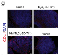
Application: WB Species: Mouse Sample:

Application: WB Species: mouse Sample: liver
Restrictive clause
Affinity Biosciences tests all products strictly. Citations are provided as a resource for additional applications that have not been validated by Affinity Biosciences. Please choose the appropriate format for each application and consult Materials and Methods sections for additional details about the use of any product in these publications.
For Research Use Only.
Not for use in diagnostic or therapeutic procedures. Not for resale. Not for distribution without written consent. Affinity Biosciences will not be held responsible for patent infringement or other violations that may occur with the use of our products. Affinity Biosciences, Affinity Biosciences Logo and all other trademarks are the property of Affinity Biosciences LTD.