Product: Estrogen Receptor-beta Antibody
Catalog: AF6469
Description: Rabbit polyclonal antibody to Estrogen Receptor-beta
Application: WB IHC IF/ICC
Cited expt.: WB, IHC, IF/ICC
Reactivity: Human, Mouse, Rat, Monkey
Prediction: Horse, Sheep, Dog, Chicken
Mol.Wt.: 59kDa; 59kD(Calculated).
Uniprot: Q92731
RRID: AB_2835288

View similar products>>

   Size Price Inventory
 100ul $280 In stock
 200ul $350 In stock

Lead Time: Same day delivery

For pricing and ordering contact:
Local distributors

Product Info

Source:
Rabbit
Application:
WB 1:500-1:2000, IHC 1:50-1:200, IF/ICC 1:100-1:500
*The optimal dilutions should be determined by the end user.
*Tips:

WB: For western blot detection of denatured protein samples. IHC: For immunohistochemical detection of paraffin sections (IHC-p) or frozen sections (IHC-f) of tissue samples. IF/ICC: For immunofluorescence detection of cell samples. ELISA(peptide): For ELISA detection of antigenic peptide.

Reactivity:
Human,Mouse,Rat,Monkey
Prediction:
Horse(100%), Sheep(89%), Dog(100%), Chicken(86%)
Clonality:
Polyclonal
Specificity:
Estrogen Receptor-beta Antibody detects endogenous levels of total Estrogen Receptor-beta.
RRID:
AB_2835288
Cite Format: Affinity Biosciences Cat# AF6469, RRID:AB_2835288.
Conjugate:
Unconjugated.
Purification:
The antiserum was purified by peptide affinity chromatography using SulfoLink™ Coupling Resin (Thermo Fisher Scientific).
Storage:
Rabbit IgG in phosphate buffered saline , pH 7.4, 150mM NaCl, 0.02% sodium azide and 50% glycerol. Store at -20 °C. Stable for 12 months from date of receipt.
Alias:

Fold/Unfold

ER BETA; ER-beta; Erb; ESR B; ESR BETA; ESR2; ESR2_HUMAN; ESRB; ESTRB; estrogen nuclear receptor beta variant a; estrogen nuclear receptor beta variant b; estrogen receptor 2 (ER beta); Estrogen receptor 2; estrogen receptor beta 4; Estrogen receptor beta; NR3A2; Nuclear receptor subfamily 3 group A member 2;

Immunogens

Immunogen:

A synthesized peptide derived from human Estrogen Receptor-beta, corresponding to a region within N-terminal amino acids.

Uniprot:
Gene(ID):
Expression:
Q92731 ESR2_HUMAN:

Isoform 1 is expressed in testis and ovary, and at a lower level in heart, brain, placenta, liver, skeletal muscle, spleen, thymus, prostate, colon, bone marrow, mammary gland and uterus. Also found in uterine bone, breast, and ovarian tumor cell lines, but not in colon and liver tumors. Isoform 2 is expressed in spleen, thymus, testis and ovary and at a lower level in skeletal muscle, prostate, colon, small intestine, leukocytes, bone marrow, mammary gland and uterus. Isoform 4 is found in testis. Isoform 5 is expressed in testis, and at a lower level in spleen, thymus, ovary, mammary gland and uterus. Isoform 6 is expressed in testis, placenta, skeletal muscle, spleen and leukocytes, and at a lower level in heart, lung, liver, kidney, pancreas, thymus, prostate, colon, small intestine, bone marrow, mammary gland and uterus. Not expressed in brain.

Description:
Estrogen receptor-beta (ESR2) is a member of the superfamily of nuclear receptors, which can transduce extracellular signals into transcriptional responses.
Sequence:
MDIKNSPSSLNSPSSYNCSQSILPLEHGSIYIPSSYVDSHHEYPAMTFYSPAVMNYSIPSNVTNLEGGPGRQTTSPNVLWPTPGHLSPLVVHRQLSHLYAEPQKSPWCEARSLEHTLPVNRETLKRKVSGNRCASPVTGPGSKRDAHFCAVCSDYASGYHYGVWSCEGCKAFFKRSIQGHNDYICPATNQCTIDKNRRKSCQACRLRKCYEVGMVKCGSRRERCGYRLVRRQRSADEQLHCAGKAKRSGGHAPRVRELLLDALSPEQLVLTLLEAEPPHVLISRPSAPFTEASMMMSLTKLADKELVHMISWAKKIPGFVELSLFDQVRLLESCWMEVLMMGLMWRSIDHPGKLIFAPDLVLDRDEGKCVEGILEIFDMLLATTSRFRELKLQHKEYLCVKAMILLNSSMYPLVTATQDADSSRKLAHLLNAVTDALVWVIAKSGISSQQQSMRLANLLMLLSHVRHASNKGMEHLLNMKCKNVVPVYDLLLEMLNAHVLRGCKSSITGSECSPAEDSKSKEGSQNPQSQ

Predictions

Predictions:

Score>80(red) has high confidence and is suggested to be used for WB detection. *The prediction model is mainly based on the alignment of immunogen sequences, the results are for reference only, not as the basis of quality assurance.

Species
Results
Score
Horse
100
Dog
100
Sheep
89
Chicken
86
Pig
78
Bovine
78
Rabbit
67
Xenopus
0
Zebrafish
0
Model Confidence:
High(score>80) Medium(80>score>50) Low(score<50) No confidence

Research Backgrounds

Function:

Nuclear hormone receptor. Binds estrogens with an affinity similar to that of ESR1, and activates expression of reporter genes containing estrogen response elements (ERE) in an estrogen-dependent manner. Isoform beta-cx lacks ligand binding ability and has no or only very low ere binding activity resulting in the loss of ligand-dependent transactivation ability. DNA-binding by ESR1 and ESR2 is rapidly lost at 37 degrees Celsius in the absence of ligand while in the presence of 17 beta-estradiol and 4-hydroxy-tamoxifen loss in DNA-binding at elevated temperature is more gradual.

PTMs:

Phosphorylation at Ser-87 and Ser-105 recruits NCOA1.

Subcellular Location:

Nucleus.

Extracellular region or secreted Cytosol Plasma membrane Cytoskeleton Lysosome Endosome Peroxisome ER Golgi apparatus Nucleus Mitochondrion Manual annotation Automatic computational assertionSubcellular location
Tissue Specificity:

Isoform 1 is expressed in testis and ovary, and at a lower level in heart, brain, placenta, liver, skeletal muscle, spleen, thymus, prostate, colon, bone marrow, mammary gland and uterus. Also found in uterine bone, breast, and ovarian tumor cell lines, but not in colon and liver tumors. Isoform 2 is expressed in spleen, thymus, testis and ovary and at a lower level in skeletal muscle, prostate, colon, small intestine, leukocytes, bone marrow, mammary gland and uterus. Isoform 4 is found in testis. Isoform 5 is expressed in testis, and at a lower level in spleen, thymus, ovary, mammary gland and uterus. Isoform 6 is expressed in testis, placenta, skeletal muscle, spleen and leukocytes, and at a lower level in heart, lung, liver, kidney, pancreas, thymus, prostate, colon, small intestine, bone marrow, mammary gland and uterus. Not expressed in brain.

Family&Domains:

Composed of three domains: a modulating N-terminal domain, a DNA-binding domain and a C-terminal ligand-binding domain.

Belongs to the nuclear hormone receptor family. NR3 subfamily.

Research Fields

· Human Diseases > Drug resistance: Antineoplastic > Endocrine resistance.

· Human Diseases > Cancers: Overview > Pathways in cancer.   (View pathway)

· Human Diseases > Cancers: Specific types > Breast cancer.   (View pathway)

· Organismal Systems > Endocrine system > Estrogen signaling pathway.   (View pathway)

· Organismal Systems > Endocrine system > Prolactin signaling pathway.   (View pathway)

References

1). Genistin: A Novel Estrogen Analogue Targeting ERβ to Alleviate Thrombocytopenia. International journal of biological sciences, 2024 (PubMed: 38617546) [IF=8.2]

Application: WB    Species: human    Sample: Meg-01 cells

Figure 7. Genistin interacts with ERβ to regulate megakaryocyte differentiation. (A) ERα or ERβ and ligands (genistin) by molecular docking. (B) Lysates from Meg-01 cells were incubated with or without genistin (200 μM) for 2 h, and different concentrations of pronase E (1:500, 1:1000, or 1500) were added for 10 min at 40°C, ERβ content was detected using western blot analysis. (C) Lysates from Meg-01 cells were incubated with genistin (100, 200, 400 μM) for 2 h, and a final concentration of pronase E (1:1000) was added for 10 min at 40 °C. ERβ expression was detected by western blot analysis. (D) Lysates from Meg-01 cells were incubated with genistin (200 μM) for 9 h, then reacted at different temperatures (45, 48, 51, 54, 57 °C) for 5 min, and finally the ERβ content was detected by western blot. (E) Lysates from Meg-01 cells containing or without different doses of genistin (100, 200, 400 μM) were incubated for 9 h, and then reacted at 48 °C for 5 minutes, and finally the ERβ content was detected by western blot. (F-G) Meg-01 cells were detected by flow cytometry using genistin (20 μM), PHTPP (20 μM), and both, respectively. The histogram shows the percentage of CD41+CD42b+ cells for each group. (H-I) PI staining in Meg‑01 cells was used to analyze the DNA content (2, 4 and ≥ 8 N) by flow cytometry after genistin (20 μM), PHTPP (20 μM), and both interventions, respectively. All data represents the mean±SD of three independent experiments; *p < 0.05, **p < 0.01, ***p < 0.001, vs the Ctrl group; #p < 0.05, ##p < 0.01, ###p < 0.001, vs the Gen group; Gen: genistin, Ctrl: control.

Application: IF/ICC    Species: human    Sample: Meg-01 cells

Figure 8. Genistin can promote PI3K/AKT and MEK/ERK expression. (A-G) The expression levels of ERβ/GAPDH, HSP90/GAPDH, p-PI3K/PI3K, p-AKT/AKT, p-MEK/MEK, and p-ERK/ERK were detected by western blot after Meg-01 cells were treated with genistin for 5 days. (H) Representative immunofluorescence images of ERβ fluorescently double-stained with p-ERK and p-AKT, respectively, on bone marrow cells after 10 days of treatment with genistin in a mouse model of radiation thrombocytopenia. All data represents the mean ± SD of three independent experiments; *p < 0.05, **p < 0.01, ***p < 0.001, vs the Ctrl group; Gen: genistin, Ctrl: control.

2). Low-dose phthalates promote breast cancer stem cell properties via the oncogene ΔNp63α and the Sonic hedgehog pathway. Ecotoxicology and Environmental Safety, 2023 (PubMed: 36753971) [IF=6.2]

3). Crocetin Alleviates Ovariectomy-Induced Metabolic Dysfunction through Regulating Estrogen Receptor β. Journal of agricultural and food chemistry, 2021 (PubMed: 34851635) [IF=5.7]

4). Estrogen receptor β/substance P signaling in spinal cord mediates antinociceptive effect in a mouse model of discogenic low back pain. Frontiers in cellular neuroscience, 2023 (PubMed: 36756381) [IF=5.3]

Application: IF/ICC    Species: Mouse    Sample: spinal cord

Figure 3. Spatial colocalization of substance P with ERβ in the spinal cord was investigated using dual-label confocal immunofluorescence examination. The specificities of the immunopositive staining of ERβ (green) and substance P (red) were demonstrated. Substance P colocalized with ERβ in the dorsal horn of the spinal cord, mainly in the laminae I and II, a connection site of pain transmission. (A) Coronal sections through the spinal cord of DLBP mice. Scale bar: 200 μm. (B) The magnified dorsal horn areas of the spinal cord of DLBP mice. Scale bar: 20 μm.

5). Genistein accelerates glucose catabolism via activation the GPER-mediated cAMP/PKA-AMPK signaling pathway in broiler chickens. Life Sciences, 2022 (PubMed: 35640778) [IF=5.2]

6). Jiawei Shoutai Pill promotes decidualization by regulating the SGK1/ENaC pathway in recurrent spontaneous abortion. Journal of ethnopharmacology, 2024 (PubMed: 37479068) [IF=4.8]

Application: IHC    Species: Human    Sample: decidua tissue

Fig. 3. The expression of SGK1, ENaC-α, ERβ and PR in decidua tissue was detected by immunohistochemistry. Note: vs Control group, ***P < 0.001. Scale bars: a 100 μm, b 50 μm (n = 10).

7). Rasd2 Mediates Acute Fasting-Induced Antidepressant-Like Effects via Dopamine D2 Receptor Activation in Ovariectomized Mice. The international journal of neuropsychopharmacology, 2023 (PubMed: 36566472) [IF=4.5]

Application: WB    Species: Mouse    Sample:

Figure 6.Effect of sulpiride (SUL) on fasting-induced changes in protein expression. Figures represent the changes in the protein expression of RASD Family Member 2 (RASD2), brain-derived neurotrophic factor (BDNF), cAMP-response element binding protein (CREB), phospho-CREB (p-CREB), protein kinase B (Akt), dopamine D2 receptor (DRD2), estrogen receptor α (ERα), and estrogen receptor β (ERβ) in the hippocampus (HP) (A, a–h), prefrontal cortex (PFC) (B, i–p), and striatum (C, q–x) in ovariectomized mice. The data are expressed as mean ± SEM (n = 3–5). Two-way ANOVA with Tukey’s honestly significant difference (HSD), *P < .05 vs ovariectomy (OV), **P < .01 vs vehicle (VEH):non-fasting; #P < .05, ##P < .01 vs VEH:fasting.

8). Ormeloxifene, a selective estrogen receptor modulator, protects against pulmonary hypertension. European Journal of Pharmacology, 2023 (PubMed: 36731722) [IF=4.2]

9). TLR4 inhibition suppresses growth in oestrogen-induced prolactinoma models. Endocrine-Related Cancer, 2022 (PubMed: 36219868) [IF=4.1]

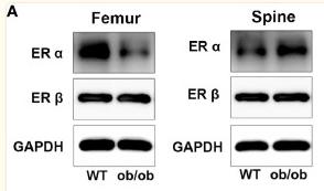
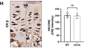
10). The Role of Estrogen Receptor α in Response to Longitudinal Bone Growth in ob/ob Mice. Frontiers in Endocrinology, 2021 (PubMed: 34925230) [IF=3.9]

Application: WB    Species: Mice    Sample: femoral and spine

Figure 5 The expression level of estrogen receptors (ERs) in femur and spine. (A) Western blotting results of expression level of ERα and ERβ in femoral and spine growth plate (GP), n = 3. (B–E) Semiquantitative analysis of ER expression levels. (F) Quantitative real-time PCR results of ER genes in femoral and vertebral GP, n = 4. (G–J) Immunohistochemistry (IHC) results of expression level of ERα and ERβ in femoral and vertebral GP, n = 6. Scale bar: 20 μm. (K) Comparison of ERα HSCORE between femoral and spine GP (FGP, femoral growth plate; SGP, spine growth plate). (L, M) Results of Spearman’s analysis between ERα HSCORE and femur/spine length. *p < 0.05, ***p < 0.001 and ****p < 0.0001; ns denotes not significant.

Application: IHC    Species: Mice    Sample: femoral and vertebral

Figure 5 The expression level of estrogen receptors (ERs) in femur and spine. (A) Western blotting results of expression level of ERα and ERβ in femoral and spine growth plate (GP), n = 3. (B–E) Semiquantitative analysis of ER expression levels. (F) Quantitative real-time PCR results of ER genes in femoral and vertebral GP, n = 4. (G–J) Immunohistochemistry (IHC) results of expression level of ERα and ERβ in femoral and vertebral GP, n = 6. Scale bar: 20 μm. (K) Comparison of ERα HSCORE between femoral and spine GP (FGP, femoral growth plate; SGP, spine growth plate). (L, M) Results of Spearman’s analysis between ERα HSCORE and femur/spine length. *p < 0.05, ***p < 0.001 and ****p < 0.0001; ns denotes not significant.

Load more

Restrictive clause


Affinity Biosciences tests all products strictly. Citations are provided as a resource for additional applications that have not been validated by Affinity Biosciences. Please choose the appropriate format for each application and consult Materials and Methods sections for additional details about the use of any product in these publications.

For Research Use Only.
Not for use in diagnostic or therapeutic procedures. Not for resale. Not for distribution without written consent. Affinity Biosciences will not be held responsible for patent infringement or other violations that may occur with the use of our products. Affinity Biosciences, Affinity Biosciences Logo and all other trademarks are the property of Affinity Biosciences LTD.