.")
.")


, using AMPK alpha Antibody at 1/1000 dilution.
5ug/NC membrane strip.
Exposure for 5s with Affinity™ ECL Kit(#KF8003).
Bands result from membrane strip incubation.")


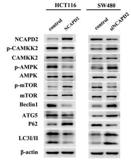
 Knockdown efficiency was confirmed by western blot. GAPDH was
used to verify equivalent loading. (B) AMPK-α siRNA abrogated irisin-induced
phosphorylation of AMPK-α. (C) Knockdown of AMPK-α caused a decrement of
CD206-APC expression in irisin-treated macrophages. (D-F) Knockdown of AMPK-α
reversed the inductive effect of irisin on the expression of ARG-1 and TGF-β1, but increased TNF-α expression. (G-H) Knockout of AMPK-α reduced the osteogenic
ability of irisin-treated macrophage. The data are represented as the mean ± SD
from three independent experiments.
#
p < 0.05, ##p < 0.01 compared with si NC group.
Unstained, macrophages without treatment and antibody incubation; w, week.")
 Hep2 cells were mock-infected or infected with RSV (MOI = 5) in the absence or presence of Compound C (5 μM) (G) or NAC (5854 mM) (H) for 24 h. The expression of p-AMPK (Thr172), p-MTOR (Ser2448), p-ULK1 (Ser371), p-ULK1 (Ser555) and LC3Ċ was analyzed by immunoblotting with specific antibodies as described in Materials and Methods. The relative levels of targeted proteins were quantitated by densitometry and normalized to AMPK, MTOR, ULK1 and GAPDH respectively.")
 The signaling pathways involved in the downstream of circPAN3 were analyzed by Kyoto Encyclopedia of Genes and Genomes (KEGG). (B) Expression levels of key proteins in the AMPK/mTOR pathway in THP-1 and THP-1/ADM cells were detected and quantified by Western blot.")
 Representative western blot results of proteins related to the AMPK and NF-κB p65/NLRP3 signaling pathway proteins in HUVECs with or without AGEs/salidroside treatment. GAPDH was used as a loading control. The data are representative of one experiment performed in triplicate and are expressed as the mean ± S.D. **P<0.01 vs. Control group; ##P<0.01 vs. AGEs group")
 Representative images and histograms of AMPK, NF-κB p65 and NLRP3 in HUVECs determined by immunofluorescence. The data are representative of one experiment performed in triplicate and are expressed as the mean ± S.D. **P<0.01 vs. control group; ##P<0.01 vs. AGEs group.")
Irisin induced phosphorylation of AMPK-α, when compared to control medium, LPS plus IFN-γ, and IL-4 groups")
 CHP upregulation protein expression of white adipose browning gene in obese mice induced by HFD. Control group (CON): normal feed; high-fat diet group (HFD): highfat diet; high-fat diet feed supplemented orlistat (HFD + OST): HFD was fed for 12 weeks, orlistat was gavaged at 15.6 mg/kg bw/day, as a positive control group; CHP group supplemented with high-fat feed (HFD + CHP): HFD was fed for 12 weeks, CHP was gavaged 250 mg/kg bw/ day. Values are expressed as the mean ± standard deviation, n = 3. **p < 0.01, *p < 0.05.")
 Representative immunoblots and relative protein levels of AMPK,pAMPK, mTOR, and p-mTOR. (C) Hepatic expression, distribution and relative flourescent intensity of AMPK, p-AMPK and mTOR protein by immunofluorescent staining. (D, E) Representative immunoblots and relative protein levels of AMPK, pAMPK, mTOR, and p-mTOR. *p < 0.05; **p < 0.01; ***p < 0.005 vs WT-HFD;##p < 0.01; ###p < 0.005 vs ApoE−/−-HFD. n = 3 for each group.")
 The ability of Exos-26a to generate neurofilament (red fluorescent dye) in PC12 cells, which could be reversed by rapamycin. (b, c) Representative images of western blots used to determine the expression levels of NF, mTOR, p-mTOR, AMPK, p-AMPK, S6K, p-S6K, ULK1, p-ULK1, and p62 and semiquantification of the data. RAP indicates miR-26a exosome and rapamycin (100 nM) treatment for 48 h before lysis. *P < 0.05, **P < 0.01, ***P < 0.001 compared with the control group by t test or ANOVA. #P < 0.05 and ##P < 0.01 compared with the RAP group by t test. n = 3 for each group.")
.")
.Bar = 50 µm. *P < 0.05 compared to the sham group. ##P < 0.01 compared to the 5/6 Nx model group. C-E, Representative immune blots of key proteins involved in muscle atrophy and proteins associated with the AMPK/SKP2/CARM1 signalling pathway")
 AMPK activation, (B) ERK
activation, (C) JNK activation, and (D) P38 activation. The blots showed representative samples. All samples derived from the same experiment and that blots were
processed in parallel. AMPK and p-AMPK were normalized to the expression of GAPDH on the same membrane. Each phosphoprotein was normalized to the
expression of the corresponding total protein of the same sample in Fig. 5B–D. Results are expressed as mean ± S.E.M. (n = 6–8 for each group). *P < 0.05,
**P < 0.01, ***P < 0.001, ****P < 0.0001 according to one-way ANOVA followed by Bonferroni test.")
 Relative AMPK expression, the ratio of p-AMPK/AMPK, relative
SIRT1 expression and densitometric quantification. (B) Relative expression of p-NF-κB, ASC, Cleaved IL-1β, NLRP3, Cleaved caspase1, GSDMD-N and densitometric
quantification. Values are expressed as mean ± SEM. #p < 0.05 and ##p < .01 versus Ctrl; *p < 0.05 and **p < 0.01 versus DN.")
. (A) Relative AMPK expression, the ratio of p-AMPK/AMPK, relative SIRT1
expression and their density quantification results. (B) Relative expression levels of p–NF–κB, ASC, Cleave-IL-1β, NLRP3, Cleave-Caspase-1, GSDMD-N, and their
density quantification results. Values are expressed as mean ± S.E.M. #P < 0.05 and ##P < 0.01 versus Control group; *P < 0.05 and **P < 0.01 versus DN group.")
 activation following treatment with isoprenaline or high glucose in H9c2 cells. (A) Immunofluorescence detection of SIRT1 (scale bar: 50 μm). (B) Experiments were performed as in (A). The protein levels of SIRT1, PGC‐1α and glyceraldehyde 3‐phosphate dehydrogenase (GAPDH) were determined by Western blotting. (C) Experiments were performed as in (D). Quantitative analysis of SIRT1 and PGC‐1α normalized to GAPDH levels and expressed as relative fold changes versus the control group. The mRNA expressions of SIRT1 and PGC‐1α were measured by qRT‐PCR (E, F and G). Data are presented as the mean ± SEM. # P < .05, ## P < .01, ### P < .01 vs the control group; * P < .05, ** P < .01, *** P < .001 vs the isoprenaline or high‐glucose group")
. Immunoblot analyzed of AMPK phosphorylation in colon tissues of mice (B). THP-1 cells were primed with LPS, followed by GPA, Compound C or GSK621 treatment 6 h before stimulation with ATP for 30 min. Levels of the ROS was measured in THP-1 cells (C). Cell death was measured by and LDH released (D). Immunoblot analyzed of IL-1β in supernatants and cell lysate of THP-1 cells (E). IL-1β in supernatants of THP-1 cells was detected by ELISA (F). Immunoblot analyzed of mitochondrial components of NLRP3 inflammasome in THP-1 cells (G). Data are presented as mean ± SD, three independent experiments. *p < 0.05, **p < 0.01 and ***p < 0.001.")
 Adipocyte size of the mice; (B) Quantification of Fig. 2A; (C) Immunohistochemical assay of WAT; (D) Quantification of Fig. 2C; (E) Protein expression in WAT; (F) Quantification of Fig. 3E n ¼ 6 mice/group. #p < 0.05, ##p < 0.01,###p < 0.001 vs CON;
*p < 0.05,**p < 0.01, ***p < 0.001 vs DM.")
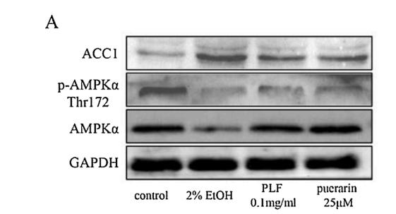
, (B) Protein expression of AMPKα, P-AMPKα, SREBP1, ACC1 in liver (n = 3), (C) inmmunofluorescence staining of ACC1, SREBP1 in Liver sections (200 ×). *p < 0.05; as compared to the control group. # p < 0.05, ## p < 0.01; as compared to the model group.")
 The protein levels of pAMPK, AMPK and their ratio. (A and E) The levels of CDX2 in the colon were detected by immunoblotting. The results were represented as mean±SEM (n= 6 each group). *P <0.05, **P <0.01, ***P<0.001, compared to HFD +Sed group; #P<0.05, compared to SD + Sed group.")
 Representative western blot for p-AMPKα
(Thr172) and AMPKα in the kidney of HFD-treated mice 12 weeks after RSPO1-RNAi lentivirus administration. (B) The levels of pro-inflammatory cytokines TNF-α
and IL-1β of HFD-treated mice 12 weeks after RSPO1-RNAi lentivirus administration. (C) As markers of oxidative stress, ROS fluorescence intensity and SOD level of
HFD-treated mice 12 weeks after RSPO1-RNAi lentivirus administration. (D) Representative western blot for Beclin-1 and p62 in the kidney of HFD-treated mice 12
weeks after RSPO1-RNAi lentivirus. n = 6 mice/group. Data was expressed as mean ± SD and analyzed by one-way ANOVA test followed by Tukey’s multiple
comparison test. *p < 0.05 vs. the indicated group. All experiments were performed at least three times.")
 Western blot analysis evaluated the protein expression levels of p-AMPK, AMPK, p-mTOR, and mTOR in rats and cells. (D, E) LO2 and HepG2 cells were treated with 0.3 mM PA, 20 μM dapagliflozin and 10 µM compound C (Comp C) for 24 h. The cells were stained with Oil Red O and intracellular TG was quantitatively analyzed. Scale bars: 20 μm. Data are expressed as the means ± SEM from three independent experiments. *p < 0.05, **p < 0.01, and ***p < 0.001.")
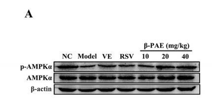
 Western blot bands. (B) The protein expression levels of AMPK-α, PPAR-α, CPT-1A, SREBP-1c, SCD-1, and
FAS. (C) The mRNA expression levels of AMPK-α, PPAR-α, CPT-1A, SREBP-1c, SCD-1, and FAS. The contents of target proteins were normalized to β-actin. The results
are presented as the mean ± SD (protein expression: n = 3, mRNA expression: n = 6). ## p < 0.01 vs. NC group; * p < 0.05, ** p < 0.01 vs. model group.")
 for 6 weeks were harvested and subjected to mRNA and immunoblotting analysis (A, B) Protein levels of p-AMPK and AMPK in DIO mice were analysed by immunoblotting. The relative protein level was shown. AMPK was used as a control for protein levels; n = 5 per group (C–E) Relative mRNA expression of hepatic genes; n = 3–5 per group. β-ACTIN was used as an internal control for normalizing the mRNA levels and protein levels. # p < 0.05, HF versus Chow group. *p < 0.05, HF versus HF + VTE group. NS, no significance (F) Hepatocellular NF-κB activity was analysed using a reporter assay. HepG2 cells were co-transfected with the p65 expression vector and NF-κBx3-LUC for 24 h and treated with the control (0.1% DMSO), VTE (250 and 500 μg ml−1) for another 18 h. Then cells were treated with TNFα (10 ng ml−1) for an additional 6 h. The relative luciferase units (RLUs) were measured by comparison to renilla luciferase activities. The results represent three independent experiments, and data are statistically analysed as means ± SEM (n = 3). *p < 0.05, versus vehicle control.")

. The phosphorylation levels of (B) p70S6K and 4E-BP1; (C) AMPK, ACC, and ULK1; (D) ERK1/2; (E) p38MAPK; and (F) eEF2 are shown. The phosphorylation is normalized to the total protein expression. The β-tubulin content in the lysate was measured as a loading control (G). The time course of these experiments is shown in the upper region. DM: differentiation medium, DMEM: Dulbecco’s modified Eagle’s medium, and w/o AA: without amino acids. Data are displayed as the means ± SD, and n = 4 for each group in all bar graphs. * p < 0.05 and ** p < 0.01 vs. the vehicle-treated group.")
 Representative images of p-AMPKα, AMPKα,SIRT1, and PGC-1α expression in the TA muscles of rats using immunohistochemical (IHC) staining (magnification: ×200 scale bars: 100 µm).")
 The expression levels of pAMPK, AMPK, and SIRT1 were measured and quantified by Western blot.")
 The hepatic mRNA expressions of AMPKα, SREBP-1c and PPARα. (B) Representative bands of p-AMPKα,
AMPKα, SREBP-1c and PPARα. (C) Quantitative results of Western blot bands densities of p-AMPKα/ AMPKα, SREBP-1c and PPARα. (D) Spearman correlation
between hepatic CD36 expression and AMPK signalling pathway-related proteins indicators, respectively. Data are presented as the mean ± SD (n = 3 ~ 6). ##P <
0.01 vs. NC group; **P < 0.01 vs. Model group.")
 To confrm that the efect of baicalin is mediated by regulating autophagy, 3-MA was used with baicalin treatment, and the levels of the autophagy-related proteins LC3II/I, Beclin1, P62, p-AMPK, AMPK, and mTOR were measured by Western blotting.")
 The protein levels of pAMPK, AMPK and their ratio. (A and E) The levels of CDX2 in the colon were detected by immunoblotting. The results were represented as mean±SEM (n= 6 each group). *P <0.05, **P <0.01, ***P<0.001, compared to HFD + Sed group; #P<0.05, compared to SD + Sed group.")
 The western blot analysis. (b) The protein expression of PI3K. (c) The protein expression of Akt. (d) The protein expression of AMPK. (e) The ratio of p‐PI3K to PI3K. (f) The ratio of p‐Akt to Akt. (g) The ratio of p‐AMPK to AMPK. All data are presented as mean ± SD (n ≥ 3) for each group. Different lowercase alphabet letters over bars indicate statistically significant differences between two groups (p < .05)")
 The protein expression of AMPK, p-AMPK, SIRT1, and PGC-1a was detected via Western blotting; (B) Quantitative evaluation of the protein expression of AMPK and p-AMPK; (C) Quantitative evaluation of the protein expression of SIRT1; (D) Quantitative evaluation of the protein expression of PGC-1a; (E) The mRNA expression of SIRT1 was detected in indicated groups; (F) The mRNA expression of PGC-1a was detected in indicated groups. * p < 0.05 vs. NSD. # p < 0.05, ## p < 0.01 vs. HSD.")
 for 4 h, and then treated with Aβ42 oligomers (1 μM) for 24 h, followed by treatment with stigmasterol (20 μM) for 4 h. (A,B) Representative western blot analysis of AMPK signaling. GAPDH immunoreactivity was used as a loading control. (C-E) Representative western blot analysis of NF-κB signaling. The cytosolic and nuclear fractions were prepared and analyzed with total NF-κB p65. α-Tubulin immunoreactivity was used as a loading control in the cytosolic fraction, Histone H3 was used as the loading control in the nuclear fraction. (F-H) Representative western blot analysis of NLRP3 signaling, including NLRP3 and Caspase-1, p20. GAPDH immunoreactivity was used as a loading control (I, J) Concentration of TNFα and IL-1β. Data were presented as the mean ± SEM from three independent experiments. One-way ANOVA with Tukey’s multiple comparison test revealed a difference between groups.")
 Representative immunofluorescence staining images of the expression of AMPK and Drp1 in the spinal dorsal horn of sham and CIBP rats. Scale bar: 20 μm. (C) Quantitative analysis of fluorescence intensity in (A) and (B). Data are expressed as the mean±SD ( n=3). * P")
 for 48 h. JWH133 elevated the expressions of p-AMPKα (Thr172), p-P53 and P21, reduced the expressions of p-mTOR and PCNA, while had no effect on the expressions of p-LKB1 (Ser428), LKB1, T-AMPKα, P53, mTOR and β-Actin. The original western blot images (A) were representative of three different experiments. (B) The statistic graphs of proteins variation. Data were relative density to that in control group. Data were mean ± SEM from three replicate experiments, *P")
 Representative Western blotting images of SREBP-1, p-Raptor, Raptor, p-AMPK, AMPK, p-Akt, Akt, FoxO1. B) Relative expression of srebp-1 **P < 0.01. C) Relative expression of mTORC1. D) PI3K-Akt signaling pathway. E) Grey value analyses of SREBP-1, p-Raptor, p-AMPK, p-Akt, and FoxO1 *P < 0.05, n = 3 rats per group. F) Directionality of the changes in mTORC1 and PI3K-Akt signaling pathway, frame with red background: proteins up-regulated in MDL, frame with claybank background: proteins down-regulated in MDL, green arrows: proteins regulated by LCF. (For interpretation of the references to color in this figure legend, the reader is referred to the web version of this article.)")
 Western blotting analysis of ColII, MMP13, IL-1β, Beclin1, P62, LC3B, AMPK, P-AMPK, PINK1, and Parkin, with GAPDH as an internal control. (B) Statistical analysis of western blotting. (C) Immunofluorescence double staining of LC3B and COXIV. Green fluorescence represents the mitochondrial marker COXIV, while red fluorescence represents the autophagosome marker LC3. Blue fluorescence indicates DAPI-stained nuclei. Double staining is indicated in yellow. (D) Transmission electron microscopy of damaged mitochondria and mitophagy in rat chondrocytes. Blue arrows indicate damaged mitochondria, and yellow arrows indicate autophagosomes with mitochondrial-like organelles. Data are presented as means ± standard deviation. #P")
 Western blot analysis was used to measure the expression levels of AMPK, pAMPK, ERK and pERK proteins following the indicated treatments; GAPDH was used as a control for the standardization of the total cellular protein. (B) Quantitative analysis of the activation of the AMPK and ERK enzymes. (C) Formation of foam cells, as detected by Oil Red O staining. Scale bar=50 µM. ***P")
. Representative western blots (A, C, E, G, K) and quantification of AMP-activated protein kinase (AMPK) and AMPK gene expression (B, I), phosphorylated AMPK (p-AMPK; Thr172) (D), PGC1-α (F, J), SIRT-1 (H) and NRF-2 (L). CON control, EMA combined therapies + compound-c, EMC combined therapies, EXE moderate-exercise, GAPDH glyceraldehyde-3-phosphate dehydrogenase, MET metformin, NRF-2 nuclear factor erythroid-2 related factor 2, PGC-1α peroxisome proliferator-activated receptor-γ coactlvator-1α, PRE prediabetes, SIRT-1 silent mating type information regulation 2 homolog-1. The data shown represent mean ± the standard error of the mean (n = 5–6/group).")
 Mitochondrial protein expression of the pathway protein was detected by western blotting. (B) The gray intensity of SIRT1 expression relative to β-actin (n=3 vs. n=3). (C) The ratio of P-AMPKα to AMPK expression (n=3 vs. n=3). The protein ratio of P-AMPK to AMPK in the cGCs of the PCOS group was significantly lower than in NOR group. (D) The gray intensity of PGC1α expression relative to β-actin (n=3 vs. n=3). The expression protein levels of the energy depletion signaling-related factors SIRT1, AMPK, p-AMPK and PGC-1α were decreased in the cGCs of the PCOS group. (E) mRNA transcription level of SIRT1 by RT-qPCR (n=5 vs. n=8). (F) mRNA transcription level of AMPK by RT-qPCR (n=8 vs. n=5). (G) mRNA transcription level of PGC-1α by RT-qPCR (n=4 vs. n=7). (H) mRNA transcription level of TFAM detected by RT-qPCR (n=6 vs. n=6). There was no statistically significant difference observed in SIRT1, AMPK and PGC-1α mRNA transcription level. *P")
 phosphorylation under proinflammatory conditions. (a) Time-course changes in phos-phorylation of p65 (p-p65) relative to p65 level. Human IVD NP cells were stimulated with 10 ng/mL IL-1β, and the levels of p-p65 and p65 were assessed at different time points, i.e., 0 (set as 1.0), 15, 30, 60, 120, and 240 min. (b,c) Expression of p-p65 relative to that of p65 and phosphorylated AMPK (p-AMPK) relative to that of AMPK in IVD NP cells treated with phosphate-buffered saline (PBS) (group C; set as 1.0), AdipoRon (2 μM, group A), IL-1β (10 ng/mL, group I), or both (group A + I) for 15 min. Data are presented as dot and box plots. Results from six independent experiments were analyzed using one-way repeated-measures ANOVA with the Tukey–Kramer post hoc test (n = 6). Significant differences were set as * p < 0.05 and ** p < 0.01.")
 for 8 h. Cells were subsequently treated for 2 h with HBSS, and then LC3-I/II, p62, p53, p-AMPK, AMPK, p-mTOR, mTOR, and S100P levels were assayed by western blot (n=3, *P0.05 vs. shS100P group); D. HL-60 and Jurkat cells were transfected with S100P shRNA or control shRNA and then pre-treated with Compound C (20 μM) for 6 h. Cells were subsequently treated for 2 h with HBSS, and then LC3-I/II, p62, p53, p-AMPK, AMPK, p-mTOR and mTOR levels were assayed by western blot. CC, Compound C (n=3, *P0.05 vs. shS100P group); E. HL-60 and Jurkat cells were transfected with S100P shRNA or control shRNA and then pre-treated with Tenovin-6 (5 μM) for 8 h. Cells were subsequently treated for 24 h with ADM (0.2 μg/ml) or 48 h with Ara-C (4 μM), then cell viability was assayed using a CCK-8 kit (n=3, *P")
 Protein synthesis related gene expression; (B) lipolysis-related gene expression; (C) Western blot analysis of p-AMPK, AMPK, PPARα, TOR, p70S6K, PI3K, and AKT protein expression; (D) relative quantification of p-AMPK, PPARα, TOR, p70S6K, PI3K, and AKT protein expression. The bars indicate the mean ± standard error (SE). Different superscripts denote significant differences (P < 0.05). CON = a control group (containing 4.95% crude fat); HFD = a high fat diet (containing 10.27% crude fat); HFDS = supplementing 1200 μg/kg sanguinarine to HFD.")
 AMPK and p-AMPK representative western blot image. Full-length blots/gels are presented in Supplementary Figs. 5 and 6. (B) p-AMPK/AMPK ratio in lung grafts (n = 3). Compared with CS group, *P")
 Hepatic p-AMPK, p-mTOR proteins expressions. (d–f) Hepatic ULK1 protein and gene expression. Results were expressed as the mean ± SD, n = 3 in (a–e) and n = 6 in (f) ∗P < 0.05 and ∗∗P < 0.01, compared with the model group.")
 PA restores the inhibition of autophagy in the mice cortex and hippocampal regions caused by Aβ1–42. Autophagy is aided by PA. PA enhanced Beclin- 1 expression, AMPK and LC3B-II protein levels, and inhibited p62 and P-mTOR expression in (B, D) and (A, C, E) respectively. (F) Effects of PA on the content of PSD95 in the cortex and hippocampus of the mouse. The amounts of AMPK, P-mTOR, Beclin 1, P62, LC3II and PSD95proteins were measured in the mice cortex and hippocampus using the Western blot method. Data are expressed as mean ± SEM (n= 4). Compared with sham control group, #p < 0.05, ##p < 0.01; compared with Aβ1–42 group, *p < 0.05, **p < 0.01, ***p < 0.001.")
 Western blot of the effect of schisandrin B on Akt and AMPK in HK2 cells. * p < 0.05 versus N + DMSO group, # p < 0.05 versus H + DMSO group. (B) Real-time PCR of KCP, TGF-β1, and PGC-1α mRNA in schisandrin B-stimulated HK2 cells treated with insulin (Ins) and compound C (CC). (C) Western blot of TGF-β1 and PGC-1α in schisandrin B-stimulated HK2 cells treated with Ins and CC. (D) ROS detection of schisandrin B-stimulated HK2 cells treated with Ins and CC. (E) ATP content detection of schisandrin B-stimulated HK2 cells treated with Ins and CC. * p < 0.05 versus H + DMSO group, # p < 0.05 versus H + Sch B group. N: normal glucose, H: high glucose.")
 Representative immunoblots showed the protein expression of AMPK and p‐AMPK in the lung tissues. (B) The quantification of western blot. (C) The ratio of p‐AMPK/AMPK in the lung tissues. ***p")

 The bands of p-AMPK, AMPK, p-mTOR, mTOR, and p-Tau protein in the hippocampus. (b) Mean OD of p-AMPK/GAPDH in the hippocampus. (c) Mean OD of AMPK/GAPDH in the hippocampus. (d) Mean OD of p-mTOR/GAPDH in the hippocampus. (e)Mean OD of mTOR/GAPDH in the hippocampus.(f) Mean OD of p-Tau/GAPDH in the hippocampus. Values are expressed as mean±SD (n = 6). *P < 0.05, **P < 0.01, ***P < 0.001.")
 and (B) quantification for p65 protein level and AMPK phosphorylation extent in the hippocampus. n = 3. p values were obtained from Tukey test.")
 and (b) Representative immunofluorescence staining images (a) and quantitative intensity analysis (b) of NLRP3 in spinal dorsal horn. Scale bar = 20 μm. (c) and (d) Representative immunofluorescence staining images (c) and quantitative intensity analysis (d) of caspase-1 in spinal dorsal horn. Scale bar = 20 μm. (e) and (f) Western blot analysis (e) and quantitative grey value analysis (f) of NLRP3 and cleaved caspase-1 levels in spinal cord of control, CIA and CIA + Xn groups. Data are presented as mean ± SD (n = 5 mice/group). *p < 0.05 versus control group, #p < 0.05 versus CIA group.")
 patients group (DKD-Exo) on SIRT3/AMPK signaling pathway. HKB20 cells were transfected with miR-516b-5p inhibitor or silence SIRT3, and exposed to DKD-Exo. A: the levels of miR-516b-5p were evaluated using quantitative real time-polymerase chain reaction (qRT-PCR). B: SIRT3 mRNA level and protein expression were determined using qRT-PCR and Western blot. C: the binding sites of miR-516b-5p on SIRT3 were anticipated by bioinformatics software and verified using dual-luciferase reporter gene assay. D: the expression of SIRT3, AMPK, and p-AMPK was examined using Western blot.")
. (A) The bands of p-AMPK, AMPK, p-mTOR, mTOR and β-actin. (B and C) Relative expression of (B) p-AMPK/AMPK and (C) p-mTOR/mTOR over WT. (D) The bands of Beclin1, LC3, P62 and β-actin. (E-G) Relative expression of (E) Beclin1, (F) LC3 II/LC3 I and (G) P62 over WT. Results are expressed as the mean ± SD (n=4). *P")
 Statistical graph of expression levels of Ampk, p-Ampk, Acly, Srebp-1c, Acaca, Fasn, Scd-1, and β-actin related proteins; (I–L) Statistical graph of expression levels of Cd36, Cpt-1, Ppar-α, and β-actin related proteins. Data are means ± standard deviations (SD, n = 3). #P < 0.05 and ##P < 0.01 compared with the ND group; *P < 0.05, **P < 0.01 and ***P < 0.001 compared with the HFD group.")


 ECARs were evaluated via a Seahorse XFe96 Extracellular Flux Analyzer. (B) The acetylation levels of PFKFB3 were examined via Western blotting. (C) After treatment with Remodelin (an inhibitor of NAT10), the acetylation levels of PFKFB3 in MLFs were examined. (D) The acetylation sites of PFKFB3 were examined by immunoprecipitation. (E) The effects of the K472Q and K473Q mutations on the distribution of PFKFB3 in MLFs were assessed via immunofluorescence staining. Scale bars, 50 μm. (F) Phosphorylation levels of PFKFB3 and AMPK were examined. (G and H) The effects of the K472Q and K473Q mutations on the phosphorylation levels of PFKFB3 were examined. (I) The effects of the K472Q and K473Q mutations on the ECAR were evaluated via the Seahorse assay.")

 Molecular docking sites. (B) RMSD of LYC-AMPK. (C) RMSF of LYC-AMPK. (D and E) AML-12 cells were pretreated with 20 μM LYC for 2 h. CETSA results in the absence or presence of LYC. (F) Western blot was performed using primary antibodies against p-AMPK and AMPK in AML-12 cells. All values are expressed as means ± SD (n = 3). *P")
 Hepatic MDA content; (b) Hepatic GSH content; (c) Hepatic T-SOD content; (d) Western blot analysis of p-AMPK(65 KDa), AMPK (64 KDa), and GAPDH (36 KDa, loading control) expression in rat liver tissues under varying conditions (lanes 1–4); (e) Densitometry of p-AMPK/AMPK. Lane 1: normal group; Lane 2: HM group; Lane 3: STZ group; Lane 4: HM+STZ group. Representative blots are shown. *P")
 Venn diagram illustrating the overlap between AR and the targets of Ginsenoside Rh1. (B) PPI network highlighting interactions among key targets, including AMPK (PRKAG1), ULK1, and FUNDC1. (C) GO analysis of the three biological ontologies. (D) The chemical structure of Ginsenoside Rh1. (E) MD of Ginsenoside Rh1 and AMPK. (F) WB analysis demonstrating protein levels of AMPK, ULK1, and FUNDC1, as well as their phosphorylated forms. (G) WB results showing the protein levels of PINK1 and Parkin. (H, I) IF detection of PINK1 and Parkin in the nasal mucosa. Data are presented as mean ± SD (n = 8). *p")
 Representative western blot results of placental tissues. (B) Representative western blot results showing HTR8/SVeno and TEV-1 expression. Data shown in the bar chart are presented as mean ± SD. Statistical analysis between two groups in (A) was performed by the student t-test, inter-group statistical analysis in (B) was performed by one-way ANOVA. ** in (A) represent p 0.05, and ** represent p")
 The protein-protein interaction (PPI) network of intersection targets. (B) GO analysis (biological process, cellular component, and molecular function) and KEGG analysis of the top 50 genes. (C-D) The expression of p-AMPK, AMPK, p-mTOR, mTOR, and INSIG1.")
. B Glycogen content in skeletal muscle was measured in the indicated groups. C qPCR analysis was performed to evaluate the expression of gluconeogenesis-related genes in the indicated groups. D, E Western blot analysis and quantitative assessment were performed to evaluate the expression levels of p-AKT, p-GSK3β, p-PI3K, p-IRS1 and p-AMPKα in the indicated groups (n = 3). F, G Western blot analysis and quantitative assessment were performed to evaluate the expression levels of p-IRE1, p-eIF2α and p-PERK, in the indicated groups (n = 3). For all statistical plots, circles represent individual mice, the data are presented as mean ± S.E.M, and statistical significance is indicated as ns (not significant)")
 for 24 h. (A) Expressions of IκB, pIκB, NF-κB p65, AMPK, p-AMPK, NLRP3, Caspase-1, and Caspase-1 p20 in the protein of macrophages after ox-LDL or stigmasterol treatment were evaluated by Western blot. (B) Quantification of NF-κB (cytosol), NF-κB (nuclear), pIκB/IκB, NLRP3, p-AMPK/AMPK, Caspase-1 p20 and Caspase-1. n = 3. (C) Expression of TNF-α and IL-6 was evaluated by using ELISA kit. n = 4. (D) The percentage of CD86+ cells in the control group, model group, stigmasterol group, and compound C group was analyzed by flow cytometry. n = 3. (E) Expression of AMPK, p-AMPK, NF-κB, NLRP3, Caspase-1, and Caspase-1 p20 in the control group, model group, stigmasterol group, and compound C group was evaluated by western blot. (F) Quantification of p-AMPK/AMPK, NF-κB (cytosol), NF-κB (nuclear), NLRP3, Caspase-1, and Caspase-1 p20 after Compound C treatment. n = 3. Multi-group comparison was measured by one-way ANOVA. The data are presented as the mean ± SD. *p")
| Product: | AMPK alpha Antibody |
| Catalog: | AF6423 |
| Description: | Rabbit polyclonal antibody to AMPK alpha |
| Application: | WB IHC IF/ICC |
| Cited expt.: | WB, IHC, IF/ICC |
| Reactivity: | Human, Mouse, Rat |
| Prediction: | Pig, Zebrafish, Bovine, Sheep, Rabbit, Dog, Chicken, Xenopus |
| Mol.Wt.: | 62kDa(Observed); 64kD,62kD(Calculated). |
| Uniprot: | Q13131 | P54646 |
| RRID: | AB_2835253 |
Control Products

Related Downloads
Protocols
Product Info
*The optimal dilutions should be determined by the end user. For optimal experimental results, antibody reuse is not recommended.
*Tips:
WB: For western blot detection of denatured protein samples. IHC: For immunohistochemical detection of paraffin sections (IHC-p) or frozen sections (IHC-f) of tissue samples. IF/ICC: For immunofluorescence detection of cell samples. ELISA(peptide): For ELISA detection of antigenic peptide.
Cite Format: Affinity Biosciences Cat# AF6423, RRID:AB_2835253.
Fold/Unfold
5 AMP activated protein kinase alpha 1catalytic subunit; 5 AMP activated protein kinase catalytic alpha 1 chain; 5' AMP activated protein kinase catalytic subunit alpha 1; 5'-AMP-activated protein kinase catalytic subunit alpha-1; AAPK1; AAPK1_HUMAN; ACACA kinase; acetyl CoA carboxylase kinase; AI194361; AI450832; AL024255; AMP -activate kinase alpha 1 subunit; AMP-activated protein kinase, catalytic, alpha -1; AMPK 1; AMPK alpha 1; AMPK alpha 1 chain; AMPK; AMPK subunit alpha-1; AMPK1; AMPKa1; AMPKalpha1; C130083N04Rik; cb116; EC 2.7.11.1; HMG CoA reductase kinase; HMGCR kinase; hormone sensitive lipase kinase; Hydroxymethylglutaryl CoA reductase kinase; im:7154392; kinase AMPK alpha1; MGC33776; MGC57364; OTTHUMP00000161795; OTTHUMP00000161796; PRKAA 1; PRKAA1; Protein kinase AMP activated alpha 1 catalytic subunit; SNF1-like protein AMPK; SNF1A; Tau protein kinase PRKAA1; wu:fa94c10; 5'-AMP-activated protein kinase catalytic subunit alpha-2; AAPK2_HUMAN; ACACA kinase; Acetyl-CoA carboxylase kinase; AMPK alpha 2 chain; AMPK subunit alpha-2; AMPK2; AMPKa2; AMPKalpha2; HMGCR kinase; Hydroxymethylglutaryl-CoA reductase kinase; PRKAA; PRKAA2; Protein kinase AMP activated alpha 2 catalytic subunit; Protein kinase AMP activated catalytic subunit alpha 2;
Immunogens
A synthesized peptide derived from human AMPK alpha, corresponding to a region within the internal amino acids.
- Q13131 AAPK1_HUMAN:
- Protein BLAST With
- NCBI/
- ExPASy/
- Uniprot
MRRLSSWRKMATAEKQKHDGRVKIGHYILGDTLGVGTFGKVKVGKHELTGHKVAVKILNRQKIRSLDVVGKIRREIQNLKLFRHPHIIKLYQVISTPSDIFMVMEYVSGGELFDYICKNGRLDEKESRRLFQQILSGVDYCHRHMVVHRDLKPENVLLDAHMNAKIADFGLSNMMSDGEFLRTSCGSPNYAAPEVISGRLYAGPEVDIWSSGVILYALLCGTLPFDDDHVPTLFKKICDGIFYTPQYLNPSVISLLKHMLQVDPMKRATIKDIREHEWFKQDLPKYLFPEDPSYSSTMIDDEALKEVCEKFECSEEEVLSCLYNRNHQDPLAVAYHLIIDNRRIMNEAKDFYLATSPPDSFLDDHHLTRPHPERVPFLVAETPRARHTLDELNPQKSKHQGVRKAKWHLGIRSQSRPNDIMAEVCRAIKQLDYEWKVVNPYYLRVRRKNPVTSTYSKMSLQLYQVDSRTYLLDFRSIDDEITEAKSGTATPQRSGSVSNYRSCQRSDSDAEAQGKSSEVSLTSSVTSLDSSPVDLTPRPGSHTIEFFEMCANLIKILAQ
- P54646 AAPK2_HUMAN:
- Protein BLAST With
- NCBI/
- ExPASy/
- Uniprot
MAEKQKHDGRVKIGHYVLGDTLGVGTFGKVKIGEHQLTGHKVAVKILNRQKIRSLDVVGKIKREIQNLKLFRHPHIIKLYQVISTPTDFFMVMEYVSGGELFDYICKHGRVEEMEARRLFQQILSAVDYCHRHMVVHRDLKPENVLLDAHMNAKIADFGLSNMMSDGEFLRTSCGSPNYAAPEVISGRLYAGPEVDIWSCGVILYALLCGTLPFDDEHVPTLFKKIRGGVFYIPEYLNRSVATLLMHMLQVDPLKRATIKDIREHEWFKQDLPSYLFPEDPSYDANVIDDEAVKEVCEKFECTESEVMNSLYSGDPQDQLAVAYHLIIDNRRIMNQASEFYLASSPPSGSFMDDSAMHIPPGLKPHPERMPPLIADSPKARCPLDALNTTKPKSLAVKKAKWHLGIRSQSKPYDIMAEVYRAMKQLDFEWKVVNAYHLRVRRKNPVTGNYVKMSLQLYLVDNRSYLLDFKSIDDEVVEQRSGSSTPQRSCSAAGLHRPRSSFDSTTAESHSLSGSLTGSLTGSTLSSVSPRLGSHTMDFFEMCASLITTLAR
Predictions
Score>80(red) has high confidence and is suggested to be used for WB detection. *The prediction model is mainly based on the alignment of immunogen sequences, the results are for reference only, not as the basis of quality assurance.
High(score>80) Medium(80>score>50) Low(score<50) No confidence
Research Backgrounds
Catalytic subunit of AMP-activated protein kinase (AMPK), an energy sensor protein kinase that plays a key role in regulating cellular energy metabolism. In response to reduction of intracellular ATP levels, AMPK activates energy-producing pathways and inhibits energy-consuming processes: inhibits protein, carbohydrate and lipid biosynthesis, as well as cell growth and proliferation. AMPK acts via direct phosphorylation of metabolic enzymes, and by longer-term effects via phosphorylation of transcription regulators. Also acts as a regulator of cellular polarity by remodeling the actin cytoskeleton; probably by indirectly activating myosin. Regulates lipid synthesis by phosphorylating and inactivating lipid metabolic enzymes such as ACACA, ACACB, GYS1, HMGCR and LIPE; regulates fatty acid and cholesterol synthesis by phosphorylating acetyl-CoA carboxylase (ACACA and ACACB) and hormone-sensitive lipase (LIPE) enzymes, respectively. Regulates insulin-signaling and glycolysis by phosphorylating IRS1, PFKFB2 and PFKFB3. AMPK stimulates glucose uptake in muscle by increasing the translocation of the glucose transporter SLC2A4/GLUT4 to the plasma membrane, possibly by mediating phosphorylation of TBC1D4/AS160. Regulates transcription and chromatin structure by phosphorylating transcription regulators involved in energy metabolism such as CRTC2/TORC2, FOXO3, histone H2B, HDAC5, MEF2C, MLXIPL/ChREBP, EP300, HNF4A, p53/TP53, SREBF1, SREBF2 and PPARGC1A. Acts as a key regulator of glucose homeostasis in liver by phosphorylating CRTC2/TORC2, leading to CRTC2/TORC2 sequestration in the cytoplasm. In response to stress, phosphorylates 'Ser-36' of histone H2B (H2BS36ph), leading to promote transcription. Acts as a key regulator of cell growth and proliferation by phosphorylating TSC2, RPTOR and ATG1/ULK1: in response to nutrient limitation, negatively regulates the mTORC1 complex by phosphorylating RPTOR component of the mTORC1 complex and by phosphorylating and activating TSC2. In response to nutrient limitation, promotes autophagy by phosphorylating and activating ATG1/ULK1. In that process also activates WDR45. In response to nutrient limitation, phosphorylates transcription factor FOXO3 promoting FOXO3 mitochondrial import (By similarity). AMPK also acts as a regulator of circadian rhythm by mediating phosphorylation of CRY1, leading to destabilize it. May regulate the Wnt signaling pathway by phosphorylating CTNNB1, leading to stabilize it. Also has tau-protein kinase activity: in response to amyloid beta A4 protein (APP) exposure, activated by CAMKK2, leading to phosphorylation of MAPT/TAU; however the relevance of such data remains unclear in vivo. Also phosphorylates CFTR, EEF2K, KLC1, NOS3 and SLC12A1.
Ubiquitinated.
Phosphorylated at Thr-183 by STK11/LKB1 in complex with STE20-related adapter-alpha (STRADA) pseudo kinase and CAB39. Also phosphorylated at Thr-183 by CAMKK2; triggered by a rise in intracellular calcium ions, without detectable changes in the AMP/ATP ratio. CAMKK1 can also phosphorylate Thr-183, but at a much lower level. Dephosphorylated by protein phosphatase 2A and 2C (PP2A and PP2C). Phosphorylated by ULK1 and ULK2; leading to negatively regulate AMPK activity and suggesting the existence of a regulatory feedback loop between ULK1, ULK2 and AMPK. Dephosphorylated by PPM1A and PPM1B.
Cytoplasm. Nucleus.
Note: In response to stress, recruited by p53/TP53 to specific promoters.
The AIS (autoinhibitory sequence) region shows some sequence similarity with the ubiquitin-associated domains and represses kinase activity.
Belongs to the protein kinase superfamily. CAMK Ser/Thr protein kinase family. SNF1 subfamily.
Catalytic subunit of AMP-activated protein kinase (AMPK), an energy sensor protein kinase that plays a key role in regulating cellular energy metabolism. In response to reduction of intracellular ATP levels, AMPK activates energy-producing pathways and inhibits energy-consuming processes: inhibits protein, carbohydrate and lipid biosynthesis, as well as cell growth and proliferation. AMPK acts via direct phosphorylation of metabolic enzymes, and by longer-term effects via phosphorylation of transcription regulators. Also acts as a regulator of cellular polarity by remodeling the actin cytoskeleton; probably by indirectly activating myosin. Regulates lipid synthesis by phosphorylating and inactivating lipid metabolic enzymes such as ACACA, ACACB, GYS1, HMGCR and LIPE; regulates fatty acid and cholesterol synthesis by phosphorylating acetyl-CoA carboxylase (ACACA and ACACB) and hormone-sensitive lipase (LIPE) enzymes, respectively. Regulates insulin-signaling and glycolysis by phosphorylating IRS1, PFKFB2 and PFKFB3. Involved in insulin receptor/INSR internalization. AMPK stimulates glucose uptake in muscle by increasing the translocation of the glucose transporter SLC2A4/GLUT4 to the plasma membrane, possibly by mediating phosphorylation of TBC1D4/AS160. Regulates transcription and chromatin structure by phosphorylating transcription regulators involved in energy metabolism such as CRTC2/TORC2, FOXO3, histone H2B, HDAC5, MEF2C, MLXIPL/ChREBP, EP300, HNF4A, p53/TP53, SREBF1, SREBF2 and PPARGC1A. Acts as a key regulator of glucose homeostasis in liver by phosphorylating CRTC2/TORC2, leading to CRTC2/TORC2 sequestration in the cytoplasm. In response to stress, phosphorylates 'Ser-36' of histone H2B (H2BS36ph), leading to promote transcription. Acts as a key regulator of cell growth and proliferation by phosphorylating TSC2, RPTOR and ATG1/ULK1: in response to nutrient limitation, negatively regulates the mTORC1 complex by phosphorylating RPTOR component of the mTORC1 complex and by phosphorylating and activating TSC2. In response to nutrient limitation, promotes autophagy by phosphorylating and activating ATG1/ULK1. In that process also activates WDR45. AMPK also acts as a regulator of circadian rhythm by mediating phosphorylation of CRY1, leading to destabilize it. May regulate the Wnt signaling pathway by phosphorylating CTNNB1, leading to stabilize it. Also phosphorylates CFTR, EEF2K, KLC1, NOS3 and SLC12A1. Plays an important role in the differential regulation of pro-autophagy (composed of PIK3C3, BECN1, PIK3R4 and UVRAG or ATG14) and non-autophagy (composed of PIK3C3, BECN1 and PIK3R4) complexes, in response to glucose starvation. Can inhibit the non-autophagy complex by phosphorylating PIK3C3 and can activate the pro-autophagy complex by phosphorylating BECN1 (By similarity).
Ubiquitinated.
Phosphorylated at Thr-172 by STK11/LKB1 in complex with STE20-related adapter-alpha (STRADA) pseudo kinase and CAB39. Also phosphorylated at Thr-172 by CAMKK2; triggered by a rise in intracellular calcium ions, without detectable changes in the AMP/ATP ratio. CAMKK1 can also phosphorylate Thr-172, but at much lower level. Dephosphorylated by protein phosphatase 2A and 2C (PP2A and PP2C). Phosphorylated by ULK1; leading to negatively regulate AMPK activity and suggesting the existence of a regulatory feedback loop between ULK1 and AMPK. Dephosphorylated by PPM1A and PPM1B at Thr-172 (mediated by STK11/LKB1).
Cytoplasm. Nucleus.
Note: In response to stress, recruited by p53/TP53 to specific promoters.
The AIS (autoinhibitory sequence) region shows some sequence similarity with the ubiquitin-associated domains and represses kinase activity.
Belongs to the protein kinase superfamily. CAMK Ser/Thr protein kinase family. SNF1 subfamily.
Research Fields
· Cellular Processes > Transport and catabolism > Autophagy - animal. (View pathway)
· Cellular Processes > Cellular community - eukaryotes > Tight junction. (View pathway)
· Environmental Information Processing > Signal transduction > FoxO signaling pathway. (View pathway)
· Environmental Information Processing > Signal transduction > mTOR signaling pathway. (View pathway)
· Environmental Information Processing > Signal transduction > PI3K-Akt signaling pathway. (View pathway)
· Environmental Information Processing > Signal transduction > AMPK signaling pathway. (View pathway)
· Environmental Information Processing > Signal transduction > Apelin signaling pathway. (View pathway)
· Human Diseases > Endocrine and metabolic diseases > Insulin resistance.
· Human Diseases > Endocrine and metabolic diseases > Non-alcoholic fatty liver disease (NAFLD).
· Human Diseases > Cardiovascular diseases > Hypertrophic cardiomyopathy (HCM).
· Organismal Systems > Aging > Longevity regulating pathway. (View pathway)
· Organismal Systems > Aging > Longevity regulating pathway - multiple species. (View pathway)
· Organismal Systems > Environmental adaptation > Circadian rhythm. (View pathway)
· Organismal Systems > Endocrine system > Insulin signaling pathway. (View pathway)
· Organismal Systems > Endocrine system > Adipocytokine signaling pathway.
· Organismal Systems > Endocrine system > Oxytocin signaling pathway.
· Organismal Systems > Endocrine system > Glucagon signaling pathway.
References

Application: WB Species: human Sample: HepG2 cells
Application: WB Species: Human Sample: CRC cells
Restrictive clause
Affinity Biosciences tests all products strictly. Citations are provided as a resource for additional applications that have not been validated by Affinity Biosciences. Please choose the appropriate format for each application and consult Materials and Methods sections for additional details about the use of any product in these publications.
For Research Use Only.
Not for use in diagnostic or therapeutic procedures. Not for resale. Not for distribution without written consent. Affinity Biosciences will not be held responsible for patent infringement or other violations that may occur with the use of our products. Affinity Biosciences, Affinity Biosciences Logo and all other trademarks are the property of Affinity Biosciences LTD.