RUNX1 / AML1 Antibody - #AF6379
| Product: | RUNX1 / AML1 Antibody |
| Catalog: | AF6379 |
| Description: | Rabbit polyclonal antibody to RUNX1 / AML1 |
| Application: | WB IHC IF/ICC |
| Cited expt.: | WB, IHC |
| Reactivity: | Human, Mouse, Rat |
| Prediction: | Zebrafish, Bovine, Horse, Sheep, Rabbit, Dog, Chicken, Xenopus |
| Mol.Wt.: | 50kDa; 49kD(Calculated). |
| Uniprot: | Q01196 |
| RRID: | AB_2835220 |
Product Info
*The optimal dilutions should be determined by the end user. For optimal experimental results, antibody reuse is not recommended.
*Tips:
WB: For western blot detection of denatured protein samples. IHC: For immunohistochemical detection of paraffin sections (IHC-p) or frozen sections (IHC-f) of tissue samples. IF/ICC: For immunofluorescence detection of cell samples. ELISA(peptide): For ELISA detection of antigenic peptide.
Cite Format: Affinity Biosciences Cat# AF6379, RRID:AB_2835220.
Fold/Unfold
Acute myeloid leukemia 1; Acute myeloid leukemia 1 protein; alpha subunit core binding factor; AML 1; AML1; AML1 EVI 1; AML1 EVI 1 fusion protein; Aml1 oncogene; AMLCR 1; AMLCR1; CBF alpha 2; CBF-alpha-2; CBFA 2; CBFA2; Core binding factor alpha 2 subunit; Core binding factor runt domain alpha subunit 2; Core-binding factor subunit alpha-2; EVI 1; EVI1; HGNC; Oncogene AML 1; Oncogene AML-1; OTTHUMP00000108696; OTTHUMP00000108697; OTTHUMP00000108699; OTTHUMP00000108700; OTTHUMP00000108702; PEA2 alpha B; PEA2-alpha B; PEBP2 alpha B; PEBP2-alpha B; PEBP2A2; PEBP2aB; Polyomavirus enhancer binding protein 2 alpha B subunit; Polyomavirus enhancer-binding protein 2 alpha B subunit; Run1; Runt related transcription factor 1; Runt-related transcription factor 1; RUNX 1; Runx1; RUNX1_HUMAN; SL3 3 enhancer factor 1 alpha B subunit; SL3-3 enhancer factor 1 alpha B subunit; SL3/AKV core binding factor alpha B subunit; SL3/AKV core-binding factor alpha B subunit;
Immunogens
A synthesized peptide derived from human RUNX1 / AML1, corresponding to a region within the internal amino acids.
Expressed in all tissues examined except brain and heart. Highest levels in thymus, bone marrow and peripheral blood.
- Q01196 RUNX1_HUMAN:
- Protein BLAST With
- NCBI/
- ExPASy/
- Uniprot
MRIPVDASTSRRFTPPSTALSPGKMSEALPLGAPDAGAALAGKLRSGDRSMVEVLADHPGELVRTDSPNFLCSVLPTHWRCNKTLPIAFKVVALGDVPDGTLVTVMAGNDENYSAELRNATAAMKNQVARFNDLRFVGRSGRGKSFTLTITVFTNPPQVATYHRAIKITVDGPREPRRHRQKLDDQTKPGSLSFSERLSELEQLRRTAMRVSPHHPAPTPNPRASLNHSTAFNPQPQSQMQDTRQIQPSPPWSYDQSYQYLGSIASPSVHPATPISPGRASGMTTLSAELSSRLSTAPDLTAFSDPRQFPALPSISDPRMHYPGAFTYSPTPVTSGIGIGMSAMGSATRYHTYLPPPYPGSSQAQGGPFQASSPSYHLYYGASAGSYQFSMVGGERSPPRILPPCTNASTGSALLNPSLPNQSDVVEAEGSHSNSPTNMAPSARLEEAVWRPY
Predictions
Score>80(red) has high confidence and is suggested to be used for WB detection. *The prediction model is mainly based on the alignment of immunogen sequences, the results are for reference only, not as the basis of quality assurance.
High(score>80) Medium(80>score>50) Low(score<50) No confidence
Research Backgrounds
Forms the heterodimeric complex core-binding factor (CBF) with CBFB. RUNX members modulate the transcription of their target genes through recognizing the core consensus binding sequence 5'-TGTGGT-3', or very rarely, 5'-TGCGGT-3', within their regulatory regions via their runt domain, while CBFB is a non-DNA-binding regulatory subunit that allosterically enhances the sequence-specific DNA-binding capacity of RUNX. The heterodimers bind to the core site of a number of enhancers and promoters, including murine leukemia virus, polyomavirus enhancer, T-cell receptor enhancers, LCK, IL3 and GM-CSF promoters (Probable). Essential for the development of normal hematopoiesis. Acts synergistically with ELF4 to transactivate the IL-3 promoter and with ELF2 to transactivate the BLK promoter. Inhibits KAT6B-dependent transcriptional activation (By similarity). Involved in lineage commitment of immature T cell precursors. CBF complexes repress ZBTB7B transcription factor during cytotoxic (CD8+) T cell development. They bind to RUNX-binding sequence within the ZBTB7B locus acting as transcriptional silencer and allowing for cytotoxic T cell differentiation. CBF complexes binding to the transcriptional silencer is essential for recruitment of nuclear protein complexes that catalyze epigenetic modifications to establish epigenetic ZBTB7B silencing (By similarity). Controls the anergy and suppressive function of regulatory T-cells (Treg) by associating with FOXP3. Activates the expression of IL2 and IFNG and down-regulates the expression of TNFRSF18, IL2RA and CTLA4, in conventional T-cells. Positively regulates the expression of RORC in T-helper 17 cells (By similarity).
Isoform AML-1G shows higher binding activities for target genes and binds TCR-beta-E2 and RAG-1 target site with threefold higher affinity than other isoforms. It is less effective in the context of neutrophil terminal differentiation.
Isoform AML-1L interferes with the transactivation activity of RUNX1.
Phosphorylated in its C-terminus upon IL-6 treatment. Phosphorylation enhances interaction with KAT6A.
Methylated.
Phosphorylated in Ser-249 Thr-273 and Ser-276 by HIPK2 when associated with CBFB and DNA. This phosphorylation promotes subsequent EP300 phosphorylation.
Nucleus.
Expressed in all tissues examined except brain and heart. Highest levels in thymus, bone marrow and peripheral blood.
A proline/serine/threonine rich region at the C-terminus is necessary for transcriptional activation of target genes.
Research Fields
· Cellular Processes > Cellular community - eukaryotes > Tight junction. (View pathway)
· Human Diseases > Cancers: Overview > Pathways in cancer. (View pathway)
· Human Diseases > Cancers: Overview > Transcriptional misregulation in cancer.
· Human Diseases > Cancers: Specific types > Chronic myeloid leukemia. (View pathway)
· Human Diseases > Cancers: Specific types > Acute myeloid leukemia. (View pathway)
· Organismal Systems > Immune system > Th17 cell differentiation. (View pathway)
References
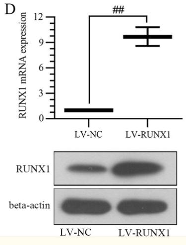
Application: WB Species: Rat Sample: BMSCs
Application: IHC Species: Rat Sample: BMSCs
Restrictive clause
Affinity Biosciences tests all products strictly. Citations are provided as a resource for additional applications that have not been validated by Affinity Biosciences. Please choose the appropriate format for each application and consult Materials and Methods sections for additional details about the use of any product in these publications.
For Research Use Only.
Not for use in diagnostic or therapeutic procedures. Not for resale. Not for distribution without written consent. Affinity Biosciences will not be held responsible for patent infringement or other violations that may occur with the use of our products. Affinity Biosciences, Affinity Biosciences Logo and all other trademarks are the property of Affinity Biosciences LTD.


