
| Product: | Paxillin Antibody |
| Catalog: | AF6332 |
| Description: | Rabbit polyclonal antibody to Paxillin |
| Application: | WB IHC |
| Cited expt.: | WB |
| Reactivity: | Human, Mouse, Rat |
| Prediction: | Pig, Bovine, Sheep, Rabbit, Dog, Chicken |
| Mol.Wt.: | 68kDa(Observed); 65kD(Calculated). |
| Uniprot: | P49023 |
| RRID: | AB_2835188 |
Control Products

Product Info
*The optimal dilutions should be determined by the end user. For optimal experimental results, antibody reuse is not recommended.
*Tips:
WB: For western blot detection of denatured protein samples. IHC: For immunohistochemical detection of paraffin sections (IHC-p) or frozen sections (IHC-f) of tissue samples. IF/ICC: For immunofluorescence detection of cell samples. ELISA(peptide): For ELISA detection of antigenic peptide.
Cite Format: Affinity Biosciences Cat# AF6332, RRID:AB_2835188.
Fold/Unfold
FLJ16691; FLJ23042; Paired box protein Pax 1; PAX 1; PAX1; PAXI_HUMAN; Paxillin alpha; Paxillin; PXN; PXN protein;
Immunogens
A synthesized peptide derived from human Paxillin, corresponding to a region within N-terminal amino acids.
- P49023 PAXI_HUMAN:
- Protein BLAST With
- NCBI/
- ExPASy/
- Uniprot
MDDLDALLADLESTTSHISKRPVFLSEETPYSYPTGNHTYQEIAVPPPVPPPPSSEALNGTILDPLDQWQPSSSRFIHQQPQSSSPVYGSSAKTSSVSNPQDSVGSPCSRVGEEEHVYSFPNKQKSAEPSPTVMSTSLGSNLSELDRLLLELNAVQHNPPGFPADEANSSPPLPGALSPLYGVPETNSPLGGKAGPLTKEKPKRNGGRGLEDVRPSVESLLDELESSVPSPVPAITVNQGEMSSPQRVTSTQQQTRISASSATRELDELMASLSDFKIQGLEQRADGERCWAAGWPRDGGRSSPGGQDEGGFMAQGKTGSSSPPGGPPKPGSQLDSMLGSLQSDLNKLGVATVAKGVCGACKKPIAGQVVTAMGKTWHPEHFVCTHCQEEIGSRNFFERDGQPYCEKDYHNLFSPRCYYCNGPILDKVVTALDRTWHPEHFFCAQCGAFFGPEGFHEKDGKAYCRKDYFDMFAPKCGGCARAILENYISALNTLWHPECFVCRECFTPFVNGSFFEHDGQPYCEVHYHERRGSLCSGCQKPITGRCITAMAKKFHPEHFVCAFCLKQLNKGTFKEQNDKPYCQNCFLKLFC
Predictions
Score>80(red) has high confidence and is suggested to be used for WB detection. *The prediction model is mainly based on the alignment of immunogen sequences, the results are for reference only, not as the basis of quality assurance.
High(score>80) Medium(80>score>50) Low(score<50) No confidence
Research Backgrounds
Cytoskeletal protein involved in actin-membrane attachment at sites of cell adhesion to the extracellular matrix (focal adhesion).
Phosphorylated by MAPK1/ERK2 (By similarity). Phosphorylated on tyrosine residues during integrin-mediated cell adhesion, embryonic development, fibroblast transformation and following stimulation of cells by mitogens. Phosphorylation at Ser-244 by CDK5 reduces its interaction with PTK2/FAK1 in matrix-cell focal adhesions (MCFA) during oligodendrocytes (OLs) differentiation. Phosphorylation at Tyr-31 and Tyr-118 by PTK6 promote the activation of RAC1 via CRK/CrKII, thereby promoting migration and invasion. Phosphorylation at Ser-250 by SLK is required for PXN redistribution and cell motility.
Cytoplasm>Cytoskeleton. Cell junction>Focal adhesion. Cytoplasm>Cell cortex.
Note: Colocalizes with integrins at the cell periphery. Colocalize with PXN to membrane ruffles and the leading edge of migrating cells (PubMed:23128389).
Belongs to the paxillin family.
Research Fields
· Cellular Processes > Cellular community - eukaryotes > Focal adhesion. (View pathway)
· Cellular Processes > Cell motility > Regulation of actin cytoskeleton. (View pathway)
· Human Diseases > Infectious diseases: Bacterial > Bacterial invasion of epithelial cells.
· Human Diseases > Infectious diseases: Viral > Human papillomavirus infection.
· Human Diseases > Cancers: Overview > Viral carcinogenesis.
· Human Diseases > Cancers: Overview > Proteoglycans in cancer.
· Organismal Systems > Immune system > Chemokine signaling pathway. (View pathway)
· Organismal Systems > Immune system > Leukocyte transendothelial migration. (View pathway)
References
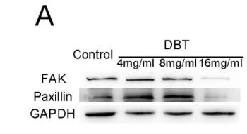
Application: WB Species: mouse Sample: BMSCs
Restrictive clause
Affinity Biosciences tests all products strictly. Citations are provided as a resource for additional applications that have not been validated by Affinity Biosciences. Please choose the appropriate format for each application and consult Materials and Methods sections for additional details about the use of any product in these publications.
For Research Use Only.
Not for use in diagnostic or therapeutic procedures. Not for resale. Not for distribution without written consent. Affinity Biosciences will not be held responsible for patent infringement or other violations that may occur with the use of our products. Affinity Biosciences, Affinity Biosciences Logo and all other trademarks are the property of Affinity Biosciences LTD.