, using Cathepsin Z Antibody. The lane on the left was treated with blocking peptide.")


 and mouse anti-beta tubulin Ab(#T0023) for 1 hour at 37°C. An AlexaFluor594 conjugated goat anti-rabbit IgG Ab(Red) and an AlexaFluor488 conjugated goat anti-mouse IgG Ab(Green) were used as the secondary antibody.
The nuclear counter stain is DAPI (blue).")
Control Products

Related Downloads
Protocols
Product Info
*The optimal dilutions should be determined by the end user. For optimal experimental results, antibody reuse is not recommended.
*Tips:
WB: For western blot detection of denatured protein samples. IHC: For immunohistochemical detection of paraffin sections (IHC-p) or frozen sections (IHC-f) of tissue samples. IF/ICC: For immunofluorescence detection of cell samples. ELISA(peptide): For ELISA detection of antigenic peptide.
Fold/Unfold
Carboxypeptidase LB; Cathepsin B2; Cathepsin IV; Cathepsin P; Cathepsin X; Cathepsin X precursor; Cathepsin Y; Cathepsin Z; Cathepsin Z precursor; Cathepsin Z1; CathepsinP; CathepsinX; CathepsinZ; CATZ_HUMAN; CTSX; CTSZ; Cysteine type carboxypeptidase; FLJ17088; Lysosomal carboxypeptidase B; Preprocathepsin P; PreprocathepsinP;
Immunogens
A synthesized peptide derived from Human Cathepsin Z.
- Q9UBR2 CATZ_HUMAN:
- Protein BLAST With
- NCBI/
- ExPASy/
- Uniprot
MARRGPGWRPLLLLVLLAGAAQGGLYFRRGQTCYRPLRGDGLAPLGRSTYPRPHEYLSPADLPKSWDWRNVDGVNYASITRNQHIPQYCGSCWAHASTSAMADRINIKRKGAWPSTLLSVQNVIDCGNAGSCEGGNDLSVWDYAHQHGIPDETCNNYQAKDQECDKFNQCGTCNEFKECHAIRNYTLWRVGDYGSLSGREKMMAEIYANGPISCGIMATERLANYTGGIYAEYQDTTYINHVVSVAGWGISDGTEYWIVRNSWGEPWGERGWLRIVTSTYKDGKGARYNLAIEEHCTFGDPIV
Research Backgrounds
Exhibits carboxy-monopeptidase as well as carboxy-dipeptidase activity. Capable of producing kinin potentiating peptides (By similarity).
Lysosome.
Widely expressed.
Belongs to the peptidase C1 family.
Research Fields
· Cellular Processes > Transport and catabolism > Lysosome. (View pathway)
· Cellular Processes > Cell growth and death > Apoptosis. (View pathway)
References
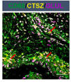
Application: IF/ICC Species: Human Sample: CRC tissues
Restrictive clause
Affinity Biosciences tests all products strictly. Citations are provided as a resource for additional applications that have not been validated by Affinity Biosciences. Please choose the appropriate format for each application and consult Materials and Methods sections for additional details about the use of any product in these publications.
For Research Use Only.
Not for use in diagnostic or therapeutic procedures. Not for resale. Not for distribution without written consent. Affinity Biosciences will not be held responsible for patent infringement or other violations that may occur with the use of our products. Affinity Biosciences, Affinity Biosciences Logo and all other trademarks are the property of Affinity Biosciences LTD.