CD31 Antibody - #AF6191
| Product: | CD31 Antibody |
| Catalog: | AF6191 |
| Description: | Rabbit polyclonal antibody to CD31 |
| Application: | WB IHC IF/ICC |
| Cited expt.: | WB, IHC, IF/ICC |
| Reactivity: | Human, Rat |
| Prediction: | Mouse, Pig, Bovine, Horse, Sheep, Rabbit, Dog |
| Mol.Wt.: | 82kDa,130Kd(Glycosylation); 83kD(Calculated). |
| Uniprot: | P16284 |
| RRID: | AB_2835074 |
Product Info
*The optimal dilutions should be determined by the end user. For optimal experimental results, antibody reuse is not recommended.
*Tips:
WB: For western blot detection of denatured protein samples. IHC: For immunohistochemical detection of paraffin sections (IHC-p) or frozen sections (IHC-f) of tissue samples. IF/ICC: For immunofluorescence detection of cell samples. ELISA(peptide): For ELISA detection of antigenic peptide.
Cite Format: Affinity Biosciences Cat# AF6191, RRID:AB_2835074.
Fold/Unfold
Adhesion molecule; CD31; CD31 antigen; CD31 EndoCAM; EndoCAM; FLJ34100; FLJ58394; GPIIA; GPIIA'; PECA1; PECA1_HUMAN; Pecam 1; PECAM 1 CD31 EndoCAM; PECAM; PECAM-1; Pecam1; Platelet and endothelial cell adhesion molecule 1; Platelet endothelial cell adhesion molecule; Platelet/endothelial cell adhesion molecule 1;
Immunogens
A synthesized peptide derived from human CD31, corresponding to a region within C-terminal amino acids.
Expressed on platelets and leukocytes and is primarily concentrated at the borders between endothelial cells (PubMed:18388311, PubMed:21464369). Expressed in human umbilical vein endothelial cells (HUVECs) (at protein level) (PubMed:19342684, PubMed:17580308). Expressed on neutrophils (at protein level) (PubMed:17580308). Isoform Long predominates in all tissues examined (PubMed:12433657). Isoform Delta12 is detected only in trachea (PubMed:12433657). Isoform Delta14-15 is only detected in lung (PubMed:12433657). Isoform Delta14 is detected in all tissues examined with the strongest expression in heart (PubMed:12433657). Isoform Delta15 is expressed in brain, testis, ovary, cell surface of platelets, human umbilical vein endothelial cells (HUVECs), Jurkat T-cell leukemia, human erythroleukemia (HEL) and U-937 histiocytic lymphoma cell lines (at protein level) (PubMed:12433657, PubMed:18388311).
- P16284 PECA1_HUMAN:
- Protein BLAST With
- NCBI/
- ExPASy/
- Uniprot
MQPRWAQGATMWLGVLLTLLLCSSLEGQENSFTINSVDMKSLPDWTVQNGKNLTLQCFADVSTTSHVKPQHQMLFYKDDVLFYNISSMKSTESYFIPEVRIYDSGTYKCTVIVNNKEKTTAEYQVLVEGVPSPRVTLDKKEAIQGGIVRVNCSVPEEKAPIHFTIEKLELNEKMVKLKREKNSRDQNFVILEFPVEEQDRVLSFRCQARIISGIHMQTSESTKSELVTVTESFSTPKFHISPTGMIMEGAQLHIKCTIQVTHLAQEFPEIIIQKDKAIVAHNRHGNKAVYSVMAMVEHSGNYTCKVESSRISKVSSIVVNITELFSKPELESSFTHLDQGERLNLSCSIPGAPPANFTIQKEDTIVSQTQDFTKIASKSDSGTYICTAGIDKVVKKSNTVQIVVCEMLSQPRISYDAQFEVIKGQTIEVRCESISGTLPISYQLLKTSKVLENSTKNSNDPAVFKDNPTEDVEYQCVADNCHSHAKMLSEVLRVKVIAPVDEVQISILSSKVVESGEDIVLQCAVNEGSGPITYKFYREKEGKPFYQMTSNATQAFWTKQKASKEQEGEYYCTAFNRANHASSVPRSKILTVRVILAPWKKGLIAVVIIGVIIALLIIAAKCYFLRKAKAKQMPVEMSRPAVPLLNSNNEKMSDPNMEANSHYGHNDDVRNHAMKPINDNKEPLNSDVQYTEVQVSSAESHKDLGKKDTETVYSEVRKAVPDAVESRYSRTEGSLDGT
Predictions
Score>80(red) has high confidence and is suggested to be used for WB detection. *The prediction model is mainly based on the alignment of immunogen sequences, the results are for reference only, not as the basis of quality assurance.
High(score>80) Medium(80>score>50) Low(score<50) No confidence
Research Backgrounds
Cell adhesion molecule which is required for leukocyte transendothelial migration (TEM) under most inflammatory conditions. Tyr-690 plays a critical role in TEM and is required for efficient trafficking of PECAM1 to and from the lateral border recycling compartment (LBRC) and is also essential for the LBRC membrane to be targeted around migrating leukocytes. Trans-homophilic interaction may play a role in endothelial cell-cell adhesion via cell junctions. Heterophilic interaction with CD177 plays a role in transendothelial migration of neutrophils. Homophilic ligation of PECAM1 prevents macrophage-mediated phagocytosis of neighboring viable leukocytes by transmitting a detachment signal. Promotes macrophage-mediated phagocytosis of apoptotic leukocytes by tethering them to the phagocytic cells; PECAM1-mediated detachment signal appears to be disabled in apoptotic leukocytes. Modulates bradykinin receptor BDKRB2 activation. Regulates bradykinin- and hyperosmotic shock-induced ERK1/2 activation in endothelial cells. Induces susceptibility to atherosclerosis (By similarity).
Does not protect against apoptosis.
Phosphorylated on Ser and Tyr residues after cellular activation by src kinases. Upon activation, phosphorylated on Ser-729 which probably initiates the dissociation of the membrane-interaction segment (residues 709-729) from the cell membrane allowing the sequential phosphorylation of Tyr-713 and Tyr-690. Constitutively phosphorylated on Ser-734 in resting platelets. Phosphorylated on tyrosine residues by FER and FES in response to FCER1 activation (By similarity). In endothelial cells Fyn mediates mechanical-force (stretch or pull) induced tyrosine phosphorylation.
Palmitoylation by ZDHHC21 is necessary for cell surface expression in endothelial cells and enrichment in membrane rafts.
Cell membrane>Single-pass type I membrane protein.
Note: Cell surface expression on neutrophils is down-regulated upon fMLP or CXCL8/IL8-mediated stimulation.
Cell membrane>Single-pass type I membrane protein. Membrane raft. Cell junction.
Note: Localizes to the lateral border recycling compartment (LBRC) and recycles from the LBRC to the junction in resting endothelial cells.
Cell junction.
Note: Localizes to the lateral border recycling compartment (LBRC) and recycles from the LBRC to the junction in resting endothelial cells.
Expressed on platelets and leukocytes and is primarily concentrated at the borders between endothelial cells. Expressed in human umbilical vein endothelial cells (HUVECs) (at protein level). Expressed on neutrophils (at protein level). Isoform Long predominates in all tissues examined. Isoform Delta12 is detected only in trachea. Isoform Delta14-15 is only detected in lung. Isoform Delta14 is detected in all tissues examined with the strongest expression in heart. Isoform Delta15 is expressed in brain, testis, ovary, cell surface of platelets, human umbilical vein endothelial cells (HUVECs), Jurkat T-cell leukemia, human erythroleukemia (HEL) and U-937 histiocytic lymphoma cell lines (at protein level).
The Ig-like C2-type domains 2 and 3 contribute to formation of the complex with BDKRB2 and in regulation of its activity.
Research Fields
· Environmental Information Processing > Signaling molecules and interaction > Cell adhesion molecules (CAMs). (View pathway)
· Human Diseases > Infectious diseases: Parasitic > Malaria.
· Organismal Systems > Immune system > Leukocyte transendothelial migration. (View pathway)
References
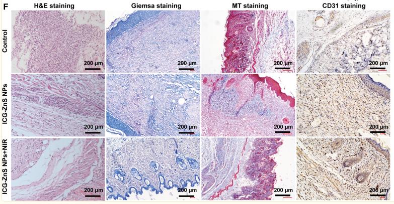
Application: IF/ICC Species: Rat Sample: bone tissue
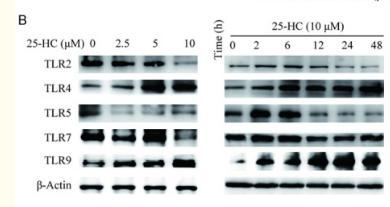
Application: WB Species: Rat Sample:
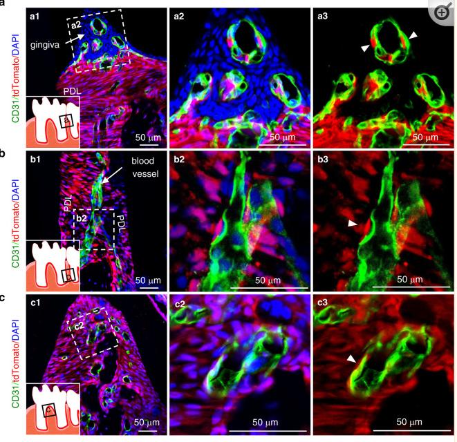
Application: IF/ICC Species: Rat Sample:
Restrictive clause
Affinity Biosciences tests all products strictly. Citations are provided as a resource for additional applications that have not been validated by Affinity Biosciences. Please choose the appropriate format for each application and consult Materials and Methods sections for additional details about the use of any product in these publications.
For Research Use Only.
Not for use in diagnostic or therapeutic procedures. Not for resale. Not for distribution without written consent. Affinity Biosciences will not be held responsible for patent infringement or other violations that may occur with the use of our products. Affinity Biosciences, Affinity Biosciences Logo and all other trademarks are the property of Affinity Biosciences LTD.


