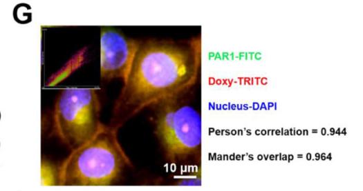
Product: Thrombin Receptor Antibody
Catalog: AF0263
Description: Rabbit polyclonal antibody to Thrombin Receptor
Application: WB IHC IF/ICC
Cited expt.: WB, IHC, IF/ICC
Reactivity: Human, Mouse, Rat
Mol.Wt.: 48kDa; 47kD(Calculated).
Uniprot: P25116
RRID: AB_2833437

View similar products>>

   Size Price Inventory
 100ul $280 In stock
 200ul $350 In stock

Lead Time: Same day delivery

For pricing and ordering contact:
Local distributors

Product Info

Source:
Rabbit
Application:
WB 1:500-1:3000, IF/ICC 1:100-1:500, IHC 1:50-1:200
*The optimal dilutions should be determined by the end user.
*Tips:

WB: For western blot detection of denatured protein samples. IHC: For immunohistochemical detection of paraffin sections (IHC-p) or frozen sections (IHC-f) of tissue samples. IF/ICC: For immunofluorescence detection of cell samples. ELISA(peptide): For ELISA detection of antigenic peptide.

Reactivity:
Human,Mouse,Rat
Clonality:
Polyclonal
Specificity:
Thrombin Receptor Antibody detects endogenous levels of total Thrombin Receptor.
RRID:
AB_2833437
Cite Format: Affinity Biosciences Cat# AF0263, RRID:AB_2833437.
Conjugate:
Unconjugated.
Purification:
The antiserum was purified by peptide affinity chromatography using SulfoLink™ Coupling Resin (Thermo Fisher Scientific).
Storage:
Rabbit IgG in phosphate buffered saline , pH 7.4, 150mM NaCl, 0.02% sodium azide and 50% glycerol. Store at -20 °C. Stable for 12 months from date of receipt.
Alias:

Fold/Unfold

CF2R; Coagulation factor II (thrombin) receptor; Coagulation factor II receptor; F2R; HTR; PAR 1; PAR-1; PAR1; PAR1_HUMAN; Protease activated receptor 1; Proteinase activated receptor 1; Proteinase-activated receptor 1; Thrombin receptor; TR;

Immunogens

Immunogen:

A synthesized peptide derived from human Thrombin Receptor, corresponding to a region within N-terminal amino acids.

Uniprot:
Gene(ID):
Expression:
P25116 PAR1_HUMAN:

Platelets and vascular endothelial cells.

Description:
PAR1 a G-protein coupled high-affinity receptor for activated thrombin or trypsin. Coupled to G proteins that stimulate phosphoinositide hydrolysis. Coupled to G proteins that stimulate phosphoinositide hydrolysis. May play a role in platelet activation and in vascular development.
Sequence:
MGPRRLLLVAACFSLCGPLLSARTRARRPESKATNATLDPRSFLLRNPNDKYEPFWEDEEKNESGLTEYRLVSINKSSPLQKQLPAFISEDASGYLTSSWLTLFVPSVYTGVFVVSLPLNIMAIVVFILKMKVKKPAVVYMLHLATADVLFVSVLPFKISYYFSGSDWQFGSELCRFVTAAFYCNMYASILLMTVISIDRFLAVVYPMQSLSWRTLGRASFTCLAIWALAIAGVVPLLLKEQTIQVPGLNITTCHDVLNETLLEGYYAYYFSAFSAVFFFVPLIISTVCYVSIIRCLSSSAVANRSKKSRALFLSAAVFCIFIICFGPTNVLLIAHYSFLSHTSTTEAAYFAYLLCVCVSSISCCIDPLIYYYASSECQRYVYSILCCKESSDPSSYNSSGQLMASKMDTCSSNLNNSIYKKLLT

Research Backgrounds

Function:

High affinity receptor for activated thrombin coupled to G proteins that stimulate phosphoinositide hydrolysis. May play a role in platelets activation and in vascular development.

PTMs:

A proteolytic cleavage generates a new N-terminus that functions as a tethered ligand.

Phosphorylated in the C-terminal tail; probably mediating desensitization prior to the uncoupling and internalization of the receptor.

Subcellular Location:

Cell membrane>Multi-pass membrane protein.

Extracellular region or secreted Cytosol Plasma membrane Cytoskeleton Lysosome Endosome Peroxisome ER Golgi apparatus Nucleus Mitochondrion Manual annotation Automatic computational assertionSubcellular location
Tissue Specificity:

Platelets and vascular endothelial cells.

Family&Domains:

The cleaved signal peptide may not be degraded and may function as an intracellular angiogenesis inhibitor peptide known as parstatin.

Belongs to the G-protein coupled receptor 1 family.

Research Fields

· Cellular Processes > Cell motility > Regulation of actin cytoskeleton.   (View pathway)

· Environmental Information Processing > Signal transduction > Rap1 signaling pathway.   (View pathway)

· Environmental Information Processing > Signal transduction > Calcium signaling pathway.   (View pathway)

· Environmental Information Processing > Signal transduction > cAMP signaling pathway.   (View pathway)

· Environmental Information Processing > Signal transduction > Phospholipase D signaling pathway.   (View pathway)

· Environmental Information Processing > Signaling molecules and interaction > Neuroactive ligand-receptor interaction.

· Environmental Information Processing > Signal transduction > PI3K-Akt signaling pathway.   (View pathway)

· Human Diseases > Cancers: Overview > Pathways in cancer.   (View pathway)

· Organismal Systems > Immune system > Complement and coagulation cascades.   (View pathway)

· Organismal Systems > Immune system > Platelet activation.   (View pathway)

References

1). Thrombin receptor activating peptide-6 decreases acute graft-versus-host disease through activating GPR15. Leukemia, 2024 (PubMed: 38459169) [IF=12.8]

2). Gut microbiome and serum short-chain fatty acids are associated with responses to chemo- or targeted therapies in Chinese patients with lung cancer. Frontiers in microbiology, 2023 (PubMed: 37564290) [IF=5.2]

Application: IF/ICC    Species: human    Sample: LC cells

Figure 5. Isobutyric acid regulated G protein-coupled receptor (GPCR) expression and histone acetyltransferase (HAT) activity. For the expression of GPCRs, the results of RT-qPCR (A), Western blot (B), and immunocytochemical staining (C) showed that GPR41, GPR43, and GPRC5A expressions were significantly higher, while PAR1 expression was significantly lower in isobutyric acid treatment group (1.85 mM) than the control groups (control and NC). Immunofluorescence staining (D) identified the expression location of GPCRs. For the expression of acetyl-histones and histones, the results of Western blot (E) and immunocytochemical staining (F) showed that acetyl-histone H3 and H4 expressions were significantly higher in the isobutyric acid treatment group (1.85 mM) than the control groups (control and NC), whereas Western blot results showed that there was no significant difference of the expression of histone H3 and H4 between isobutyric acid treatment group and the control groups. Immunofluorescence staining (G) identified the expression location of acetyl-histone H3 and H4. HAT activity assay (H) results showed that HAT activity was increased in the isobutyric acid treatment group (1.85mM) than in the control groups (control and NC). P-values were calculated using Student's t-tests. ns: no significance; *p < 0.05; **p < 0.01; ***p < 0.001. All experiments were repeated three times. NC, negative control (0mM).

Application: IHC    Species: human    Sample: LC cells

Figure 5. Isobutyric acid regulated G protein-coupled receptor (GPCR) expression and histone acetyltransferase (HAT) activity. For the expression of GPCRs, the results of RT-qPCR (A), Western blot (B), and immunocytochemical staining (C) showed that GPR41, GPR43, and GPRC5A expressions were significantly higher, while PAR1 expression was significantly lower in isobutyric acid treatment group (1.85 mM) than the control groups (control and NC). Immunofluorescence staining (D) identified the expression location of GPCRs. For the expression of acetyl-histones and histones, the results of Western blot (E) and immunocytochemical staining (F) showed that acetyl-histone H3 and H4 expressions were significantly higher in the isobutyric acid treatment group (1.85 mM) than the control groups (control and NC), whereas Western blot results showed that there was no significant difference of the expression of histone H3 and H4 between isobutyric acid treatment group and the control groups. Immunofluorescence staining (G) identified the expression location of acetyl-histone H3 and H4. HAT activity assay (H) results showed that HAT activity was increased in the isobutyric acid treatment group (1.85mM) than in the control groups (control and NC). P-values were calculated using Student's t-tests. ns: no significance; *p < 0.05; **p < 0.01; ***p < 0.001. All experiments were repeated three times. NC, negative control (0mM).

Application: WB    Species: human    Sample: LC cells

Figure 5. Isobutyric acid regulated G protein-coupled receptor (GPCR) expression and histone acetyltransferase (HAT) activity. For the expression of GPCRs, the results of RT-qPCR (A), Western blot (B), and immunocytochemical staining (C) showed that GPR41, GPR43, and GPRC5A expressions were significantly higher, while PAR1 expression was significantly lower in isobutyric acid treatment group (1.85 mM) than the control groups (control and NC). Immunofluorescence staining (D) identified the expression location of GPCRs. For the expression of acetyl-histones and histones, the results of Western blot (E) and immunocytochemical staining (F) showed that acetyl-histone H3 and H4 expressions were significantly higher in the isobutyric acid treatment group (1.85 mM) than the control groups (control and NC), whereas Western blot results showed that there was no significant difference of the expression of histone H3 and H4 between isobutyric acid treatment group and the control groups. Immunofluorescence staining (G) identified the expression location of acetyl-histone H3 and H4. HAT activity assay (H) results showed that HAT activity was increased in the isobutyric acid treatment group (1.85mM) than in the control groups (control and NC). P-values were calculated using Student's t-tests. ns: no significance; *p < 0.05; **p < 0.01; ***p < 0.001. All experiments were repeated three times. NC, negative control (0mM).

3). TREM2 improves coagulopathy and lung inflammation in sepsis through the AKT-mTOR pathway. International immunopharmacology, 2025 (PubMed: 39970715) [IF=4.8]

4). Inhibiting trophoblast PAR-1 overexpression suppresses sFlt-1-induced anti-angiogenesis and abnormal vascular remodeling: a possible therapeutic approach for preeclampsia. MOLECULAR HUMAN REPRODUCTION, 2018 (PubMed: 29325127) [IF=3.6]

5). pHLIP(Var7)-P1AP suppresses tumor cell proliferation in MDA-MB-231 triple-negative breast cancer by targeting protease activated receptor 1. BREAST CANCER RESEARCH AND TREATMENT, 2020 (PubMed: 32034579) [IF=3.0]

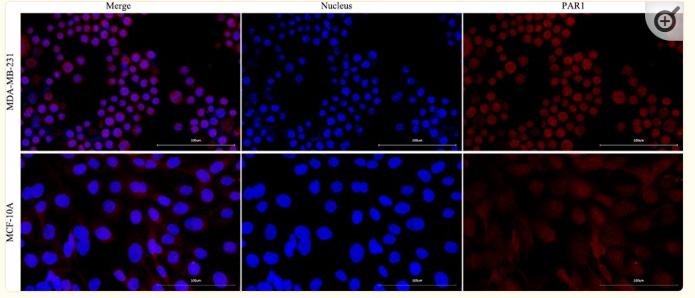
Application: IF/ICC    Species: Human    Sample: MDA-MB-231 and MCF-10A cells

Fig. 1 PAR1 expression was positive in MDA-MB-231 cells, and PAR1 expression was negative in MCF-10A cells (400 ×)

6). Expression and clinical implications of PARs in the stenotic tissue of ureteropelvic junction obstruction. Frontiers in pediatrics, 2023 (PubMed: 38161438) [IF=2.1]

Application: IF/ICC    Species: human    Sample:

Figure 1. Immunofluorescence double staining was conducted on PAR1 with various types of SIP syncytium. (A) Immunofluorescence double staining of PAR1 (red) and c-kit (green) in UPJ control; (B) immunofluorescence double staining of PAR1 (red) and c-kit (green) in UPJO; (C) immunofluorescence double staining of PAR1 (red) and phalloidin (green) in UPJ control; (D) immunofluorescence double staining of PAR1 (red) and phalloidin (green) in UPJO; (E) immunofluorescence double staining of PAR1 (red) and PDGFRα (green) in UPJ control; (F) immunofluorescence double staining of PAR1 (red) and PDGFRα (green) in UPJO. The blue areas in all images represent DAPI-labelled nuclei. Nuclei were counterstained with DAPI (blue) in all panels. Bar = 50 μm in all panels.

7). Doxycycline directly targets PAR1 to suppress tumor progression. Oncotarget, 2017 (PubMed: 28187433)

Application: IF/ICC    Species: human    Sample:

(G) Confocal immunofluorescence imaging was performed to visualize PAR1 and doxycycline. PAR1 and doxycycline co-localize, with a Pearson’s correlation coeffcient of 0.944 and a Mander’s overlap of 0.964.

Restrictive clause


Affinity Biosciences tests all products strictly. Citations are provided as a resource for additional applications that have not been validated by Affinity Biosciences. Please choose the appropriate format for each application and consult Materials and Methods sections for additional details about the use of any product in these publications.

For Research Use Only.
Not for use in diagnostic or therapeutic procedures. Not for resale. Not for distribution without written consent. Affinity Biosciences will not be held responsible for patent infringement or other violations that may occur with the use of our products. Affinity Biosciences, Affinity Biosciences Logo and all other trademarks are the property of Affinity Biosciences LTD.