Product: PDGF Receptor beta Antibody
Catalog: AF6133
Description: Rabbit polyclonal antibody to PDGF Receptor beta
Application: WB IHC IF/ICC
Reactivity: Human, Mouse, Rat
Prediction: Pig, Horse, Sheep, Rabbit, Dog, Chicken, Xenopus
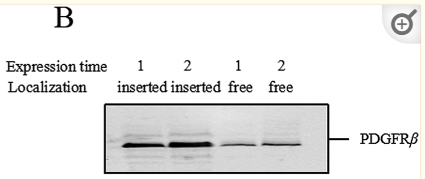
Mol.Wt.: 123kDa; 124kD(Calculated).
Uniprot: P09619
RRID: AB_2835016

View similar products>>

   Size Price Inventory
 100ul $280 In stock
 200ul $350 In stock

Lead Time: Same day delivery

For pricing and ordering contact:
Local distributors

Product Info

Source:
Rabbit
Application:
WB 1:500-1:2000, IHC 1:50-1:200, IF/ICC 1:100-1:500
*The optimal dilutions should be determined by the end user.
*Tips:

WB: For western blot detection of denatured protein samples. IHC: For immunohistochemical detection of paraffin sections (IHC-p) or frozen sections (IHC-f) of tissue samples. IF/ICC: For immunofluorescence detection of cell samples. ELISA(peptide): For ELISA detection of antigenic peptide.

Reactivity:
Human,Mouse,Rat
Prediction:
Pig(100%), Horse(100%), Sheep(100%), Rabbit(100%), Dog(100%), Chicken(89%), Xenopus(88%)
Clonality:
Polyclonal
Specificity:
PDGF Receptor beta Antibody detects endogenous levels of total PDGF Receptor beta.
RRID:
AB_2835016
Cite Format: Affinity Biosciences Cat# AF6133, RRID:AB_2835016.
Conjugate:
Unconjugated.
Purification:
The antiserum was purified by peptide affinity chromatography using SulfoLink™ Coupling Resin (Thermo Fisher Scientific).
Storage:
Rabbit IgG in phosphate buffered saline , pH 7.4, 150mM NaCl, 0.02% sodium azide and 50% glycerol. Store at -20 °C. Stable for 12 months from date of receipt.
Alias:

Fold/Unfold

Beta platelet derived growth factor receptor; Beta-type platelet-derived growth factor receptor; CD 140B; CD140 antigen-like family member B; CD140b; CD140b antigen; IBGC4; IMF1; JTK12; OTTHUMP00000160528; PDGF R beta; PDGF-R-beta; PDGFR 1; PDGFR; PDGFR beta; PDGFR1; PDGFRB; PGFRB_HUMAN; Platelet derived growth factor receptor 1; Platelet derived growth factor receptor beta; Platelet derived growth factor receptor beta polypeptide;

Immunogens

Immunogen:
Uniprot:
Gene(ID):
Description:
This gene encodes a cell surface tyrosine kinase receptor for members of the platelet-derived growth factor family. These growth factors are mitogens for cells of mesenchymal origin. The identity of the growth factor bound to a receptor monomer determines whether the functional receptor is a homodimer or a heterodimer, composed of both platelet-derived growth factor receptor alpha and beta polypeptides.
Sequence:
MRLPGAMPALALKGELLLLSLLLLLEPQISQGLVVTPPGPELVLNVSSTFVLTCSGSAPVVWERMSQEPPQEMAKAQDGTFSSVLTLTNLTGLDTGEYFCTHNDSRGLETDERKRLYIFVPDPTVGFLPNDAEELFIFLTEITEITIPCRVTDPQLVVTLHEKKGDVALPVPYDHQRGFSGIFEDRSYICKTTIGDREVDSDAYYVYRLQVSSINVSVNAVQTVVRQGENITLMCIVIGNEVVNFEWTYPRKESGRLVEPVTDFLLDMPYHIRSILHIPSAELEDSGTYTCNVTESVNDHQDEKAINITVVESGYVRLLGEVGTLQFAELHRSRTLQVVFEAYPPPTVLWFKDNRTLGDSSAGEIALSTRNVSETRYVSELTLVRVKVAEAGHYTMRAFHEDAEVQLSFQLQINVPVRVLELSESHPDSGEQTVRCRGRGMPQPNIIWSACRDLKRCPRELPPTLLGNSSEEESQLETNVTYWEEEQEFEVVSTLRLQHVDRPLSVRCTLRNAVGQDTQEVIVVPHSLPFKVVVISAILALVVLTIISLIILIMLWQKKPRYEIRWKVIESVSSDGHEYIYVDPMQLPYDSTWELPRDQLVLGRTLGSGAFGQVVEATAHGLSHSQATMKVAVKMLKSTARSSEKQALMSELKIMSHLGPHLNVVNLLGACTKGGPIYIITEYCRYGDLVDYLHRNKHTFLQHHSDKRRPPSAELYSNALPVGLPLPSHVSLTGESDGGYMDMSKDESVDYVPMLDMKGDVKYADIESSNYMAPYDNYVPSAPERTCRATLINESPVLSYMDLVGFSYQVANGMEFLASKNCVHRDLAARNVLICEGKLVKICDFGLARDIMRDSNYISKGSTFLPLKWMAPESIFNSLYTTLSDVWSFGILLWEIFTLGGTPYPELPMNEQFYNAIKRGYRMAQPAHASDEIYEIMQKCWEEKFEIRPPFSQLVLLLERLLGEGYKKKYQQVDEEFLRSDHPAILRSQARLPGFHGLRSPLDTSSVLYTAVQPNEGDNDYIIPLPDPKPEVADEGPLEGSPSLASSTLNEVNTSSTISCDSPLEPQDEPEPEPQLELQVEPEPELEQLPDSGCPAPRAEAEDSFL

Predictions

Predictions:

Score>80(red) has high confidence and is suggested to be used for WB detection. *The prediction model is mainly based on the alignment of immunogen sequences, the results are for reference only, not as the basis of quality assurance.

Species
Results
Score
Pig
100
Horse
100
Sheep
100
Dog
100
Rabbit
100
Chicken
89
Xenopus
88
Zebrafish
71
Bovine
0
Model Confidence:
High(score>80) Medium(80>score>50) Low(score<50) No confidence

PTMs - P09619 As Substrate

Site PTM Type Enzyme
N89 N-Glycosylation
N103 N-Glycosylation
S212 Phosphorylation
S213 Phosphorylation
N215 N-Glycosylation
N230 N-Glycosylation
N292 N-Glycosylation
N307 N-Glycosylation
Y562 Phosphorylation P09619 (PDGFRB)
Y579 Phosphorylation P09619 (PDGFRB)
Y581 Phosphorylation P09619 (PDGFRB)
T605 Phosphorylation
S608 Phosphorylation
K645 Ubiquitination
Y678 Phosphorylation
Y683 Phosphorylation
Y686 Phosphorylation P42684 (ABL2) , P00519 (ABL1)
Y692 Phosphorylation
S705 Phosphorylation
K707 Ubiquitination
S712 Phosphorylation
Y716 Phosphorylation P09619 (PDGFRB)
S717 Phosphorylation
Y740 Phosphorylation P09619 (PDGFRB)
Y751 Phosphorylation P09619 (PDGFRB)
Y763 Phosphorylation P09619 (PDGFRB)
Y771 Phosphorylation P09619 (PDGFRB)
Y775 Phosphorylation P09619 (PDGFRB)
Y778 Phosphorylation P09619 (PDGFRB)
Y857 Phosphorylation P09619 (PDGFRB)
Y921 Phosphorylation
S930 Phosphorylation
Y934 Phosphorylation P00519 (ABL1)
Y970 Phosphorylation P00519 (ABL1)
Y1009 Phosphorylation P09619 (PDGFRB)
Y1021 Phosphorylation P09619 (PDGFRB)
S1104 Phosphorylation

PTMs - P09619 As Enzyme

Substrate Site Source
P06241-3 (FYN) Y28 Uniprot
P09619 (PDGFRB) Y562 Uniprot
P09619 (PDGFRB) Y579 Uniprot
P09619 (PDGFRB) Y581 Uniprot
P09619-1 (PDGFRB) Y716 Uniprot
P09619 (PDGFRB) Y740 Uniprot
P09619 (PDGFRB) Y751 Uniprot
P09619-1 (PDGFRB) Y763 Uniprot
P09619-1 (PDGFRB) Y771 Uniprot
P09619 (PDGFRB) Y775 Uniprot
P09619 (PDGFRB) Y778 Uniprot
P09619-1 (PDGFRB) Y857 Uniprot
P09619 (PDGFRB) Y1009 Uniprot
P09619 (PDGFRB) Y1021 Uniprot
P12931 (SRC) Y139 Uniprot
P12931 (SRC) Y419 Uniprot
P15941 (MUC1) Y1203 Uniprot
P15941 (MUC1) Y1218 Uniprot
P16234 (PDGFRA) Y754 Uniprot
P27986 (PIK3R1) Y508 Uniprot
P34947 (GRK5) Y90 Uniprot
P34947 (GRK5) Y109 Uniprot
P34947 (GRK5) Y309 Uniprot
P34947 (GRK5) Y368 Uniprot
P42684 (ABL2) Y139 Uniprot
P42684 (ABL2) Y161 Uniprot
P42684 (ABL2) Y272 Uniprot
Q05397 (PTK2) Y5 Uniprot
Q05397 (PTK2) Y194 Uniprot
Q05655 (PRKCD) Y313 Uniprot
Q05655 (PRKCD) Y334 Uniprot
Q06124-2 (PTPN11) Y542 Uniprot
Q06124 (PTPN11) Y546 Uniprot
Q07912 (TNK2) Y635 Uniprot
Q13480 (GAB1) Y627 Uniprot
Q13480 (GAB1) Y659 Uniprot
Q13769 (THOC5) Y225 Uniprot

Research Backgrounds

Function:

Tyrosine-protein kinase that acts as cell-surface receptor for homodimeric PDGFB and PDGFD and for heterodimers formed by PDGFA and PDGFB, and plays an essential role in the regulation of embryonic development, cell proliferation, survival, differentiation, chemotaxis and migration. Plays an essential role in blood vessel development by promoting proliferation, migration and recruitment of pericytes and smooth muscle cells to endothelial cells. Plays a role in the migration of vascular smooth muscle cells and the formation of neointima at vascular injury sites. Required for normal development of the cardiovascular system. Required for normal recruitment of pericytes (mesangial cells) in the kidney glomerulus, and for normal formation of a branched network of capillaries in kidney glomeruli. Promotes rearrangement of the actin cytoskeleton and the formation of membrane ruffles. Binding of its cognate ligands - homodimeric PDGFB, heterodimers formed by PDGFA and PDGFB or homodimeric PDGFD -leads to the activation of several signaling cascades; the response depends on the nature of the bound ligand and is modulated by the formation of heterodimers between PDGFRA and PDGFRB. Phosphorylates PLCG1, PIK3R1, PTPN11, RASA1/GAP, CBL, SHC1 and NCK1. Activation of PLCG1 leads to the production of the cellular signaling molecules diacylglycerol and inositol 1,4,5-trisphosphate, mobilization of cytosolic Ca(2+) and the activation of protein kinase C. Phosphorylation of PIK3R1, the regulatory subunit of phosphatidylinositol 3-kinase, leads to the activation of the AKT1 signaling pathway. Phosphorylation of SHC1, or of the C-terminus of PTPN11, creates a binding site for GRB2, resulting in the activation of HRAS, RAF1 and down-stream MAP kinases, including MAPK1/ERK2 and/or MAPK3/ERK1. Promotes phosphorylation and activation of SRC family kinases. Promotes phosphorylation of PDCD6IP/ALIX and STAM. Receptor signaling is down-regulated by protein phosphatases that dephosphorylate the receptor and its down-stream effectors, and by rapid internalization of the activated receptor.

PTMs:

Autophosphorylated on tyrosine residues upon ligand binding. Autophosphorylation occurs in trans, i.e. one subunit of the dimeric receptor phosphorylates tyrosine residues on the other subunit. Phosphorylation at Tyr-579, and to a lesser degree, at Tyr-581, is important for interaction with SRC family kinases. Phosphorylation at Tyr-740 and Tyr-751 is important for interaction with PIK3R1. Phosphorylation at Tyr-751 is important for interaction with NCK1. Phosphorylation at Tyr-771 and Tyr-857 is important for interaction with RASA1/GAP. Phosphorylation at Tyr-857 is important for efficient phosphorylation of PLCG1 and PTPN11, resulting in increased phosphorylation of AKT1, MAPK1/ERK2 and/or MAPK3/ERK1, PDCD6IP/ALIX and STAM, and in increased cell proliferation. Phosphorylation at Tyr-1009 is important for interaction with PTPN11. Phosphorylation at Tyr-1009 and Tyr-1021 is important for interaction with PLCG1. Phosphorylation at Tyr-1021 is important for interaction with CBL; PLCG1 and CBL compete for the same binding site. Dephosphorylated by PTPRJ at Tyr-751, Tyr-857, Tyr-1009 and Tyr-1021. Dephosphorylated by PTPN2 at Tyr-579 and Tyr-1021.

N-glycosylated.

Ubiquitinated. After autophosphorylation, the receptor is polyubiquitinated, leading to its degradation.

Subcellular Location:

Cell membrane>Single-pass type I membrane protein. Cytoplasmic vesicle. Lysosome lumen.
Note: After ligand binding, the autophosphorylated receptor is ubiquitinated and internalized, leading to its degradation.

Extracellular region or secreted Cytosol Plasma membrane Cytoskeleton Lysosome Endosome Peroxisome ER Golgi apparatus Nucleus Mitochondrion Manual annotation Automatic computational assertionSubcellular location
Subunit Structure:

Interacts with homodimeric PDGFB and PDGFD, and with heterodimers formed by PDGFA and PDGFB. May also interact with homodimeric PDGFC. Monomer in the absence of bound ligand. Interaction with homodimeric PDGFB, heterodimers formed by PDGFA and PDGFB or homodimeric PDGFD, leads to receptor dimerization, where both PDGFRA homodimers and heterodimers with PDGFRB are observed. Interacts with SH2B2/APS. Interacts directly (tyrosine phosphorylated) with SHB. Interacts (tyrosine phosphorylated) with PIK3R1 and RASA1. Interacts (tyrosine phosphorylated) with CBL. Interacts (tyrosine phosphorylated) with SRC and SRC family kinases. Interacts (tyrosine phosphorylated) with PIK3C2B, maybe indirectly. Interacts (tyrosine phosphorylated) with SHC1, GRB7, GRB10 and NCK1. Interaction with GRB2 is mediated by SHC1. Interacts (via C-terminus) with SLC9A3R1.

Family&Domains:

Belongs to the protein kinase superfamily. Tyr protein kinase family. CSF-1/PDGF receptor subfamily.

Research Fields

· Cellular Processes > Cellular community - eukaryotes > Focal adhesion.   (View pathway)

· Cellular Processes > Cellular community - eukaryotes > Gap junction.   (View pathway)

· Cellular Processes > Cell motility > Regulation of actin cytoskeleton.   (View pathway)

· Environmental Information Processing > Signal transduction > MAPK signaling pathway.   (View pathway)

· Environmental Information Processing > Signal transduction > Ras signaling pathway.   (View pathway)

· Environmental Information Processing > Signal transduction > Rap1 signaling pathway.   (View pathway)

· Environmental Information Processing > Signal transduction > Calcium signaling pathway.   (View pathway)

· Environmental Information Processing > Signaling molecules and interaction > Cytokine-cytokine receptor interaction.   (View pathway)

· Environmental Information Processing > Signal transduction > Phospholipase D signaling pathway.   (View pathway)

· Environmental Information Processing > Signal transduction > PI3K-Akt signaling pathway.   (View pathway)

· Environmental Information Processing > Signal transduction > Jak-STAT signaling pathway.   (View pathway)

· Human Diseases > Drug resistance: Antineoplastic > EGFR tyrosine kinase inhibitor resistance.

· Human Diseases > Infectious diseases: Viral > Human papillomavirus infection.

· Human Diseases > Infectious diseases: Viral > HTLV-I infection.

· Human Diseases > Cancers: Overview > Pathways in cancer.   (View pathway)

· Human Diseases > Cancers: Overview > MicroRNAs in cancer.

· Human Diseases > Cancers: Specific types > Glioma.   (View pathway)

· Human Diseases > Cancers: Specific types > Prostate cancer.   (View pathway)

· Human Diseases > Cancers: Specific types > Melanoma.   (View pathway)

· Human Diseases > Cancers: Overview > Central carbon metabolism in cancer.   (View pathway)

· Human Diseases > Cancers: Overview > Choline metabolism in cancer.   (View pathway)

References

1). In situ synthesis and unidirectional insertion of membrane proteins in liposome-immobilized silica stationary phase for rapid preparation of microaffinity chromatography. Acta Pharmaceutica Sinica B (PubMed: 36176904) [IF=14.5]

Application: WB    Species: Mouse    Sample:

Figure 2 Optimization of expression conditions: (A) investigation of added silica gel in per 50 μL system, (B) investigation of expression time: observe the amount of inserted and not-inserted PDGFRβ at 1 or 2 h expression time, (C) investigation of insertion time given to PDGFRβ, (D) investigation of maximum proportion of DOPE, (E) quantification analysis of bands in (C), (F) quantification analysis of bands in (D).

2). PDGFRβ targeted innovative imaging probe for pancreatic adenocarcinoma detection. Talanta (PubMed: 36587427) [IF=6.1]

Application: IF/ICC    Species: Mouse    Sample:

The precisely detection and dissection of pancreatic adenocarcinoma by the probes based on a novel PDGFRβ targeting peptide.

3). Overexpression of Corin Ameliorates Kidney Fibrosis through Inhibition of Wnt/β-Catenin Signaling in Mice. The American journal of pathology (PubMed: 37827215) [IF=6.0]

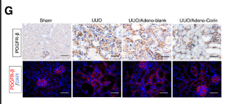
Application: IHC    Species: Mouse    Sample: kidney tissues

Figure 5Adenovirus-mediated overexpression of corin ameliorates kidney fibrosis after unilateral ureteral obstruction (UUO). A: Experimental design. Green arrow indicates the time point of UUO and injection of corin-overexpressing adenovirus (Adeno-corin) or blank adenovirus (Adeno-blank). Fluorescence activity was detected 48 hours after UUO and adenovirus infection to confirm adenovirus gene delivery. B: Representative micrographs of kidney tissues in different groups at day 7 after UUO as indicated. Kidney sections were subjected to Sirius red and Masson trichrome staining. C: Graphic presentation showing the area of Masson trichrome and Sirius red staining in kidney tissues among the indicated groups. D: Representative immunohistochemical micrographs of α-smooth muscle actin (α-SMA) and fibronectin at day 7 after UUO. Paraffin-embedded kidney sections were stained with α-SMA and fibronectin antibodies. Arrows indicate positive staining. E and F: Western blot analysis of α-SMA, fibronectin, and collagen I in kidney tissues at day 7 after UUO. Representative gel images (E) and quantitative data (F) are shown. G and H: Immunohistochemical and immunofluorescence staining of platelet-derived growth factor receptor β (PDGFR-β) in kidney tissues of different groups as indicated at day 7 after UUO. PDGFR-β is a pericyte marker. Representative micrographs (G) and quantitative data (H) are presented. I and J: Immunohistochemical and immunofluorescence staining of CD31 in kidney tissues of different groups as indicated at day 7 after UUO. CD31 is the marker of mature endothelial cells. Representative micrographs (I) and quantitative data (J) are presented. Data are the means ± SEM (C, F, H, and J). n = 5 animals per group (C, F, H, and J). ∗P < 0.05, ∗∗P < 0.01, and ∗∗∗P < 0.001. Scale bars = 50 μm (B, D, G, and I). GAPDH, glyceraldehyde-3-phosphate dehydrogenase.

Application: IF/ICC    Species: Mouse    Sample: kidney tissues

Figure 5Adenovirus-mediated overexpression of corin ameliorates kidney fibrosis after unilateral ureteral obstruction (UUO). A: Experimental design. Green arrow indicates the time point of UUO and injection of corin-overexpressing adenovirus (Adeno-corin) or blank adenovirus (Adeno-blank). Fluorescence activity was detected 48 hours after UUO and adenovirus infection to confirm adenovirus gene delivery. B: Representative micrographs of kidney tissues in different groups at day 7 after UUO as indicated. Kidney sections were subjected to Sirius red and Masson trichrome staining. C: Graphic presentation showing the area of Masson trichrome and Sirius red staining in kidney tissues among the indicated groups. D: Representative immunohistochemical micrographs of α-smooth muscle actin (α-SMA) and fibronectin at day 7 after UUO. Paraffin-embedded kidney sections were stained with α-SMA and fibronectin antibodies. Arrows indicate positive staining. E and F: Western blot analysis of α-SMA, fibronectin, and collagen I in kidney tissues at day 7 after UUO. Representative gel images (E) and quantitative data (F) are shown. G and H: Immunohistochemical and immunofluorescence staining of platelet-derived growth factor receptor β (PDGFR-β) in kidney tissues of different groups as indicated at day 7 after UUO. PDGFR-β is a pericyte marker. Representative micrographs (G) and quantitative data (H) are presented. I and J: Immunohistochemical and immunofluorescence staining of CD31 in kidney tissues of different groups as indicated at day 7 after UUO. CD31 is the marker of mature endothelial cells. Representative micrographs (I) and quantitative data (J) are presented. Data are the means ± SEM (C, F, H, and J). n = 5 animals per group (C, F, H, and J). ∗P < 0.05, ∗∗P < 0.01, and ∗∗∗P < 0.001. Scale bars = 50 μm (B, D, G, and I). GAPDH, glyceraldehyde-3-phosphate dehydrogenase.

4). Total flavonoids of Rhizoma Drynariae enhances CD31hiEmcnhi vessel formation and subsequent bone regeneration in rat models of distraction osteogenesis by activating PDGF‑BB/VEGF/RUNX2/OSX signaling axis. International Journal of Molecular Medicine (PubMed: 35795995) [IF=5.4]

Application: WB    Species: Rat    Sample:

Figure 6. - TFRD promotes CD31hiEmcnhi vessel formation in angiogenic-osteogenic coupling during distraction osteogenesis via the PDGF-BB/PDGFR-β pathway. (A) Representative immunofluorescence images and (B) quantification of RUNX2, OSX, CD31 and PDGF-BB in the distracted tibias after distraction for 4 weeks. (C) Representative western blotting images and (D) semi-quantitative analyses of PDGF-BB, VEGF, RUNX2, OSX as well as the phosphorylation of AKT and ERK1/2 in the distracted tibias at 4 weeks post-distraction. (E) Quantification of mRNA expression levels of PDGF-BB, VEGF, RUNX2 and OSX. Data represent the mean ± SD. n=3 rats in each group from three independent experiments. *P

5). Anticancer Effect of Puerarin on Ovarian Cancer Progression Contributes to the Tumor Suppressor Gene Expression and Gut Microbiota Modulation. Journal of Immunology Research (PubMed: 35935578) [IF=4.1]

6). ProS/Mer alleviates sepsis-induced neuromuscular dysfunction by inhibiting TLR4/MyD88/NF-κB signals. Research Square

Restrictive clause


Affinity Biosciences tests all products strictly. Citations are provided as a resource for additional applications that have not been validated by Affinity Biosciences. Please choose the appropriate format for each application and consult Materials and Methods sections for additional details about the use of any product in these publications.

For Research Use Only.
Not for use in diagnostic or therapeutic procedures. Not for resale. Not for distribution without written consent. Affinity Biosciences will not be held responsible for patent infringement or other violations that may occur with the use of our products. Affinity Biosciences, Affinity Biosciences Logo and all other trademarks are the property of Affinity Biosciences LTD.