


 and mouse anti-beta tubulin Ab(T0023) for 1 hour at 37°C. An AlexaFluor594 conjugated goat anti-rabbit IgG(H+L) Ab(Red) and an AlexaFluor488 conjugated goat anti-mouse IgG(H+L) Ab(Green) were used as the secondary antibody.
The nuclear counter stain is DAPI(blue).")
 microscopy at x200 magnification was used to assess cell morphology. The A549 cells (parental cells) had an epithelioid, rounded cobblestone appearance and there was limited formation of pseudopodia. A549/PTX and A549/DDP cells exhibited a spindle-shaped morphology and an increased formation of pseudopodia, indicating a loss of cell polarity. (B) E-cadherin, β-catenin, vimentin, MMP-2 and MMP-9 which are EMT-related proteins, were assessed in terms of expression levels. EMT-related transcription factors (Snail, Slug, Twist and ZEB1) were measured in A549/PTX and A549/DDP cells using western blot analysis. (C) The expression changes were confirmed at the mRNA level by qRT-PCR. Expression was standardized to the expression of GAPDH and normalized to 1.0 in the parental cells (compared with the parental A549 cells, means ± SEM, n=3, * P<0.05)")
 Western blot analysis was used to detect the expression of E-cadherin, β-catenin, vimentin, MMP-9, MMP-2, Snail, Slug,Twist and ZEB1 after transfection.")


(B) Protein strips and quantitative analysis: significant decreases in the SNAIL1, ZEB1 and vimentin and a significant increase in the E-cadherin of metformin and TGF-β-treated samples compared with only TGF-β-treated samples for both SW480 and HCT116 cells; a significant increase in the ZEB2 of metformin and TGF-β-treated samples compared with only TGF-β-treated samples for SW480 cells; significant decreases in the SNAIL2, ZEB1 and ZEB2 of metformin-treated samples compared with negative control samples for HCT116 cells. *P<0.05")


 Overexpression of KRT7 increased Snail, vimentin and N-cadherin expression, and decreased E-cadherin expression in HEY cells as detected by western blotting. (C and D) Knockdown of KRT7 resulted in increased E-cadherin expression and reduced vimentin and Snail expression in OVCAR433 cells as detected by western blotting. All experiments were performed at least three times. Results are presented as the mean ± standard deviation. **P<0.01. KRT7, keratin 7; sh, short hairpin RNA; NC, negative control.")
. Immunohistochemical staining of the EMT markers E-cadherin, N-cadherin, Vimentin, Twist, Snail, and Slug in tumor tissues
from the xenograft model demonstrated the contribution of GGCT to the EMT process in vivo (C). *p < 0.05.")
. Immunohistochemical staining of the EMT markers E-cadherin, N-cadherin, Vimentin, Twist, Snail, and Slug in tumor tissues
from the xenograft model demonstrated the contribution of GGCT to the EMT process in vivo (C). *p < 0.05.")
. Immunohistochemical staining of the EMT markers E-cadherin, N-cadherin, Vimentin, Twist, Snail, and Slug in tumor tissues
from the xenograft model demonstrated the contribution of GGCT to the EMT process in vivo (C). *p < 0.05.")
. Immunohistochemical staining of the EMT markers E-cadherin, N-cadherin, Vimentin, Twist, Snail, and Slug in tumor tissues
from the xenograft model demonstrated the contribution of GGCT to the EMT process in vivo (C). *p < 0.05.")
 Relative LINP1 expression in EC9706 sh1, sh2, sh3, and sh4 was verified by qRT-PCR. The LINP1 sh2 cell line had the most significant knockdown effect compared with the control cell line (61.75%, P<0.001). (B) The Alamar Blue proliferation assay indicated that the proliferation of shRNA-LINP1 cells was significantly inhibited compared with that of the corresponding control cells and shRNA-scr cells (P<0.001). (C,D) Colony formation assays showing that the number of colonies formed was significantly lower in shRNA-LINP1 cells than in NC and shRNA-scr cells in macroscopic view (71.7 vs. 73.3 vs. 24.7, P<0.001). (E,F) Wound healing assays showed that the migration rate was slower in shRNA-LINP1 cells than in NC and shRNA-scr cells after 48 h (19.1% vs. 46.4% vs. 46.6%; P<0.001) (magnification 100×). (G,H) Transwell migration assays demonstrated that the number of migratory cells was lower in shRNA-LINP1 than in the two control cells (60.3 vs. 236.3 vs. 238.7; P<0.001) (magnification 100×). (I,J,K) qRT-PCR and western blot analyses of the expression of EMT markers. E-cadherin was significantly upregulated, whereas N-cadherin, vimentin, snail and slug were significantly downregulated in shRNA-LINP1 cells compared with NC and shRNA-scr cells (all P<0.05). **, P<0.01; ***, P<0.001.")
 The negative control or siTP53INP2 were transfected into the BIU87 and EJ cells. The cells were incubated for 48 h, and the changes in the expressions of EMT molecular markers and β-catenin were detected by Western blot. β-actin was used as an internal control. (C, D) The cellular location of active β-catenin and Snail1 in EJ cells after interference in TP53INP2. Scale bar, 50 μm. (E) The abundance of the active forms of β-catenin and Snail1 in nuclear and cytosolic fractions were detected by Western blot. GAPDH and H3 were used as cytosolic or nuclear controls.
Abbreviations: EMT, epithelial-to-mesenchymal transition; GAPDH, glyceraldehyde 3-phosphate dehydrogenase.")
 NSE overexpression promoted the EMT process of SCLC. (A) Western blot assay was performed to measure the protein expression levels of the EMT-related markers (Snail, N-cadherin and E-cadherin).")
 NSE overexpression promoted the EMT process of SCLC. (A) Western blot assay was performed to measure the protein expression levels of the EMT-related markers (Snail, N-cadherin and E-cadherin). (B) qRT-PCR was performed to measure the mRNA expression levels of the EMT markers. (C, D) NSE knockdown represses the EMT process of SCLC. Protein levels (C) and mRNA levels (D) of EMT-related markers were measured using western blot or qRT-PCR, respectively. These results were repeated of three independent experiments.")
 NSE overexpression promoted the EMT process of SCLC. (A) Western blot assay was performed to measure the protein expression levels of the EMT-related markers (Snail, N-cadherin and E-cadherin). (B) qRT-PCR was performed to measure the mRNA expression levels of the EMT markers. (C, D) NSE knockdown represses the EMT process of SCLC. Protein levels (C) and mRNA levels (D) of EMT-related markers were measured using western blot or qRT-PCR, respectively. These results were repeated of three independent experiments.")
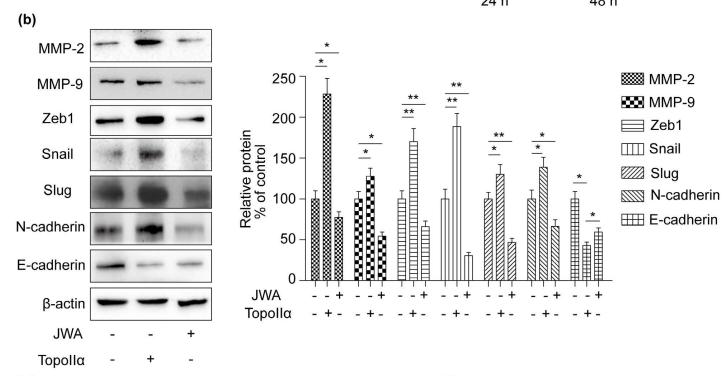
 EMT-related protein expression; representative blots of the related proteins. (B) Quantitative data of the related protein expression. (C) EMT-related gene expression at mRNA level. *P<0.05 vs. control; #P<0.05 vs. AOPPs. AOPPs; EMT, epithelial-mesenchymal transition.")
 Samples were considered negative for both LSD1 and SNAIL. (c,d) Samples considered moderate or high positive for both LSD1 and SNAIL.")
 Levels of α-SMA, Col I SHH, Gli1, and snail1 were measured by Western blot. (b) Quantitative analysis. (c) Levels of α-SMA, Col I SHH, Gli1, and snail1 were measured by qPCR. Data were expressed as mean ± SEM. ∗p < 0.05 compared with the Sham group, #p < 0.05 compared with the Model group, △p < 0.05 compared with the CPN group, ☆p < 0.05 compared with the S + C group, ▲p > 0.05 compared with the CPN group, and ★p > 0.05 compared with the C + S group.")

 Cell morphology of A2780 and SKOV3 after CA and EGF treatment. (B) Expression of E-cadherin, N-cadherin, vimentin, and Snail were detected by Western blotting in A2780 and SKOV3 cells after CA and EGF treatment. Representative fluorescence images of E-cadherin (C) and N-cadherin (D) in A2780 and SKOV3 cells. At least three independent experiments were performed.")
. The protein-protein interaction (PPI) network of the KRT17 from STRING online database (https://string-db.org) was constructed (b). The protein levels in AMC-HN-8 cells were evaluated using western blot (c). The protein levels in TU177 cells were estimated using western blot. ∗A significant difference compared with the sh-NC group. ∗∗P < 0.01 and ∗∗∗P < 0.001.")
 Morphology of cells observed under an inverted light microscope (scale bar, 100 µm). (B) The ultrastructure of cells observed under a transmission electron microscope (x10,000 magnification). (C) Cell sphere-forming rate evaluated using the sphere formation assay (x100 magnification). (D) Number of spheres measured using the colony-formation assay (x200 magnification). (E) Expression levels of CD44, ALDH1, Nanog and Snail determined using western blotting assays. *P")
 Expression levels of CD44, ALDH1, Nanog and Snail as determined via western blotting. (B) Expression level of CD44, ALDH1, Nanog and Snail in tumor tissues measured via immunohistochemistry (x200 magnification). (C) Expression levels of vimentin and E-cadherin as determined via western blotting. (D) Expression levels of vimentin and E-cadherin in tumor tissues measured via immunohistochemistry (x200 magnification). *P")
 NSE overexpression promoted the EMT process of SCLC. (A) Western blot assay was performed to measure the protein expression levels of the EMT-related markers (Snail, N-cadherin and E-cadherin). (B) qRT-PCR was performed to measure the mRNA expression levels of the EMT markers. (C, D) NSE knockdown represses the EMT process of SCLC. Protein levels (C) and mRNA levels (D) of EMT-related markers were measured using western blot or qRT-PCR, respectively. These results were repeated of three independent experiments.")
 and stroma (lower row) of the tumor; from left to right: high, low, and absence expression; IHC staining with antibodies against Snail, 400x; b Expression of HIF-1α in the parenchyma (upper row) and stroma (lower row) of the tumor; from left to right: high, low and absence")
 The effect of SEC on the migration of BC cells (Wound healing assay, 100 × ). (B) The effect of SEC on the migration of BC cells (Transwell assay, 100 × ). (C) The effect of SEC on the invasion of BC cells (Matrigel-coated transwell assay, 100 × ). (D) The effect of SEC on the protein levels of EMT markers and MMPs of BC cells (Western blot). Data are shown as mean ± SD from three independent experiments.")
.")
. (A) Gene Ontology (GO) enrichment analysis of LINC00909 based on the NGS database. (B) Top 15-enriched Kyoto Encyclopedia of Genes and Genomes (KEGG) pathways of LINC00909 based on the NGS database. (C) Gene set enrichment analysis (GSEA) of the NGS database. (D) The expression of factors associated with stemness and metastasis was analyzed by NGS using PANC-1/Vector and PANC-1/LINC00909-OE cells. These factors are shown in the heatmap. (E–G) Western blotting analysis was conducted to detect the protein expression of JNK phospho-JNK, c-JUN, phospho-JUN, TWIST, and SNAIL in LINC00909-overexpressing cells (E) and LINC00909-knockdown cells (F, G). All *P")
 EMT pathway detection after HLF knockdown in CAL62. (B) Quantitative analysis of protein expression as shown in (A). EMT, Epithelial-mesenchymal transition.")
 Histograms of mRNA expression levels. (B) WB strip. (C) Protein expression histograms. *p < 0.05 vs. Control, #p < 0.05 vs. sh-HOXB9, ! p < 0.05 vs. sh-MMP12.")
 GO and KEGG enrichment analyses showed that SULF1 was involved in CRC progression via the FAK/PI3K/AKT/mTor pathway. (B) Western blotting showed that SULF1 knockdown could decrease the protein expression of p-FAK, p-PI3K, p-AKT, and p-mTor. (C) Western blotting showed that SULF1 knockdown could decrease the protein expression of EMT markers, N-cadherin and Snail, but increase E-cadherin. All experiments were repeated three times and the data are represented as mean ± SD. *p")
 Down-regulation of GLI2 is accompanied by down-regulation of PD-L1, TGF-beta, IL6 and snail1. (E–H) Up-regulation of GLI2 is accompanied with increased expression of PD-L1, TGF-beta, and IL6. (I) Co-culture process. (J–L) Down-regulation of tumor-derived GLI2 decreases tumor-derived TGF-beta, PDL1 and IL6, likewise the NK-derived TIM-3 and PD1, while NK-derived IFN-gamma is increased. TGF-beta active protein powder restores the expression of tumor-derived PDL1 and IL6, and NK-derived TIM-3 and PD1. (M) Down-regulation of tumor-derived GIL2 leads increased secretion of NK-derived IFN-gamma, detected by Elisa.")
 Western blot showing expression levels of EMT-related proteins E-cadherin, N-cadherin, Vimentin, Snail, Slug, and Twist1 after ZDHHC9 knockdown in 143B and U2OS cells. (B) Western blot analysis of apoptosis-related proteins Cleaved Caspase-3, Cleaved Caspase-9, Bcl-2, Bax, and cell cycle-related proteins CDK4 and Cyclin D1 after ZDHHC9 knockdown in 143B and U2OS cells. (C) Flow cytometry analysis showing apoptosis rates of 143B and U2OS cells after ZDHHC9 knockdown. (D) Flow cytometry analysis of cell cycle distribution in 143B and U2OS cells after ZDHHC9 knockdown. (*P")
 assays. b Quantitation of the number of invaded cells. *** indicates P")
 The wound-healing assay was performed to evaluate the effect of SMC4 overexpression/knockdown on the migratory capacity of U-251MG and LN229 cells. Panels A and B show representative images of the wound-healing assay. Statistical analysis revealed that SMC4 overexpression enhanced the migratory capacity of U-251MG and LN229 cells by 96% and 103%, respectively, while SMC4 knockdown reduced their migratory capacity by 39% and 41%, respectively. (E-G) The Transwell assay was conducted to examine the effects of SMC4 overexpression/knockdown on the invasive capabilities of U-251MG and LN229 cells. Figure E presents a representative image of the Transwell assay. Statistical results demonstrated that SMC4 overexpression increased the invasive abilities of U-251MG and LN229 cells by 85% and 94% respectively, whereas SMC4 knockdown decreased the invasive abilities of these two cell lines by 54% and 43% respectively. (H) The WB assay was used to detect the changes in the expression levels of the core members of the TGF-β/SMAD signaling pathway in U-251MG and LN229 cells after SMC4 overexpression/knockdown, aiming to confirm the activating effect of SMC4 on the TGF-β/SMAD signaling pathway. (I) A tail vein metastasis model was established in BALB/c nude mice using LN229 cells with stable SMC4 overexpression/knockdown, and an in vivo animal imaging system was used to detect the colonization and growth of tumor cells in the lungs of mice. The images show the distribution of tumor cells in mice at the 6th week after modeling. (J) Six weeks after modeling, the mice were euthanized, and lung tissues were collected for HE staining to observe the size and number of metastatic foci in the lung tissues.")
 EMT, PI3K/AKT/mTOR pathways, and EMT transcription factor protein bands. (B-H) Expression levels of E-cadherin, N-cadherin, p-PI3K, p-AKT, p-mTOR, Snail, and Twistl in each group. E-cadherin protein: oeKRT23 vs. oeNC: MGC803, P=0.04; SGC7901, P=0.005. N-cadherin, Snail, Twist1, p-PI3K, p-AKT, and p-mTOR proteins: oeKRT23 vs. NC: MGC803, N-cadherin (P=0.004), Snail (P=0.003), Twist1 (P=0.009), p-PI3K (P=0.009), p-AKT (P=0.008), and p-mTOR (P=0.005); SGC7901, N-cadherin (P=0.006), Snail (P=0.004), Twist1 (P=0.001), p-PI3K (P=0.009), p-AKT (P=0.009), and p-mTOR (P=0.008). AKT, protein kinase B; EMT, epithelial-mesenchymal transition; KRT23, keratin 23; mTOR, mammalian target of rapamycin; NC, normal control; oeKRT23, KRT23 overexpression; oeNC, NC overexpression; p-, phospho-; PI3K, phosphatidylinositol 3-kinase.")
| Product: | SNAIL Antibody |
| Catalog: | AF6032 |
| Description: | Rabbit polyclonal antibody to SNAIL |
| Application: | WB IHC IF/ICC |
| Cited expt.: | WB, IHC, IF/ICC |
| Reactivity: | Human, Mouse, Rat |
| Prediction: | Pig, Bovine, Horse, Rabbit, Dog, Chicken, Xenopus |
| Mol.Wt.: | 29kDa(Observed); 29kD(Calculated). |
| Uniprot: | O95863 |
| RRID: | AB_2834965 |
Control Products

Product Info
*The optimal dilutions should be determined by the end user. For optimal experimental results, antibody reuse is not recommended.
*Tips:
WB: For western blot detection of denatured protein samples. IHC: For immunohistochemical detection of paraffin sections (IHC-p) or frozen sections (IHC-f) of tissue samples. IF/ICC: For immunofluorescence detection of cell samples. ELISA(peptide): For ELISA detection of antigenic peptide.
Cite Format: Affinity Biosciences Cat# AF6032, RRID:AB_2834965.
Fold/Unfold
dJ710H13.1; Protein sna; Protein snail homolog 1; Protein snail homolog; SLUGH2; SNA; Sna protein; SNAH; SNAI; snai1; SNAI1_HUMAN; Snail 1 homolog; Snail 1 zinc finger protein; SNAIL; Snail homolog 1 (Drosophila); SNAIL, Drosophila, homolog of, 1; SNAIL1; Zinc finger protein SNAI1;
Immunogens
A synthesized peptide derived from human SNAIL, corresponding to a region within C-terminal amino acids.
Expressed in a variety of tissues with the highest expression in kidney. Expressed in mesenchymal and epithelial cell lines.
- O95863 SNAI1_HUMAN:
- Protein BLAST With
- NCBI/
- ExPASy/
- Uniprot
MPRSFLVRKPSDPNRKPNYSELQDSNPEFTFQQPYDQAHLLAAIPPPEILNPTASLPMLIWDSVLAPQAQPIAWASLRLQESPRVAELTSLSDEDSGKGSQPPSPPSPAPSSFSSTSVSSLEAEAYAAFPGLGQVPKQLAQLSEAKDLQARKAFNCKYCNKEYLSLGALKMHIRSHTLPCVCGTCGKAFSRPWLLQGHVRTHTGEKPFSCPHCSRAFADRSNLRAHLQTHSDVKKYQCQACARTFSRMSLLHKHQESGCSGCPR
Predictions
Score>80(red) has high confidence and is suggested to be used for WB detection. *The prediction model is mainly based on the alignment of immunogen sequences, the results are for reference only, not as the basis of quality assurance.
High(score>80) Medium(80>score>50) Low(score<50) No confidence
Research Backgrounds
Involved in induction of the epithelial to mesenchymal transition (EMT), formation and maintenance of embryonic mesoderm, growth arrest, survival and cell migration. Binds to 3 E-boxes of the E-cadherin/CDH1 gene promoter and to the promoters of CLDN7 and KRT8 and, in association with histone demethylase KDM1A which it recruits to the promoters, causes a decrease in dimethylated H3K4 levels and represses transcription. The N-terminal SNAG domain competes with histone H3 for the same binding site on the histone demethylase complex formed by KDM1A and RCOR1, and thereby inhibits demethylation of histone H3 at 'Lys-4' (in vitro). During EMT, involved with LOXL2 in negatively regulating pericentromeric heterochromatin transcription (By similarity). SNAI1 recruits LOXL2 to pericentromeric regions to oxidize histone H3 and repress transcription which leads to release of heterochromatin component CBX5/HP1A, enabling chromatin reorganization and acquisition of mesenchymal traits (By similarity). Associates with EGR1 and SP1 to mediate tetradecanoyl phorbol acetate (TPA)-induced up-regulation of CDKN2B, possibly by binding to the CDKN2B promoter region 5'-TCACA-3. In addition, may also activate the CDKN2B promoter by itself.
Phosphorylated by GSK3B. Once phosphorylated, it becomes a target for BTRC ubiquitination. Phosphorylation by CSNK1E, probably at Ser-104, provides the priming site for the subsequent phosphorylation by GSK3B, probably at Ser-100 and Ser-96. Phosphorylation by PAK1 may modulate its transcriptional activity by promoting increased accumulation in the nucleus. Phosphorylation at Ser-11 and Ser-92 positively regulates its functions in induction of EMT and cell survival, respectively. Phosphorylation by LATS2, upon mitotic stress, oncogenic stress or Hippo pathway activation, occurs in the nucleus and promotes nuclear retention and stabilization of total cellular protein level.
Ubiquitinated on Lys-98, Lys-137 and Lys-146 by FBXL14 and BTRC leading to degradation. BTRC-triggered ubiquitination requires previous GSK3B-mediated SNAI1 phosphorylation. Ubiquitination induced upon interaction with NOTCH1 or TP53/p53 is mediated by MDM2.
O-GlcNAcylation at Ser-112 is enhanced in hyperglycaemic conditions, it opposes phosphorylation by GSK3B, and stabilizes the protein.
ADP-ribosylation by PARP1 increases protein half-life and may be involved in TGFB-induced SNAI1 up-regulation.
Nucleus. Cytoplasm.
Note: Once phosphorylated (probably on Ser-107, Ser-111, Ser-115 and Ser-119) it is exported from the nucleus to the cytoplasm where subsequent phosphorylation of the destruction motif and ubiquitination involving BTRC occurs.
Expressed in a variety of tissues with the highest expression in kidney. Expressed in mesenchymal and epithelial cell lines.
Belongs to the snail C2H2-type zinc-finger protein family.
Research Fields
· Cellular Processes > Cellular community - eukaryotes > Adherens junction. (View pathway)
References
Application: WB Species: Human Sample: HCT116 cells
Application: WB Species: Human Sample: OS cells
Application: WB Species: human Sample: NSCLC cells
Application: WB Species: Human Sample: NSCLC cells

Application: WB Species: human Sample: Tu177/CDDPTNFAIP2-/- cells

Application: WB Species: Human Sample: AGS and MGC803 cells
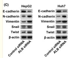
Application: WB Species: Human Sample: HepG2 and Huh7 cells
Restrictive clause
Affinity Biosciences tests all products strictly. Citations are provided as a resource for additional applications that have not been validated by Affinity Biosciences. Please choose the appropriate format for each application and consult Materials and Methods sections for additional details about the use of any product in these publications.
For Research Use Only.
Not for use in diagnostic or therapeutic procedures. Not for resale. Not for distribution without written consent. Affinity Biosciences will not be held responsible for patent infringement or other violations that may occur with the use of our products. Affinity Biosciences, Affinity Biosciences Logo and all other trademarks are the property of Affinity Biosciences LTD.