Product: SLC40A1 Antibody
Catalog: DF13561
Description: Rabbit polyclonal antibody to SLC40A1
Application: WB IHC
Reactivity: Human, Mouse, Rat
Prediction: Pig, Zebrafish, Bovine, Horse, Sheep, Rabbit, Chicken, Xenopus
Mol.Wt.: 62kDa; 63kD(Calculated).
Uniprot: Q9NP59
RRID: AB_2846580

View similar products>>

   Size Price Inventory
 100ul $280 In stock
 200ul $350 In stock

Lead Time: Same day delivery

For pricing and ordering contact:
Local distributors

Product Info

Source:
Rabbit
Application:
WB 1:500-1:2000, IHC 1:50-1:200
*The optimal dilutions should be determined by the end user.
*Tips:

WB: For western blot detection of denatured protein samples. IHC: For immunohistochemical detection of paraffin sections (IHC-p) or frozen sections (IHC-f) of tissue samples. IF/ICC: For immunofluorescence detection of cell samples. ELISA(peptide): For ELISA detection of antigenic peptide.

Reactivity:
Human,Mouse,Rat
Prediction:
Pig(100%), Zebrafish(88%), Bovine(100%), Horse(100%), Sheep(100%), Rabbit(100%), Chicken(100%), Xenopus(100%)
Clonality:
Polyclonal
Specificity:
SLC40A1 Antibody detects endogenous levels of total SLC40A1.
RRID:
AB_2846580
Cite Format: Affinity Biosciences Cat# DF13561, RRID:AB_2846580.
Conjugate:
Unconjugated.
Purification:
The antiserum was purified by peptide affinity chromatography using SulfoLink™ Coupling Resin (Thermo Fisher Scientific).
Storage:
Rabbit IgG in phosphate buffered saline , pH 7.4, 150mM NaCl, 0.02% sodium azide and 50% glycerol. Store at -20 °C. Stable for 12 months from date of receipt.
Alias:

Fold/Unfold

Ferroportin 1; Ferroportin; Ferroportin-1; FPN1; HFE4; IREG1; iron regulated gene 1; Iron regulated transporter 1; Iron-regulated transporter 1; MST079; MSTP079; MTP1; putative ferroportin 1 variant IIIA; putative ferroportin 1 variant IIIB; S40A1_HUMAN; SLC11A3; SLC11A3, formerly; SLC40A1; solute carrier family 11 (proton-coupled divalent metal ion transporters), member 3; solute carrier family 11 (proton-coupled divalent metal ion transporters), member 3, formerly; solute carrier family 40 (iron-regulated transporter), member 1; Solute carrier family 40 member 1;

Immunogens

Immunogen:
Uniprot:
Gene(ID):
Expression:
Q9NP59 S40A1_HUMAN:

Detected in erythrocytes (at protein level). Expressed in placenta, intestine, muscle and spleen.

Sequence:
MTRAGDHNRQRGCCGSLADYLTSAKFLLYLGHSLSTWGDRMWHFAVSVFLVELYGNSLLLTAVYGLVVAGSVLVLGAIIGDWVDKNARLKVAQTSLVVQNVSVILCGIILMMVFLHKHELLTMYHGWVLTSCYILIITIANIANLASTATAITIQRDWIVVVAGEDRSKLANMNATIRRIDQLTNILAPMAVGQIMTFGSPVIGCGFISGWNLVSMCVEYVLLWKVYQKTPALAVKAGLKEEETELKQLNLHKDTEPKPLEGTHLMGVKDSNIHELEHEQEPTCASQMAEPFRTFRDGWVSYYNQPVFLAGMGLAFLYMTVLGFDCITTGYAYTQGLSGSILSILMGASAITGIMGTVAFTWLRRKCGLVRTGLISGLAQLSCLILCVISVFMPGSPLDLSVSPFEDIRSRFIQGESITPTKIPEITTEIYMSNGSNSANIVPETSPESVPIISVSLLFAGVIAARIGLWSFDLTVTQLLQENVIESERGIINGVQNSMNYLLDLLHFIMVILAPNPEAFGLLVLISVSFVAMGHIMYFRFAQNTLGNKLFACGPDAKEVRKENQANTSVV

Predictions

Predictions:

Score>80(red) has high confidence and is suggested to be used for WB detection. *The prediction model is mainly based on the alignment of immunogen sequences, the results are for reference only, not as the basis of quality assurance.

Species
Results
Score
Pig
100
Horse
100
Bovine
100
Sheep
100
Xenopus
100
Chicken
100
Rabbit
100
Zebrafish
88
Dog
0
Model Confidence:
High(score>80) Medium(80>score>50) Low(score<50) No confidence

PTMs - Q9NP59 As Substrate

Site PTM Type Enzyme
Y20 Phosphorylation
K240 Ubiquitination
K247 Ubiquitination
Y302 Phosphorylation
Y303 Phosphorylation
T419 Phosphorylation
T421 Phosphorylation
Y538 Phosphorylation

Research Backgrounds

Function:

May be involved in iron export from duodenal epithelial cell and also in transfer of iron between maternal and fetal circulation. Mediates iron efflux in the presence of a ferroxidase (hephaestin and/or ceruloplasmin).

Subcellular Location:

Cell membrane>Multi-pass membrane protein.
Note: Localized to the basolateral membrane of polarized epithelial cells.

Extracellular region or secreted Cytosol Plasma membrane Cytoskeleton Lysosome Endosome Peroxisome ER Golgi apparatus Nucleus Mitochondrion Manual annotation Automatic computational assertionSubcellular location
Tissue Specificity:

Detected in erythrocytes (at protein level). Expressed in placenta, intestine, muscle and spleen.

Subunit Structure:

Identified in a complex with STOM.

Family&Domains:

Belongs to the ferroportin (FP) (TC 2.A.100) family. SLC40A subfamily.

Research Fields

· Cellular Processes > Cell growth and death > Ferroptosis.   (View pathway)

· Organismal Systems > Digestive system > Mineral absorption.

References

1). Biochanin A protects against iron overload associated knee osteoarthritis via regulating iron levels and NRF2/System xc-/GPX4 axis. Biomedicine & Pharmacotherapy (PubMed: 36379122) [IF=7.5]

2). GSK-3β-dependent Nrf2 antioxidant response modulates ferroptosis of lens epithelial cells in age-related cataract. Free Radical Biology and Medicine (PubMed: 37156294) [IF=7.4]

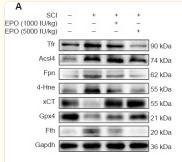
3). Erythropoietin inhibits ferroptosis and ameliorates neurological function after spinal cord injury. Neural Regeneration Research (PubMed: 36204858) [IF=6.1]

Application: WB    Species: Rat    Sample: spinal cord

Figure 3 EPO regulates the expression of ferroptotic biomarkers after SCI. (A) Western blot of the indicated proteins in injured tissues from the treatment groups. (B–H) Quantitative analysis of Tfr, Fpn, Fth, Acsl4 and 4-Hne protein expression at 14 dpi. Gapdh was used as the reference protein. (I, J) Quantitative reverse transcription polymerase chain reaction results of xCT and Gpx4 mRNA extracted from injured tissue at 7 dpi. β-Actin mRNA was used as the reference gene. (K) Quantitative analysis of reduced GSH from injured tissue at 7 dpi. All data are expressed as the mean ± SD (n = 5 in each group). *P < 0.05, **P < 0.01, ***P < 0.001 (one-way analysis of variance followed by Dunnett’s multiple comparisons test). 4-Hne: 4-Hydroxynonenal; Acsl4: acyl-coenzyme A synthetase long chain family member 4; dpi: day(s) post injury; EPO: erythropoietin; Fpn: ferroportin or solute carrier family 40 member 1; Fth: ferritin heavy chain; Gapdh: glyceraldehyde-3-phosphate dehydrogenase; Gpx4: glutathione peroxidase 4; GSH: glutathione; ns: not significant; SCI: spinal cord injury; Tfr: transferrin receptor; xCT: the solute carrier family 7 member 11.

4). Betaine Alleviates High-Fat Diet-Induced Disruptionof Hepatic Lipid and Iron Homeostasis in Mice. INTERNATIONAL JOURNAL OF MOLECULAR SCIENCES (PubMed: 35682942) [IF=5.6]

Application: WB    Species: Mice    Sample: liver tissue

Figure 3 Hepatic genes expression involved in iron metabolism. (a) Hepatic mRNA expression of iron metabolism (n = 6). (b) Protein content of ZIP14 in the liver (n = 6). (c) Protein content of DMT1 in the liver (n = 6). (d) Protein content of FTL in the liver (n = 6). (e) Protein content of FPN in the liver (n = 6). (f) Hepatic mRNA expression of HAMP and the upstream regulators (n = 6). Values are means ± SEM. # p < 0.05 and ## p < 0.01 compared with the CON group. * p < 0.05 and ** p < 0.01 compared with the HF group.

5). Ferroptosis contributes to cyclophosphamide-induced hemorrhagic cystitis. Chemico-Biological Interactions (PubMed: 37690746) [IF=5.1]

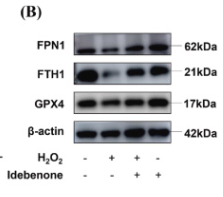
6). Idebenone attenuates ferroptosis by inhibiting excessive autophagy via the ROS-AMPK-mTOR pathway to preserve cardiac function after myocardial infarction. European Journal of Pharmacology (PubMed: 36740037) [IF=5.0]

Application: WB    Species: Rat    Sample: H9c2 cells

Fig. 2. Idebenone inhibited H2O2-induced ferroptosis. (A) The effect of deferoxamine (DFO, 100 μM) on the viability of H9c2 cells treated with 100 μM H2O2 for 4 h as detected by CCK8 assay (n = 3). (B) The GPX4, FTH1 and FPN1 protein expressions were detected via western blotting. (C-E) Quantitative analysis of GPX4, FTH1and FPN1 protein expressions (n = 3). (F) ROS level in H9c2 cells was assessed by DCFH-DA probe (scale bar:50 μm). (G) Quantitative analysis of ROS level in H9c2 cells (n = 3). (H) Quantitative analysis of MDA level in H9c2 cells (n = 3). (I) Quantitative analysis of ATP level in H9c2 cells (n = 3). (J) Representative images of JC-1 fluorescence (scale bar:50 μm). (K) Quantitative analysis of MMP level (n = 3). (L) Representative images of intracellular Fe2+ levels detected by FerroOrange (scale bar:100 μm). (M) Quantitative analysis of Fe2+ level (n = 3). The data are expressed as means ± SD. *P < 0.05, **P < 0.01 and && P < 0.01 compared with control group; #P < 0.05, ##P < 0.01 compared with H2O2 group.

7). Paraquat-induced ferroptosis suppression via NRF2 expression regulation. Toxicology in Vitro (PubMed: 37507096) [IF=3.2]

Restrictive clause


Affinity Biosciences tests all products strictly. Citations are provided as a resource for additional applications that have not been validated by Affinity Biosciences. Please choose the appropriate format for each application and consult Materials and Methods sections for additional details about the use of any product in these publications.

For Research Use Only.
Not for use in diagnostic or therapeutic procedures. Not for resale. Not for distribution without written consent. Affinity Biosciences will not be held responsible for patent infringement or other violations that may occur with the use of our products. Affinity Biosciences, Affinity Biosciences Logo and all other trademarks are the property of Affinity Biosciences LTD.