
 and mouse anti-beta tubulin Ab(T0023 1:200) for 1 hour at 37°C. An AlexaFluor594 conjugated goat anti-rabbit IgG(H+L) Ab(Red) and an AlexaFluor488 conjugated goat anti-mouse IgG(H+L) Ab(Green) were used as the secondary antibody.
The nuclear counter stain is DAPI(blue).")
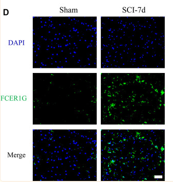
| Product: | FCER1G Antibody |
| Catalog: | DF13263 |
| Description: | Rabbit polyclonal antibody to FCER1G |
| Application: | WB IF/ICC |
| Cited expt.: | WB, IF/ICC |
| Reactivity: | Human, Mouse, Rat |
| Prediction: | Pig, Bovine, Horse, Sheep, Dog |
| Mol.Wt.: | 9kDa(Observed); 10kD(Calculated). |
| Uniprot: | P30273 |
| RRID: | AB_2846282 |
Control Products

Related Downloads
Protocols
Product Info
*The optimal dilutions should be determined by the end user. For optimal experimental results, antibody reuse is not recommended.
*Tips:
WB: For western blot detection of denatured protein samples. IHC: For immunohistochemical detection of paraffin sections (IHC-p) or frozen sections (IHC-f) of tissue samples. IF/ICC: For immunofluorescence detection of cell samples. ELISA(peptide): For ELISA detection of antigenic peptide.
Cite Format: Affinity Biosciences Cat# DF13263, RRID:AB_2846282.
Fold/Unfold
Fc fragment of IgE, high affinity I, receptor for; gamma polypeptide; Fc receptor gamma chain; Fc-epsilon RI-gamma; FCER1G; FCERG_HUMAN; FceRI gamma; FCRG; FcRgamma; High affinity immunoglobulin epsilon receptor subunit gamma; IgE Fc receptor subunit gamma; Immunoglobulin E receptor, high affinity, gamma chain;
Immunogens
A synthesized peptide derived from human FCER1G, corresponding to a region within C-terminal amino acids.
- P30273 FCERG_HUMAN:
- Protein BLAST With
- NCBI/
- ExPASy/
- Uniprot
MIPAVVLLLLLLVEQAAALGEPQLCYILDAILFLYGIVLTLLYCRLKIQVRKAAITSYEKSDGVYTGLSTRNQETYETLKHEKPPQ
Predictions
Score>80(red) has high confidence and is suggested to be used for WB detection. *The prediction model is mainly based on the alignment of immunogen sequences, the results are for reference only, not as the basis of quality assurance.
High(score>80) Medium(80>score>50) Low(score<50) No confidence
Research Backgrounds
Adapter protein containing an immunoreceptor tyrosine-based activation motif (ITAM) that transduces activation signals from various immunoreceptors. As a component of the high-affinity immunoglobulin E (IgE) receptor, mediates allergic inflammatory signaling in mast cells. As a constitutive component of interleukin-3 receptor complex, selectively mediates interleukin 4/IL4 production by basophils, priming T-cells toward effector T-helper 2 subset. Associates with pattern recognition receptors CLEC4D and CLEC4E to form a functional signaling complex in myeloid cells. Binding of mycobacterial trehalose 6,6'-dimycolate (TDM) to this receptor complex leads to phosphorylation of ITAM, triggering activation of SYK, CARD9 and NF-kappa-B, consequently driving maturation of antigen-presenting cells and shaping antigen-specific priming of T-cells toward effector T-helper 1 and T-helper 17 cell subtypes. May function cooperatively with other activating receptors. Functionally linked to integrin beta-2/ITGB2-mediated neutrophil activation. Also involved in integrin alpha-2/ITGA2-mediated platelet activation.
Cell membrane>Single-pass type I membrane protein.
Belongs to the CD3Z/FCER1G family.
Research Fields
· Environmental Information Processing > Signal transduction > Sphingolipid signaling pathway. (View pathway)
· Environmental Information Processing > Signal transduction > Phospholipase D signaling pathway. (View pathway)
· Human Diseases > Infectious diseases: Bacterial > Tuberculosis.
· Human Diseases > Immune diseases > Asthma.
· Organismal Systems > Immune system > Platelet activation. (View pathway)
· Organismal Systems > Immune system > Natural killer cell mediated cytotoxicity. (View pathway)
· Organismal Systems > Immune system > Fc epsilon RI signaling pathway. (View pathway)
References

Application: WB Species: Rat Sample:
Application: IF/ICC Species: Rat Sample:
Restrictive clause
Affinity Biosciences tests all products strictly. Citations are provided as a resource for additional applications that have not been validated by Affinity Biosciences. Please choose the appropriate format for each application and consult Materials and Methods sections for additional details about the use of any product in these publications.
For Research Use Only.
Not for use in diagnostic or therapeutic procedures. Not for resale. Not for distribution without written consent. Affinity Biosciences will not be held responsible for patent infringement or other violations that may occur with the use of our products. Affinity Biosciences, Affinity Biosciences Logo and all other trademarks are the property of Affinity Biosciences LTD.