Product: Phospho-ASK1 (Ser966) Antibody
Catalog: AF3477
Description: Rabbit polyclonal antibody to Phospho-ASK1 (Ser966)
Application: WB IHC IF/ICC
Reactivity: Human, Mouse, Rat
Prediction: Pig, Zebrafish, Bovine, Horse, Sheep, Rabbit, Dog, Chicken
Mol.Wt.: 155kDa; 155kD(Calculated).
Uniprot: Q99683
RRID: AB_2834915

View similar products>>

   Size Price Inventory
 100ul $280 In stock
 200ul $350 In stock

Lead Time: Same day delivery

For pricing and ordering contact:
Local distributors

Product Info

Source:
Rabbit
Application:
WB 1:500-1:2000, IHC 1:50-1:200, IF/ICC 1:100-1:500
*The optimal dilutions should be determined by the end user.
*Tips:

WB: For western blot detection of denatured protein samples. IHC: For immunohistochemical detection of paraffin sections (IHC-p) or frozen sections (IHC-f) of tissue samples. IF/ICC: For immunofluorescence detection of cell samples. ELISA(peptide): For ELISA detection of antigenic peptide.

Reactivity:
Human,Mouse,Rat
Prediction:
Pig(100%), Zebrafish(100%), Bovine(100%), Horse(100%), Sheep(100%), Rabbit(100%), Dog(100%), Chicken(100%)
Clonality:
Polyclonal
Specificity:
Phospho-ASK1 (Ser966) Antibody detects endogenous levels of ASK1 only when phosphorylated at Serine 966.
RRID:
AB_2834915
Cite Format: Affinity Biosciences Cat# AF3477, RRID:AB_2834915.
Conjugate:
Unconjugated.
Purification:
The antibody is from purified rabbit serum by affinity purification via sequential chromatography on phospho-peptide and non-phospho-peptide affinity columns.
Storage:
Rabbit IgG in phosphate buffered saline , pH 7.4, 150mM NaCl, 0.02% sodium azide and 50% glycerol. Store at -20 °C. Stable for 12 months from date of receipt.
Alias:

Fold/Unfold

Apoptosis signal regulating kinase 1; Apoptosis signal-regulating kinase 1; ASK 1; ASK-1; ASK1; M3K5; M3K5_HUMAN; MAP/ERK kinase kinase 5; MAP3K5; MAPK/ERK kinase kinase 5; MAPKKK5; MEK kinase 5; MEKK 5; MEKK5; Mitogen activated protein kinase kinase kinase 5; Mitogen-activated protein kinase kinase kinase 5;

Immunogens

Immunogen:
Uniprot:
Gene(ID):
Expression:
Q99683 M3K5_HUMAN:

Abundantly expressed in heart and pancreas.

Description:
Mitogen-activated protein kinase (MAPK) signaling cascades include MAPK or extracellular signal-regulated kinase (ERK), MAPK kinase (MKK or MEK), and MAPK kinase kinase (MAPKKK or MEKK). MAPKK kinase/MEKK phosphorylates and activates its downstream protein kinase, MAPK kinase/MEK, which in turn activates MAPK.
Sequence:
MSTEADEGITFSVPPFAPSGFCTIPEGGICRRGGAAAVGEGEEHQLPPPPPGSFWNVESAAAPGIGCPAATSSSSATRGRGSSVGGGSRRTTVAYVINEASQGQLVVAESEALQSLREACETVGATLETLHFGKLDFGETTVLDRFYNADIAVVEMSDAFRQPSLFYHLGVRESFSMANNIILYCDTNSDSLQSLKEIICQKNTMCTGNYTFVPYMITPHNKVYCCDSSFMKGLTELMQPNFELLLGPICLPLVDRFIQLLKVAQASSSQYFRESILNDIRKARNLYTGKELAAELARIRQRVDNIEVLTADIVINLLLSYRDIQDYDSIVKLVETLEKLPTFDLASHHHVKFHYAFALNRRNLPGDRAKALDIMIPMVQSEGQVASDMYCLVGRIYKDMFLDSNFTDTESRDHGASWFKKAFESEPTLQSGINYAVLLLAAGHQFESSFELRKVGVKLSSLLGKKGNLEKLQSYWEVGFFLGASVLANDHMRVIQASEKLFKLKTPAWYLKSIVETILIYKHFVKLTTEQPVAKQELVDFWMDFLVEATKTDVTVVRFPVLILEPTKIYQPSYLSINNEVEEKTISIWHVLPDDKKGIHEWNFSASSVRGVSISKFEERCCFLYVLHNSDDFQIYFCTELHCKKFFEMVNTITEEKGRSTEEGDCESDLLEYDYEYDENGDRVVLGKGTYGIVYAGRDLSNQVRIAIKEIPERDSRYSQPLHEEIALHKHLKHKNIVQYLGSFSENGFIKIFMEQVPGGSLSALLRSKWGPLKDNEQTIGFYTKQILEGLKYLHDNQIVHRDIKGDNVLINTYSGVLKISDFGTSKRLAGINPCTETFTGTLQYMAPEIIDKGPRGYGKAADIWSLGCTIIEMATGKPPFYELGEPQAAMFKVGMFKVHPEIPESMSAEAKAFILKCFEPDPDKRACANDLLVDEFLKVSSKKKKTQPKLSALSAGSNEYLRSISLPVPVLVEDTSSSSEYGSVSPDTELKVDPFSFKTRAKSCGERDVKGIRTLFLGIPDENFEDHSAPPSPEEKDSGFFMLRKDSERRATLHRILTEDQDKIVRNLMESLAQGAEEPKLKWEHITTLIASLREFVRSTDRKIIATTLSKLKLELDFDSHGISQVQVVLFGFQDAVNKVLRNHNIKPHWMFALDSIIRKAVQTAITILVPELRPHFSLASESDTADQEDLDVEDDHEEQPSNQTVRRPQAVIEDAVATSGVSTLSSTVSHDSQSAHRSLNVQLGRMKIETNRLLEELVRKEKELQALLHRAIEEKDQEIKHLKLKSQPIEIPELPVFHLNSSGTNTEDSELTDWLRVNGADEDTISRFLAEDYTLLDVLYYVTRDDLKCLRLRGGMLCTLWKAIIDFRNKQT

Predictions

Predictions:

Score>80(red) has high confidence and is suggested to be used for WB detection. *The prediction model is mainly based on the alignment of immunogen sequences, the results are for reference only, not as the basis of quality assurance.

Species
Results
Score
Pig
100
Horse
100
Bovine
100
Sheep
100
Dog
100
Zebrafish
100
Chicken
100
Rabbit
100
Xenopus
0
Model Confidence:
High(score>80) Medium(80>score>50) Low(score<50) No confidence

PTMs - Q99683 As Substrate

Site PTM Type Enzyme
S82 Phosphorylation
S83 Phosphorylation P11309 (PIM1) , P51812 (RPS6KA3) , P31749 (AKT1) , P31751 (AKT2)
R89 Methylation
S101 Phosphorylation
S115 Phosphorylation
S461 Phosphorylation
K465 Ubiquitination
T528 Phosphorylation
K535 Sumoylation
Y570 Phosphorylation
Y574 Phosphorylation
K709 Ubiquitination
Y718 Phosphorylation P23458 (JAK1) , O60674 (JAK2)
K730 Ubiquitination
K785 Ubiquitination
T813 Phosphorylation Q99683 (MAP3K5)
Y814 Phosphorylation
S821 Phosphorylation
T825 Phosphorylation
K827 Ubiquitination
T838 Phosphorylation Q14680 (MELK) , Q99683 (MAP3K5) , O95382 (MAP3K6) , Q15139 (PRKD1)
T842 Phosphorylation Q99683 (MAP3K5)
C869 S-Nitrosylation
K946 Acetylation
S958 Phosphorylation
S964 Phosphorylation
S966 Phosphorylation O15530 (PDPK1) , Q99683 (MAP3K5)
Y982 Phosphorylation
S986 Phosphorylation
S997 Phosphorylation
S1029 Phosphorylation
S1033 Phosphorylation
T1059 Phosphorylation
K1064 Ubiquitination
K1083 Sumoylation
T1109 Phosphorylation P51812 (RPS6KA3)
K1114 Sumoylation
S1240 Phosphorylation
T1326 Phosphorylation P51812 (RPS6KA3)

PTMs - Q99683 As Enzyme

Substrate Site Source
O15530 (PDPK1) S394 Uniprot
O15530 (PDPK1) S398 Uniprot
P38936 (CDKN1A) S98 Uniprot
P45379-11 (TNNT2) T191 Uniprot
P45379-6 (TNNT2) T194 Uniprot
P45379-11 (TNNT2) S195 Uniprot
P45379-6 (TNNT2) S198 Uniprot
P45379-10 (TNNT2) T201 Uniprot
P45379 (TNNT2) T204 Uniprot
P45379-10 (TNNT2) S205 Uniprot
P45379 (TNNT2) S208 Uniprot
P45983 (MAPK8) T183 Uniprot
P45983 (MAPK8) Y185 Uniprot
P45985 (MAP2K4) T261 Uniprot
P46734 (MAP2K3) S218 Uniprot
P52564 (MAP2K6) S207 Uniprot
Q16539 (MAPK14) T180 Uniprot
Q16539 (MAPK14) Y182 Uniprot
Q969S3 (ZNF622) S314 Uniprot
Q969S3 (ZNF622) T318 Uniprot
Q99683 (MAP3K5) T813 Uniprot
Q99683 (MAP3K5) T838 Uniprot
Q99683 (MAP3K5) T842 Uniprot
Q99683 (MAP3K5) S966 Uniprot
Q9UER7 (DAXX) S176 Uniprot
Q9UER7 (DAXX) S184 Uniprot
Q9Y3F4 (STRAP) T175 Uniprot
Q9Y3F4 (STRAP) S179 Uniprot

Research Backgrounds

Function:

Serine/threonine kinase which acts as an essential component of the MAP kinase signal transduction pathway. Plays an important role in the cascades of cellular responses evoked by changes in the environment. Mediates signaling for determination of cell fate such as differentiation and survival. Plays a crucial role in the apoptosis signal transduction pathway through mitochondria-dependent caspase activation. MAP3K5/ASK1 is required for the innate immune response, which is essential for host defense against a wide range of pathogens. Mediates signal transduction of various stressors like oxidative stress as well as by receptor-mediated inflammatory signals, such as the tumor necrosis factor (TNF) or lipopolysaccharide (LPS). Once activated, acts as an upstream activator of the MKK/JNK signal transduction cascade and the p38 MAPK signal transduction cascade through the phosphorylation and activation of several MAP kinase kinases like MAP2K4/SEK1, MAP2K3/MKK3, MAP2K6/MKK6 and MAP2K7/MKK7. These MAP2Ks in turn activate p38 MAPKs and c-jun N-terminal kinases (JNKs). Both p38 MAPK and JNKs control the transcription factors activator protein-1 (AP-1).

PTMs:

Phosphorylated at Thr-838 through autophosphorylation and by MAP3K6/ASK2 which leads to activation. Thr-838 is dephosphorylated by PPP5C. Ser-83 and Ser-1033 are inactivating phosphorylation sites, the former of which is phosphorylated by AKT1 and AKT2. Phosphorylated at Ser-966 which induces association of MAP3K5/ASK1 with the 14-3-3 family proteins and suppresses MAP3K5/ASK1 activity. Calcineurin (CN) dephosphorylates this site. Also dephosphorylated and activated by PGAM5.

Ubiquitinated. Tumor necrosis factor (TNF) induces TNFR2-dependent ubiquitination leading to proteasomal degradation.

Subcellular Location:

Cytoplasm. Endoplasmic reticulum.
Note: Interaction with 14-3-3 proteins alters the distribution of MAP3K5/ASK1 and restricts it to the perinuclear endoplasmic reticulum region.

Extracellular region or secreted Cytosol Plasma membrane Cytoskeleton Lysosome Endosome Peroxisome ER Golgi apparatus Nucleus Mitochondrion Manual annotation Automatic computational assertionSubcellular location
Tissue Specificity:

Abundantly expressed in heart and pancreas.

Subunit Structure:

Homodimer when inactive. Binds both upstream activators and downstream substrates in multimolecular complexes. Part of a cytoplasmic complex made of HIPK1, DAB2IP and MAP3K5 in response to TNF. This complex formation promotes MAP3K5-JNK activation and subsequent apoptosis. Interacts with SOCS1 which recognizes phosphorylation of Tyr-718 and induces MAP3K5/ASK1 degradation in endothelial cells. Interacts with the 14-3-3 family proteins such as YWHAB, YWHAE, YWHAQ, YWHAH, YWHAZ and SFN. Interacts with ARRB2, BIRC2, DAB2IP, IGF1R, MAP3K6/ASK2, PGAM5, PIM1, PPP5C, SOCS1, STUB1, TRAF2, TRAF6 and TXN. Interacts with ERN1 in a TRAF2-dependent manner. Interacts with calcineurin subunit PPP3R1 and with PPM1L (By similarity). Interacts (via N-terminus) with RAF1 and this interaction inhibits the proapoptotic function of MAP3K5. Interacts with DAB2IP (via N-terminus C2 domain); the interaction occurs in a TNF-alpha-dependent manner. Interacts with DUSP13/DUSP13A; may positively regulate apoptosis. Interacts with DAXX. Interacts with RC3H2.

(Microbial infection) Interacts with HIV-1 Nef; this interaction inhibits MAP3K5 signaling.

Family&Domains:

Belongs to the protein kinase superfamily. STE Ser/Thr protein kinase family. MAP kinase kinase kinase subfamily.

Research Fields

· Cellular Processes > Cell growth and death > Apoptosis.   (View pathway)

· Cellular Processes > Cellular community - eukaryotes > Tight junction.   (View pathway)

· Environmental Information Processing > Signal transduction > MAPK signaling pathway.   (View pathway)

· Environmental Information Processing > Signal transduction > Sphingolipid signaling pathway.   (View pathway)

· Environmental Information Processing > Signal transduction > TNF signaling pathway.   (View pathway)

· Genetic Information Processing > Folding, sorting and degradation > Protein processing in endoplasmic reticulum.   (View pathway)

· Human Diseases > Drug resistance: Antineoplastic > Platinum drug resistance.

· Human Diseases > Endocrine and metabolic diseases > Non-alcoholic fatty liver disease (NAFLD).

· Human Diseases > Neurodegenerative diseases > Amyotrophic lateral sclerosis (ALS).

· Organismal Systems > Nervous system > Neurotrophin signaling pathway.   (View pathway)

References

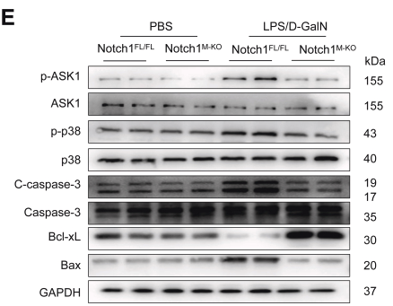
1). Targeting Notch1-YAP Circuit Reprograms Macrophage Polarization and Alleviates Acute Liver Injury in Mice. Cellular and Molecular Gastroenterology and Hepatology, 2023 (PubMed: 36706917) [IF=7.2]

Application: WB    Species: Mouse    Sample:

Figure 3 Myeloid-specific deletion of Notch1 alleviates LPS/D-GalN-induced hepatocellular apoptosis. (A) Representative TUNEL staining images and quantification of TUNEL+ cells in liver sections from the Notch1FL/FL and Notch1M-KO mice treated with PBS or LPS/D-GalN injection (n = 5 mice/group). Scale bars, 20 μm. (B) Immunohistochemistry staining and quantification of cleaved caspase-3 (C-caspase-3) positive cells in liver sections (n = 5 mice/group). Scale bars, 40 μm. ELISA analysis of serum TNF-α (C) and HMGB1 (D) levels in the Notch1FL/FL and Notch1M-KO mice (n = 5 samples/group). (E) Western blot analysis and relative density ratio of p-ASK1, ASK1, p-p38, p38, C-caspase-3, caspase-3, Bcl-xL, and Bax in the Notch1FL/FL and Notch1M-KO livers. Data are presented as the mean ± standard deviation. ∗P < .05, ∗∗P < .01.

2). Dual function of peroxiredoxin I in lipopolysaccharide-induced osteoblast apoptosis via reactive oxygen species and the apoptosis signal-regulating kinase 1 signaling pathway. Cell Death Discovery, 2018 (PubMed: 29707240) [IF=7.0]

Application: WB    Species:    Sample: Wild-type, Prx1KO, and Prx1OE cells

Fig. 6 |The overexpression of Prx1 promotes ASK1 activation by inhibiting the activity of Trx towards ASK1. a–d Wild-type, Prx1KO, and Prx1OE cells were each divided into three groups, which were control groups, groups treated with 100 ng/ml LPS, and groups treated with 100 ng/ml LPS and NQDI. Western blot analysis was then performed with antibodies against P-ASK1, ASK1, Trx and GAPDH.

3). JNK signaling pathway mediates acetaminophen-induced hepatotoxicity accompanied by changes of glutathione S-transferase A1 content and expression. Frontiers in Pharmacology, 2019 (PubMed: 31620005) [IF=5.6]

Application: WB    Species: mouse    Sample: liver

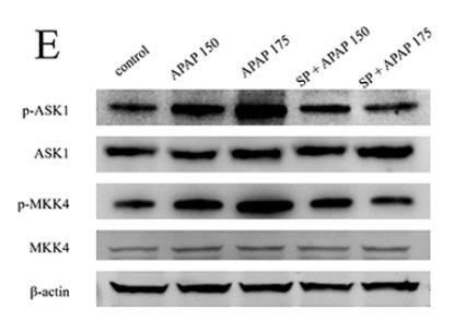
FIGURE 2 | Activation of JNK signaling pathway under different dosages of APAP. .(E) Western blot analyses of total tissue lysate for p-ASK1, ASK1, p-MKK4, MKK4, and β-actin (loading control). Values represented as means ± SD, n = 3. *Represents statistical differences caused by APAP. #Represents statistical differences caused by SP600125 under 150 mg·kg−1 APAP;§Represents statistical differences caused by SP600125 under 175 mg·kg−1 APAP. 0.05 > P > 0.01 (*, #, §). P < 0.01 (**, §§).

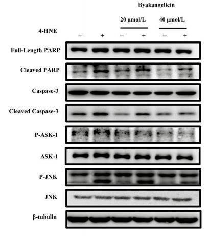
4). Byakangelicin protects against carbon tetrachloride–induced liver injury and fibrosis in mice. JOURNAL OF CELLULAR AND MOLECULAR MEDICINE, 2020 (PubMed: 32643868) [IF=5.3]

Application: WB    Species: human    Sample: LX-2 cells

FIGURE 5|Byakangelicin inhibits 4-HNE–induced hepatocyte apoptosis by inhibiting ASK1/JNK pathway. D and E, Protein full-length PARP, PARP, cleaved caspase-3, caspase-3, JNK, P-JNK, ASK-1 and P-ASK-1 were also detected and quantified using Western blot analyses.

5). CTGF Triggers Rat Astrocyte Activation and Astrocyte-Mediated Inflammatory Response in Culture Conditions. INFLAMMATION, 2019 (PubMed: 31183597) [IF=5.1]

Application: WB    Species: rat    Sample: RA cells

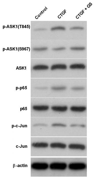
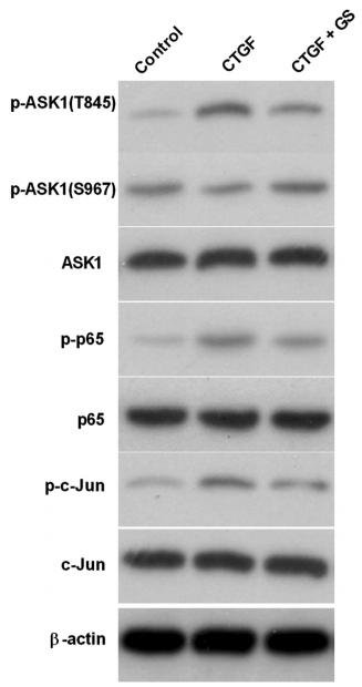
Fig. 4.| CTGF activated the NF-κB and AP-1 through ASK1-p38/JNK pathways. b RA cells were treated with either 10 μM ASK1 inhibitor GS-4997, or solvent control as indicated, for 30 min prior to stimulation with 20 ng/ml CTGF for 24 h. The expression of ASK, p65, and c-Jun and their phosphorylation states were examined by Western blotting.

6). Elaidic acid induced NLRP3 inflammasome activation via ERS-MAPK signaling pathways in Kupffer cells. BIOCHIMICA ET BIOPHYSICA ACTA-MOLECULAR AND CELL BIOLOGY OF LIPIDS, 2022 (PubMed: 34610469) [IF=4.8]

7). Allicin alleviated acrylamide-induced NLRP3 inflammasome activation via oxidative stress and endoplasmic reticulum stress in Kupffer cells and SD rats liver. FOOD AND CHEMICAL TOXICOLOGY, 2021 (PubMed: 33348049) [IF=4.3]

Application: WB    Species: rats    Sample: Kupffer cells

Fig. 4. Effects of allicin on AA-induced ERS in Kupffer cells and SD rats liver. (A) Effects of allicin on AA-induced ERS proteins levels in Kupffer cells. (B) Effects of allicin on AA-induced ERS proteins levels in SD rats liver. GRP78, CHOP, p-IRE1α, p-ASK, XBP-1s and TRAF2 proteins expression were tested by Western-blotting analysis. The values are presented as means ± of SD (n = 3). *P < 0.05, **P < 0.01, versus the control group; #P < 0.05, ##P < 0.01, versus the model group.

8). Rosmarinic acid attenuates acrylamide induced apoptosis of BRL-3A cells by inhibiting oxidative stress and endoplasmic reticulum stress. Food and Chemical Toxicology, 2021 (PubMed: 33781805) [IF=4.3]

Application: WB    Species: Mouse    Sample: BRL-3A cells

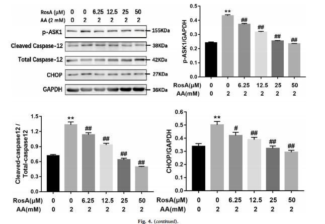
Fig. 4. The effect of AA, RosA and NAC on ERS in BRL-3A cells. DMEM basic medium, RosA (6.25, 12.5, 25, 50 μM) and 10 mM NAC were used for pretreatment for 2 h, incubated with 2 mM AA for 24 h. (A) The effect of AA and RosA on the release of calcium ions in BRL-3A cells. (B) The effect of AA and RosA on IRE1α pathway signal proteins. (C) The effect of AA and RosA on ERS-mediated apoptosis-related proteins. (D) The effect of NAC on ERS-related proteins in BRL-3A cells. Western blotting analysis showed the protein levels of p-IRE1α, IRE1α, GRP78, XBP-1s, TRAF2, p-ASK1, Caspase-12, and CHOP in BRL-3A cells. Values are expressed as mean ± standard deviation (n = 3). Compared with the control group, **P < 0.01; compared with the AA group, #P < 0.05 and ##P < 0.01.

9). Abrogating PDK4 activates autophagy-dependent ferroptosis in breast cancer via ASK1/JNK pathway. Journal of cancer research and clinical oncology, 2024 (PubMed: 38678126) [IF=3.6]

10). The PERK/ATF4/CHOP signaling branch of the unfolded protein response mediates cisplatin-induced ototoxicity in hair cells. Drug and Chemical Toxicology, 2022 (PubMed: 35172660) [IF=2.6]

Load more

Restrictive clause


Affinity Biosciences tests all products strictly. Citations are provided as a resource for additional applications that have not been validated by Affinity Biosciences. Please choose the appropriate format for each application and consult Materials and Methods sections for additional details about the use of any product in these publications.

For Research Use Only.
Not for use in diagnostic or therapeutic procedures. Not for resale. Not for distribution without written consent. Affinity Biosciences will not be held responsible for patent infringement or other violations that may occur with the use of our products. Affinity Biosciences, Affinity Biosciences Logo and all other trademarks are the property of Affinity Biosciences LTD.