. The lane on the left was treated with blocking peptide.")
, using Cleaved-Caspase 7 (Asp198) Antibody at 1/1000 dilution.
5ug/NC membrane strip.
Exposure for 5s with Affinity™ ECL Kit(#KF8003).
Bands result from membrane strip incubation.")
 The Mlg cells were incubated with TGF-β1 and/or PZ (2 or 4 μM) for 24 hours. The expression levels of caspase3, cleaved-caspase3, caspase7, cleaved-caspase7, caspase9 and cleaved-caspase9, were detected via using Western blot. The loading control was β-tubulin. (B,C) Mlg cells were exposed to Baf a1/CQ and/or PZ (2 or 4 μM) for 24 hours. The p62 expression level was evaluated by Western blot. GAPDH was used as a loading control. (D,E) The plasmid of GFP-LC3B and GFP-Cherry-LC3B were transfected to NIH-3T3 with PEI, and fluorescence microscopy was used to examine the number of autophagosomes (green puncta) and autolysosome (red puncta) after exposing to TGF-β1 and/or PZ for 12 hours. Scale bars: 50 µm. Data in (A-C) are mean ± standard deviation. ###, P")
| Product: | Cleaved-Caspase 7 (Asp198) Antibody |
| Catalog: | AF4023 |
| Description: | Rabbit polyclonal antibody to Cleaved-Caspase 7 (Asp198) |
| Application: | WB |
| Cited expt.: | WB |
| Reactivity: | Human, Mouse |
| Prediction: | Pig, Bovine, Horse, Sheep, Rabbit, Dog |
| Mol.Wt.: | 20kDa, 25 kDa(Observed); 34kD(Calculated). |
| Uniprot: | P55210 |
| RRID: | AB_2846219 |
Control Products

Product Info
*The optimal dilutions should be determined by the end user. For optimal experimental results, antibody reuse is not recommended.
*Tips:
WB: For western blot detection of denatured protein samples. IHC: For immunohistochemical detection of paraffin sections (IHC-p) or frozen sections (IHC-f) of tissue samples. IF/ICC: For immunofluorescence detection of cell samples. ELISA(peptide): For ELISA detection of antigenic peptide.
Cite Format: Affinity Biosciences Cat# AF4023, RRID:AB_2846219.
Fold/Unfold
Apoptotic protease Mch-3; Apoptotic protease MCH3; CASP-7; CASP7; CASP7_HUMAN; Caspase 7; Caspase 7 apoptosis related cysteine peptidase; Caspase-7 subunit p11; Caspase7; CMH 1; CMH-1; CMH1; ICE LAP3; ICE-LAP3; ICE-like apoptotic protease 3; LICE2; MCH3;
Immunogens
A synthesized peptide derived from human Caspase 7.
Highly expressed in lung, skeletal muscle, liver, kidney, spleen and heart, and moderately in testis. No expression in the brain.
- P55210 CASP7_HUMAN:
- Protein BLAST With
- NCBI/
- ExPASy/
- Uniprot
MADDQGCIEEQGVEDSANEDSVDAKPDRSSFVPSLFSKKKKNVTMRSIKTTRDRVPTYQYNMNFEKLGKCIIINNKNFDKVTGMGVRNGTDKDAEALFKCFRSLGFDVIVYNDCSCAKMQDLLKKASEEDHTNAACFACILLSHGEENVIYGKDGVTPIKDLTAHFRGDRCKTLLEKPKLFFIQACRGTELDDGIQADSGPINDTDANPRYKIPVEADFLFAYSTVPGYYSWRSPGRGSWFVQALCSILEEHGKDLEIMQILTRVNDRVARHFESQSDDPHFHEKKQIPCVVSMLTKELYFSQ
Predictions
Score>80(red) has high confidence and is suggested to be used for WB detection. *The prediction model is mainly based on the alignment of immunogen sequences, the results are for reference only, not as the basis of quality assurance.
High(score>80) Medium(80>score>50) Low(score<50) No confidence
Research Backgrounds
Involved in the activation cascade of caspases responsible for apoptosis execution. Cleaves and activates sterol regulatory element binding proteins (SREBPs). Proteolytically cleaves poly(ADP-ribose) polymerase (PARP) at a '216-Asp-|-Gly-217' bond. Overexpression promotes programmed cell death.
Cleavages by granzyme B or caspase-10 generate the two active subunits. Propeptide domains can also be cleaved efficiently by caspase-3. Active heterodimers between the small subunit of caspase-7 and the large subunit of caspase-3, and vice versa, also occur.
Cytoplasm.
Highly expressed in lung, skeletal muscle, liver, kidney, spleen and heart, and moderately in testis. No expression in the brain.
Belongs to the peptidase C14A family.
Research Fields
· Cellular Processes > Cell growth and death > Apoptosis. (View pathway)
· Cellular Processes > Cell growth and death > Apoptosis - multiple species. (View pathway)
· Environmental Information Processing > Signal transduction > TNF signaling pathway. (View pathway)
· Human Diseases > Endocrine and metabolic diseases > Non-alcoholic fatty liver disease (NAFLD).
· Human Diseases > Neurodegenerative diseases > Alzheimer's disease.
· Human Diseases > Infectious diseases: Bacterial > Pertussis.
· Human Diseases > Infectious diseases: Bacterial > Legionellosis.
· Human Diseases > Cancers: Overview > Pathways in cancer. (View pathway)
References
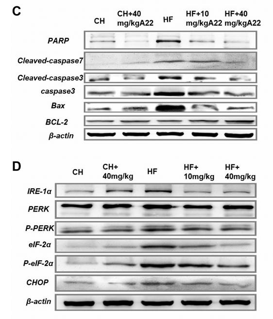
Application: WB Species: mouse Sample: liver
Application: WB Species: rat Sample: chondrocytes
Application: WB Species: Rat Sample: PC12 cells
Application: WB Species: Human Sample: HepG2 cells

Application: WB Species: human Sample:
Restrictive clause
Affinity Biosciences tests all products strictly. Citations are provided as a resource for additional applications that have not been validated by Affinity Biosciences. Please choose the appropriate format for each application and consult Materials and Methods sections for additional details about the use of any product in these publications.
For Research Use Only.
Not for use in diagnostic or therapeutic procedures. Not for resale. Not for distribution without written consent. Affinity Biosciences will not be held responsible for patent infringement or other violations that may occur with the use of our products. Affinity Biosciences, Affinity Biosciences Logo and all other trademarks are the property of Affinity Biosciences LTD.