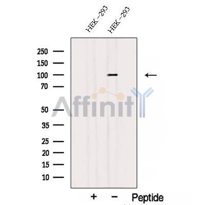
STT3A Antibody - #DF12757
| Product: | STT3A Antibody |
| Catalog: | DF12757 |
| Description: | Rabbit polyclonal antibody to STT3A |
| Application: | WB |
| Reactivity: | Human, Mouse, Rat, Monkey |
| Prediction: | Pig, Zebrafish, Bovine, Horse, Sheep, Rabbit, Dog, Chicken, Xenopus |
| Mol.Wt.: | 100 kDa; 81kD(Calculated). |
| Uniprot: | P46977 |
| RRID: | AB_2845718 |
Related Downloads
Protocols
Product Info
*The optimal dilutions should be determined by the end user. For optimal experimental results, antibody reuse is not recommended.
*Tips:
WB: For western blot detection of denatured protein samples. IHC: For immunohistochemical detection of paraffin sections (IHC-p) or frozen sections (IHC-f) of tissue samples. IF/ICC: For immunofluorescence detection of cell samples. ELISA(peptide): For ELISA detection of antigenic peptide.
Cite Format: Affinity Biosciences Cat# DF12757, RRID:AB_2845718.
Immunogens
A synthesized peptide derived from human STT3A, corresponding to a region within the internal amino acids.
Expressed at high levels in placenta, liver, muscle and pancreas, and at very low levels in brain, lung and kidney. Expressed in skin fibroblasts (at protein level).
- P46977 STT3A_HUMAN:
- Protein BLAST With
- NCBI/
- ExPASy/
- Uniprot
MTKFGFLRLSYEKQDTLLKLLILSMAAVLSFSTRLFAVLRFESVIHEFDPYFNYRTTRFLAEEGFYKFHNWFDDRAWYPLGRIIGGTIYPGLMITSAAIYHVLHFFHITIDIRNVCVFLAPLFSSFTTIVTYHLTKELKDAGAGLLAAAMIAVVPGYISRSVAGSYDNEGIAIFCMLLTYYMWIKAVKTGSICWAAKCALAYFYMVSSWGGYVFLINLIPLHVLVLMLTGRFSHRIYVAYCTVYCLGTILSMQISFVGFQPVLSSEHMAAFGVFGLCQIHAFVDYLRSKLNPQQFEVLFRSVISLVGFVLLTVGALLMLTGKISPWTGRFYSLLDPSYAKNNIPIIASVSEHQPTTWSSYYFDLQLLVFMFPVGLYYCFSNLSDARIFIIMYGVTSMYFSAVMVRLMLVLAPVMCILSGIGVSQVLSTYMKNLDISRPDKKSKKQQDSTYPIKNEVASGMILVMAFFLITYTFHSTWVTSEAYSSPSIVLSARGGDGSRIIFDDFREAYYWLRHNTPEDAKVMSWWDYGYQITAMANRTILVDNNTWNNTHISRVGQAMASTEEKAYEIMRELDVSYVLVIFGGLTGYSSDDINKFLWMVRIGGSTDTGKHIKENDYYTPTGEFRVDREGSPVLLNCLMYKMCYYRFGQVYTEAKRPPGFDRVRNAEIGNKDFELDVLEEAYTTEHWLVRIYKVKDLDNRGLSRT
Predictions
Score>80(red) has high confidence and is suggested to be used for WB detection. *The prediction model is mainly based on the alignment of immunogen sequences, the results are for reference only, not as the basis of quality assurance.
High(score>80) Medium(80>score>50) Low(score<50) No confidence
Research Backgrounds
Catalytic subunit of the oligosaccharyl transferase (OST) complex that catalyzes the initial transfer of a defined glycan (Glc(3)Man(9)GlcNAc(2) in eukaryotes) from the lipid carrier dolichol-pyrophosphate to an asparagine residue within an Asn-X-Ser/Thr consensus motif in nascent polypeptide chains, the first step in protein N-glycosylation. N-glycosylation occurs cotranslationally and the complex associates with the Sec61 complex at the channel-forming translocon complex that mediates protein translocation across the endoplasmic reticulum (ER). All subunits are required for a maximal enzyme activity. This subunit contains the active site and the acceptor peptide and donor lipid-linked oligosaccharide (LLO) binding pockets (By similarity). STT3A is present in the majority of OST complexes and mediates cotranslational N-glycosylation of most sites on target proteins, while STT3B-containing complexes are required for efficient post-translational glycosylation and mediate glycosylation of sites that have been skipped by STT3A.
Endoplasmic reticulum. Endoplasmic reticulum membrane>Multi-pass membrane protein.
Expressed at high levels in placenta, liver, muscle and pancreas, and at very low levels in brain, lung and kidney. Expressed in skin fibroblasts (at protein level).
Despite low primary sequence conservation between eukaryotic catalytic subunits and bacterial and archaeal single subunit OSTs (ssOST), structural comparison revealed several common motifs at spatially equivalent positions, like the DXD motif 1 on the external loop 1 and the DXD motif 2 on the external loop 2 involved in binding of the metal ion cofactor and the carboxamide group of the acceptor asparagine, the conserved Glu residue of the TIXE/SVSE motif on the external loop 5 involved in catalysis, as well as the WWDYG and the DK/MI motifs in the globular domain that define the binding pocket for the +2 Ser/Thr of the acceptor sequon. In bacterial ssOSTs, an Arg residue was found to interact with a negatively charged side chain at the -2 position of the sequon. This Arg is conserved in bacterial enzymes and correlates with an extended sequon requirement (Asp-X-Asn-X-Ser/Thr) for bacterial N-glycosylation.
Belongs to the STT3 family.
Research Fields
· Genetic Information Processing > Folding, sorting and degradation > Protein processing in endoplasmic reticulum. (View pathway)
· Metabolism > Glycan biosynthesis and metabolism > N-Glycan biosynthesis.
· Metabolism > Global and overview maps > Metabolic pathways.
Restrictive clause
Affinity Biosciences tests all products strictly. Citations are provided as a resource for additional applications that have not been validated by Affinity Biosciences. Please choose the appropriate format for each application and consult Materials and Methods sections for additional details about the use of any product in these publications.
For Research Use Only.
Not for use in diagnostic or therapeutic procedures. Not for resale. Not for distribution without written consent. Affinity Biosciences will not be held responsible for patent infringement or other violations that may occur with the use of our products. Affinity Biosciences, Affinity Biosciences Logo and all other trademarks are the property of Affinity Biosciences LTD.

