Product: LAP3 Antibody
Catalog: DF12651
Description: Rabbit polyclonal antibody to LAP3
Application: WB
Reactivity: Human, Mouse, Rat
Prediction: Zebrafish, Bovine, Horse, Sheep, Rabbit, Dog, Chicken, Xenopus
Mol.Wt.: 56 kDa; 56kD(Calculated).
Uniprot: P28838
RRID: AB_2845613

View similar products>>

   Size Price Inventory
 100ul $280 In stock
 200ul $350 In stock

Lead Time: Same day delivery

For pricing and ordering contact:
Local distributors

Product Info

Source:
Rabbit
Application:
WB 1:500-1:2000
*The optimal dilutions should be determined by the end user.
*Tips:

WB: For western blot detection of denatured protein samples. IHC: For immunohistochemical detection of paraffin sections (IHC-p) or frozen sections (IHC-f) of tissue samples. IF/ICC: For immunofluorescence detection of cell samples. ELISA(peptide): For ELISA detection of antigenic peptide.

Reactivity:
Human,Mouse,Rat
Prediction:
Zebrafish(100%), Bovine(100%), Horse(90%), Sheep(100%), Rabbit(100%), Dog(100%), Chicken(100%), Xenopus(90%)
Clonality:
Polyclonal
Specificity:
LAP3 Antibody detects endogenous levels of total LAP3.
RRID:
AB_2845613
Cite Format: Affinity Biosciences Cat# DF12651, RRID:AB_2845613.
Conjugate:
Unconjugated.
Purification:
The antiserum was purified by peptide affinity chromatography using SulfoLink™ Coupling Resin (Thermo Fisher Scientific).
Storage:
Rabbit IgG in phosphate buffered saline , pH 7.4, 150mM NaCl, 0.02% sodium azide and 50% glycerol. Store at -20 °C. Stable for 12 months from date of receipt.
Alias:

Fold/Unfold

AMPL_HUMAN; Cytosol aminopeptidase; epididymis secretory protein Li 106; HEL-S-106; LAP 3; LAP; LAP-3; Lap3; LAPEP; Leucine aminopeptidase 3; Leucyl aminopeptidase; PEPS; Peptidase S; Proline aminopeptidase; Prolyl aminopeptidase;

Immunogens

Immunogen:
Uniprot:
Gene(ID):
Sequence:
MFLLPLPAAGRVVVRRLAVRRFGSRSLSTADMTKGLVLGIYSKEKEDDVPQFTSAGENFDKLLAGKLRETLNISGPPLKAGKTRTFYGLHQDFPSVVLVGLGKKAAGIDEQENWHEGKENIRAAVAAGCRQIQDLELSSVEVDPCGDAQAAAEGAVLGLYEYDDLKQKKKMAVSAKLYGSGDQEAWQKGVLFASGQNLARQLMETPANEMTPTRFAEIIEKNLKSASSKTEVHIRPKSWIEEQAMGSFLSVAKGSDEPPVFLEIHYKGSPNANEPPLVFVGKGITFDSGGISIKASANMDLMRADMGGAATICSAIVSAAKLNLPINIIGLAPLCENMPSGKANKPGDVVRAKNGKTIQVDNTDAEGRLILADALCYAHTFNPKVILNAATLTGAMDVALGSGATGVFTNSSWLWNKLFEASIETGDRVWRMPLFEHYTRQVVDCQLADVNNIGKYRSAGACTAAAFLKEFVTHPKWAHLDIAGVMTNKDEVPYLRKGMTGRPTRTLIEFLLRFSQDNA

Predictions

Predictions:

Score>80(red) has high confidence and is suggested to be used for WB detection. *The prediction model is mainly based on the alignment of immunogen sequences, the results are for reference only, not as the basis of quality assurance.

Species
Results
Score
Bovine
100
Sheep
100
Dog
100
Zebrafish
100
Chicken
100
Rabbit
100
Horse
90
Xenopus
90
Pig
0
Model Confidence:
High(score>80) Medium(80>score>50) Low(score<50) No confidence

PTMs - P28838 As Substrate

Site PTM Type Enzyme
S26 Phosphorylation
S28 Phosphorylation
T29 Phosphorylation
S42 Phosphorylation
K45 Ubiquitination
S54 Phosphorylation
K61 Acetylation
K61 Ubiquitination
K103 Acetylation
K104 Acetylation
K118 Ubiquitination
S138 Phosphorylation
S139 Phosphorylation
S180 Phosphorylation
K188 Ubiquitination
S194 Phosphorylation
T211 Phosphorylation
K221 Acetylation
K221 Ubiquitination
K229 Ubiquitination
S238 Phosphorylation
S288 Phosphorylation
S296 Phosphorylation
T311 Phosphorylation
K356 Ubiquitination
T363 Phosphorylation
C445 S-Nitrosylation
K455 Acetylation
K455 Ubiquitination
Y456 Phosphorylation
S458 Phosphorylation
K469 Ubiquitination
K476 Ubiquitination
K489 Acetylation

Research Backgrounds

Function:

Presumably involved in the processing and regular turnover of intracellular proteins. Catalyzes the removal of unsubstituted N-terminal amino acids from various peptides.

Subcellular Location:

Cytoplasm.

Extracellular region or secreted Cytosol Plasma membrane Cytoskeleton Lysosome Endosome Peroxisome ER Golgi apparatus Nucleus Mitochondrion Manual annotation Automatic computational assertionSubcellular location
Subunit Structure:

Homohexamer.

Family&Domains:

Belongs to the peptidase M17 family.

Research Fields

· Metabolism > Amino acid metabolism > Arginine and proline metabolism.

· Metabolism > Metabolism of other amino acids > Glutathione metabolism.

· Metabolism > Global and overview maps > Metabolic pathways.

References

1). LAP3 contributes to IFN-γ-induced arginine depletion and malignant transformation of bovine mammary epithelial cells. BMC Cancer (PubMed: 35941558) [IF=3.8]

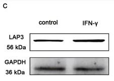
Application: WB    Species: Human    Sample: BMECs

Fig. 2IFN-γ upregulated LAP3 expression in BMECs. a qRT-PCR was used to detect the transcription level of LAP3 in BMECs after treatment with IFN-γ. b ELISA confirmed the content of LAP3 after treatment with IFN-γ in BMECs. c Western blotting was used to test the expression level of LAP3 in BMECs after treatment with 10 ng/mL IFN-γ for 24 h. Full-length blots are presented in Supplementary Fig. S5. d Leucine aminopeptidase activity was used to detect the change of LAP3 enzyme activity in BMECs before and after IFN-γ treatment. e Analysis of intracellular LAP3 expression in BMECs treated with the LAP3 inhibitor, bestatin, for 12 h using qRT-PCR. f HPLC was used to detect the changes of arginine content in BMECs pretreated with 100 mg/mL bestatin for 12 h and stimulated by IFN-γ for 24 h. g Changes in arginine content upon LAP3 overexpression in BMECs with or without IFN-γ treatment, as determined by ELISA. VC, pLenti-CMV-GFP-Puro. pLAP3, pLenti-CMV-LAP3-GFP-Puro. h-i LAP3 expression in normal and malignant BMECs by qRT-PCR (h) and western blotting analysis (i). 0, no IFN-γ treatment, normal BMECs; 30, malignant BMECs after IFN-γ-treated for 30th generations. The relative level of the target protein was estimated using densitometry and the ratio was calculated relative to the GAPDH control. Full-length blots are presented in Supplementary Fig. S6. Bars represent mean values with error bars to represent SD from three independent replicates. Differences between mean values were assessed by two-tailed Student’s t-test. *p < 0.05; **p < 0.01; ***p < 0.001, compared to control

Application: IHC    Species: Human    Sample: breast cancer cell

Fig. 8LAP3 was highly expressed in human breast cancer samples. a, c ELISA experiment was used to detected the protein expression levels of LAP3 (a) and ASS1 (c) in plasma of breast cancer patients and healthy control. Bars represent mean values with error bars to represent SD from three independent replicates. Differences between mean values were assessed by two-tailed Student’s t-test. *p < 0.05; **p < 0.01; ***p < 0.001, compared to control. b Representative images of IHC staining of LAP3 and ASS1. Scale bar, 200 μm

Restrictive clause


Affinity Biosciences tests all products strictly. Citations are provided as a resource for additional applications that have not been validated by Affinity Biosciences. Please choose the appropriate format for each application and consult Materials and Methods sections for additional details about the use of any product in these publications.

For Research Use Only.
Not for use in diagnostic or therapeutic procedures. Not for resale. Not for distribution without written consent. Affinity Biosciences will not be held responsible for patent infringement or other violations that may occur with the use of our products. Affinity Biosciences, Affinity Biosciences Logo and all other trademarks are the property of Affinity Biosciences LTD.