TREM2 Antibody - #DF12529
| Product: | TREM2 Antibody |
| Catalog: | DF12529 |
| Description: | Rabbit polyclonal antibody to TREM2 |
| Application: | WB IF/ICC |
| Cited expt.: | WB |
| Reactivity: | Human, Mouse |
| Mol.Wt.: | 25 kDa, 29 kDa; 25kD(Calculated). |
| Uniprot: | Q9NZC2 |
| RRID: | AB_2845491 |
Related Downloads
Protocols
Product Info
*The optimal dilutions should be determined by the end user. For optimal experimental results, antibody reuse is not recommended.
*Tips:
WB: For western blot detection of denatured protein samples. IHC: For immunohistochemical detection of paraffin sections (IHC-p) or frozen sections (IHC-f) of tissue samples. IF/ICC: For immunofluorescence detection of cell samples. ELISA(peptide): For ELISA detection of antigenic peptide.
Cite Format: Affinity Biosciences Cat# DF12529, RRID:AB_2845491.
Fold/Unfold
TREM 2; TREM-2; TREM2; TREM2_HUMAN; TREM2a; TREM2b; TREM2c; Trggering receptor expressed on myeloid cells 2; Trggering receptor expressed on myeloid cells 2a; Triggering receptor expressed on monocytes 2; Triggering receptor expressed on myeloid cells 2;
Immunogens
A synthesized peptide derived from human TREM2, corresponding to a region within C-terminal amino acids.
Expressed in the brain, specifically in microglia and in the fusiform gyrus (at protein level) (PubMed:28802038, PubMed:28855300, PubMed:27477018, PubMed:29752066). Expressed on macrophages and dendritic cells but not on granulocytes or monocytes (PubMed:10799849, PubMed:28855301). In the CNS strongest expression seen in the basal ganglia, corpus callosum, medulla oblongata and spinal cord (PubMed:12080485).
- Q9NZC2 TREM2_HUMAN:
- Protein BLAST With
- NCBI/
- ExPASy/
- Uniprot
MEPLRLLILLFVTELSGAHNTTVFQGVAGQSLQVSCPYDSMKHWGRRKAWCRQLGEKGPCQRVVSTHNLWLLSFLRRWNGSTAITDDTLGGTLTITLRNLQPHDAGLYQCQSLHGSEADTLRKVLVEVLADPLDHRDAGDLWFPGESESFEDAHVEHSISRSLLEGEIPFPPTSILLLLACIFLIKILAASALWAAAWHGQKPGTHPPSELDCGHDPGYQLQTLPGLRDT
Research Backgrounds
Forms a receptor signaling complex with TYROBP which mediates signaling and cell activation following ligand binding. Acts as a receptor for amyloid-beta protein 42, a cleavage product of the amyloid-beta precursor protein APP, and mediates its uptake and degradation by microglia. Binding to amyloid-beta 42 mediates microglial activation, proliferation, migration, apoptosis and expression of pro-inflammatory cytokines, such as IL6R and CCL3, and the anti-inflammatory cytokine ARG1 (By similarity). Acts as a receptor for lipoprotein particles such as LDL, VLDL, and HDL and for apolipoproteins such as APOA1, APOA2, APOB, APOE, APOE2, APOE3, APOE4, and CLU and enhances their uptake in microglia. Binds phospholipids (preferably anionic lipids) such as phosphatidylserine, phosphatidylethanolamine, phosphatidylglycerol and sphingomyelin. Regulates microglial proliferation by acting as an upstream regulator of the Wnt/beta-catenin signaling cascade (By similarity). Required for microglial phagocytosis of apoptotic neurons. Also required for microglial activation and phagocytosis of myelin debris after neuronal injury and of neuronal synapses during synapse elimination in the developing brain (By similarity). Regulates microglial chemotaxis and process outgrowth, and also the microglial response to oxidative stress and lipopolysaccharide (By similarity). It suppresses PI3K and NF-kappa-B signaling in response to lipopolysaccharide; thus promoting phagocytosis, suppressing pro-inflammatory cytokine and nitric oxide production, inhibiting apoptosis and increasing expression of IL10 and TGFB (By similarity). During oxidative stress, it promotes anti-apoptotic NF-kappa-B signaling and ERK signaling (By similarity). Plays a role in microglial MTOR activation and metabolism (By similarity). Regulates age-related changes in microglial numbers. Triggers activation of the immune responses in macrophages and dendritic cells. Mediates cytokine-induced formation of multinucleated giant cells which are formed by the fusion of macrophages (By similarity). In dendritic cells, it mediates up-regulation of chemokine receptor CCR7 and dendritic cell maturation and survival. Involved in the positive regulation of osteoclast differentiation.
Undergoes ectodomain shedding through proteolytic cleavage by ADAM10 and ADAM17 to produce a transmembrane segment, the TREM2 C-terminal fragment (TREM2-CTF), which is subsequently cleaved by gamma-secretase.
Cell membrane>Single-pass type I membrane protein.
Secreted.
Secreted.
Expressed in the brain, specifically in microglia and in the fusiform gyrus (at protein level). Expressed on macrophages and dendritic cells but not on granulocytes or monocytes. In the CNS strongest expression seen in the basal ganglia, corpus callosum, medulla oblongata and spinal cord.
Research Fields
· Organismal Systems > Development > Osteoclast differentiation. (View pathway)
References
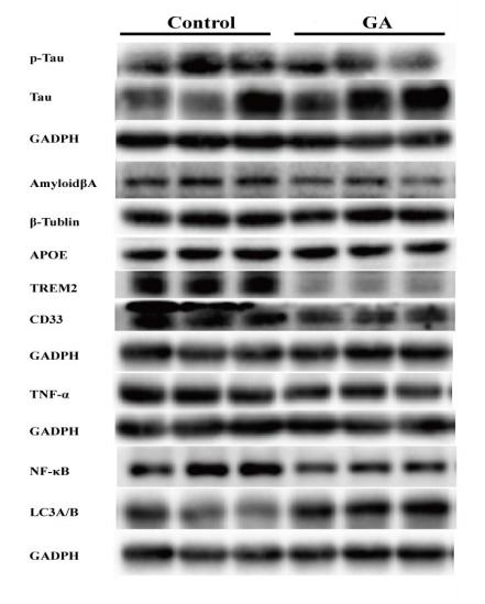
Application: WB Species: mouse Sample: brain
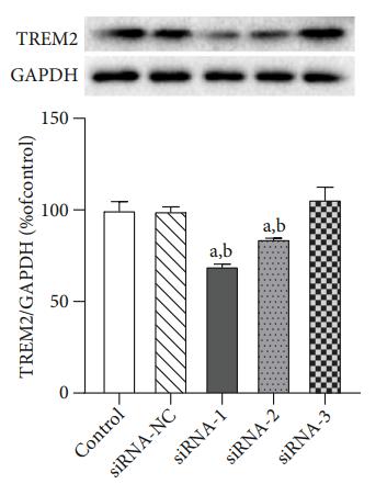
Application: WB Species: Mice Sample: MSC
Application: WB Species: mouse Sample:
Application: WB Species: Sample: RAW264.7 cells
Restrictive clause
Affinity Biosciences tests all products strictly. Citations are provided as a resource for additional applications that have not been validated by Affinity Biosciences. Please choose the appropriate format for each application and consult Materials and Methods sections for additional details about the use of any product in these publications.
For Research Use Only.
Not for use in diagnostic or therapeutic procedures. Not for resale. Not for distribution without written consent. Affinity Biosciences will not be held responsible for patent infringement or other violations that may occur with the use of our products. Affinity Biosciences, Affinity Biosciences Logo and all other trademarks are the property of Affinity Biosciences LTD.


