

 and mouse anti-beta tubulin Ab(T0023 1:200) for 1 hour at 37°C. An AlexaFluor594 conjugated goat anti-rabbit IgG(H+L) Ab(Red) and an AlexaFluor488 conjugated goat anti-mouse IgG(H+L) Ab(Green) were used as the secondary antibody.
The nuclear counter stain is DAPI(blue).")
| Product: | UQCRQ Antibody |
| Catalog: | DF12496 |
| Description: | Rabbit polyclonal antibody to UQCRQ |
| Application: | WB IHC IF/ICC |
| Cited expt.: | WB |
| Reactivity: | Human, Mouse, Rat |
| Prediction: | Pig, Bovine, Sheep, Rabbit, Dog, Chicken, Xenopus |
| Mol.Wt.: | 10 kDa(Observed); 10kD(Calculated). |
| Uniprot: | O14949 |
| RRID: | AB_2845301 |
Control Products

Related Downloads
Protocols
Product Info
*The optimal dilutions should be determined by the end user. For optimal experimental results, antibody reuse is not recommended.
*Tips:
WB: For western blot detection of denatured protein samples. IHC: For immunohistochemical detection of paraffin sections (IHC-p) or frozen sections (IHC-f) of tissue samples. IF/ICC: For immunofluorescence detection of cell samples. ELISA(peptide): For ELISA detection of antigenic peptide.
Cite Format: Affinity Biosciences Cat# DF12496, RRID:AB_2845301.
Fold/Unfold
Complex III subunit 8; Complex III subunit VIII; Cytochrome b-c1 complex subunit 8; QCR8; QCR8_HUMAN; QP-C; QPC; Ubiquinol-cytochrome c reductase complex 9.5 kDa protein; Ubiquinol-cytochrome c reductase complex ubiquinone-binding protein QP-C; Uqcrq;
Immunogens
A synthesized peptide derived from human UQCRQ, corresponding to a region within the internal amino acids.
- O14949 QCR8_HUMAN:
- Protein BLAST With
- NCBI/
- ExPASy/
- Uniprot
MGREFGNLTRMRHVISYSLSPFEQRAYPHVFTKGIPNVLRRIRESFFRVVPQFVVFYLIYTWGTEEFERSKRKNPAAYENDK
Predictions
Score>80(red) has high confidence and is suggested to be used for WB detection. *The prediction model is mainly based on the alignment of immunogen sequences, the results are for reference only, not as the basis of quality assurance.
High(score>80) Medium(80>score>50) Low(score<50) No confidence
Research Backgrounds
Component of the ubiquinol-cytochrome c oxidoreductase, a multisubunit transmembrane complex that is part of the mitochondrial electron transport chain which drives oxidative phosphorylation. The respiratory chain contains 3 multisubunit complexes succinate dehydrogenase (complex II, CII), ubiquinol-cytochrome c oxidoreductase (cytochrome b-c1 complex, complex III, CIII) and cytochrome c oxidase (complex IV, CIV), that cooperate to transfer electrons derived from NADH and succinate to molecular oxygen, creating an electrochemical gradient over the inner membrane that drives transmembrane transport and the ATP synthase. The cytochrome b-c1 complex catalyzes electron transfer from ubiquinol to cytochrome c, linking this redox reaction to translocation of protons across the mitochondrial inner membrane, with protons being carried across the membrane as hydrogens on the quinol. In the process called Q cycle, 2 protons are consumed from the matrix, 4 protons are released into the intermembrane space and 2 electrons are passed to cytochrome c.
Mitochondrion inner membrane>Single-pass membrane protein.
Belongs to the UQCRQ/QCR8 family.
Research Fields
· Human Diseases > Endocrine and metabolic diseases > Non-alcoholic fatty liver disease (NAFLD).
· Human Diseases > Neurodegenerative diseases > Alzheimer's disease.
· Human Diseases > Neurodegenerative diseases > Parkinson's disease.
· Human Diseases > Neurodegenerative diseases > Huntington's disease.
· Metabolism > Energy metabolism > Oxidative phosphorylation.
· Metabolism > Global and overview maps > Metabolic pathways.
· Organismal Systems > Circulatory system > Cardiac muscle contraction. (View pathway)
References

Application: WB Species: human Sample: HK2 cell
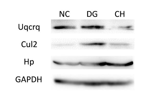
Application: WB Species: mice Sample: Liver tissues
Restrictive clause
Affinity Biosciences tests all products strictly. Citations are provided as a resource for additional applications that have not been validated by Affinity Biosciences. Please choose the appropriate format for each application and consult Materials and Methods sections for additional details about the use of any product in these publications.
For Research Use Only.
Not for use in diagnostic or therapeutic procedures. Not for resale. Not for distribution without written consent. Affinity Biosciences will not be held responsible for patent infringement or other violations that may occur with the use of our products. Affinity Biosciences, Affinity Biosciences Logo and all other trademarks are the property of Affinity Biosciences LTD.