
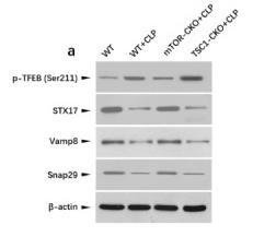
, using Syntaxin 17 Antibody at 1/1000 dilution.
5ug/NC membrane strip.
Exposure for 1min with Affinity™ ECL Kit(#KF8003).
Bands result from membrane strip incubation.")

| Product: | Syntaxin 17 Antibody |
| Catalog: | DF12483 |
| Description: | Rabbit polyclonal antibody to Syntaxin 17 |
| Application: | WB IHC |
| Cited expt.: | WB |
| Reactivity: | Human, Mouse, Rat |
| Prediction: | Pig, Bovine, Horse, Sheep, Rabbit, Dog |
| Mol.Wt.: | 33 kDa(Observed); 33kD(Calculated). |
| Uniprot: | P56962 |
| RRID: | AB_2845288 |
Control Products

Related Downloads
Protocols
Product Info
*The optimal dilutions should be determined by the end user. For optimal experimental results, antibody reuse is not recommended.
*Tips:
WB: For western blot detection of denatured protein samples. IHC: For immunohistochemical detection of paraffin sections (IHC-p) or frozen sections (IHC-f) of tissue samples. IF/ICC: For immunofluorescence detection of cell samples. ELISA(peptide): For ELISA detection of antigenic peptide.
Cite Format: Affinity Biosciences Cat# DF12483, RRID:AB_2845288.
Fold/Unfold
FLJ20651; MGC102796; MGC126613; MGC126615; Stx17; STX17_HUMAN; Syntaxin 17; Syntaxin-17;
Immunogens
A synthesized peptide derived from human Syntaxin 17, corresponding to a region within C-terminal amino acids.
- P56962 STX17_HUMAN:
- Protein BLAST With
- NCBI/
- ExPASy/
- Uniprot
MSEDEEKVKLRRLEPAIQKFIKIVIPTDLERLRKHQINIEKYQRCRIWDKLHEEHINAGRTVQQLRSNIREIEKLCLKVRKDDLVLLKRMIDPVKEEASAATAEFLQLHLESVEELKKQFNDEETLLQPPLTRSMTVGGAFHTTEAEASSQSLTQIYALPEIPQDQNAAESWETLEADLIELSQLVTDFSLLVNSQQEKIDSIADHVNSAAVNVEEGTKNLGKAAKYKLAALPVAGALIGGMVGGPIGLLAGFKVAGIAAALGGGVLGFTGGKLIQRKKQKMMEKLTSSCPDLPSQTDKKCS
Predictions
Score>80(red) has high confidence and is suggested to be used for WB detection. *The prediction model is mainly based on the alignment of immunogen sequences, the results are for reference only, not as the basis of quality assurance.
High(score>80) Medium(80>score>50) Low(score<50) No confidence
Research Backgrounds
SNAREs, soluble N-ethylmaleimide-sensitive factor-attachment protein receptors, are essential proteins for fusion of cellular membranes. SNAREs localized on opposing membranes assemble to form a trans-SNARE complex, an extended, parallel four alpha-helical bundle that drives membrane fusion. STX17 is a SNARE of the autophagosome involved in autophagy through the direct control of autophagosome membrane fusion with the lysosome membrane. May also play a role in the early secretory pathway where it may maintain the architecture of the endoplasmic reticulum-Golgi intermediate compartment/ERGIC and Golgi and/or regulate transport between the endoplasmic reticulum, the ERGIC and the Golgi.
Phosphorylated at Tyr-157 probably by ABL1. Dephosphorylation by PTPN2; regulates exit from the endoplasmic reticulum (By similarity).
Endoplasmic reticulum membrane>Multi-pass membrane protein. Smooth endoplasmic reticulum membrane>Multi-pass membrane protein. Endoplasmic reticulum-Golgi intermediate compartment membrane>Multi-pass membrane protein. Cytoplasmic vesicle>Autophagosome membrane>Multi-pass membrane protein. Cytoplasmic vesicle>COPII-coated vesicle membrane>Multi-pass membrane protein. Cytoplasm>Cytosol.
Note: Has a hairpin-like insertion into membranes. Localizes to the completed autophagosome membrane upon cell starvation (PubMed:23217709). May also localize to the mitochondria according to PubMed:23217709.
Belongs to the syntaxin family.
Research Fields
· Cellular Processes > Transport and catabolism > Autophagy - animal. (View pathway)
· Genetic Information Processing > Folding, sorting and degradation > SNARE interactions in vesicular transport.
References
Application: WB Species: Mice Sample: CD4 + T cells

Application: WB Species: mouse Sample: CD4+T cells
Restrictive clause
Affinity Biosciences tests all products strictly. Citations are provided as a resource for additional applications that have not been validated by Affinity Biosciences. Please choose the appropriate format for each application and consult Materials and Methods sections for additional details about the use of any product in these publications.
For Research Use Only.
Not for use in diagnostic or therapeutic procedures. Not for resale. Not for distribution without written consent. Affinity Biosciences will not be held responsible for patent infringement or other violations that may occur with the use of our products. Affinity Biosciences, Affinity Biosciences Logo and all other trademarks are the property of Affinity Biosciences LTD.