, using NOD2 Antibody at 1/1000 dilution.
5ug/NC membrane strip.
Exposure for 10s with Affinity™ ECL Kit(#KF8001).
Bands result from membrane strip incubation.")


 and mouse anti-beta tubulin Ab(T0023) for 1 hour at 37°C. An AlexaFluor594 conjugated goat anti-rabbit IgG(H+L) Ab(Red) and an AlexaFluor488 conjugated goat anti-mouse IgG(H+L) Ab(Green) were used as the secondary antibody.
The nuclear counter stain is DAPI (blue).")
 Ab, diluted at 1/600, was used as the secondary antibody.")
 Western blot was used to test the expressions of TNF-α, inducible nitric oxide synthase (iNOS), nuclear factor-κB (NF-κB), BAX, Bcl-2, toll-like receptor 4 (TLR4), and nucleotide-binding oligomerization domain 2 (NOD2) of intestinal tissues in rats, n = 3 in each group; ▲P < 0.05, ▲▲P < 0.01 vs. control group. ★P < 0.05, ★★P < 0.01 vs. NEC group.")
| Product: | NOD2 Antibody |
| Catalog: | DF12125 |
| Description: | Rabbit polyclonal antibody to NOD2 |
| Application: | WB IHC IF/ICC |
| Cited expt.: | WB, IHC |
| Reactivity: | Human, Mouse, Rat |
| Prediction: | Pig, Bovine, Horse, Sheep, Rabbit |
| Mol.Wt.: | 100-110 kDa(Observed); 115kD(Calculated). |
| Uniprot: | Q9HC29 |
| RRID: | AB_2844930 |
Control Products

Related Downloads
Protocols
Product Info
*The optimal dilutions should be determined by the end user. For optimal experimental results, antibody reuse is not recommended.
*Tips:
WB: For western blot detection of denatured protein samples. IHC: For immunohistochemical detection of paraffin sections (IHC-p) or frozen sections (IHC-f) of tissue samples. IF/ICC: For immunofluorescence detection of cell samples. ELISA(peptide): For ELISA detection of antigenic peptide.
Cite Format: Affinity Biosciences Cat# DF12125, RRID:AB_2844930.
Fold/Unfold
ACUG; Arthrocutaneouveal granulomatosis; BLAU; CARD15; Caspase recruitment domain family, member 15; Caspase recruitment domain protein 15; Caspase recruitment domain-containing protein 15; CD; CLR16.3; IBD1; Inflammatory bowel disease protein 1; LRR containing protein; NLR family, CARD domain containing 2; NLRC2; NOD like receptor C2; NOD2; NOD2 protein; NOD2_HUMAN; NOD2B; nucleotide binding oligomerization domain 2; Nucleotide binding oligomerization domain containing 2; Nucleotide binding oligomerization domain, leucine rich repeat and CARD domain containing 2; Nucleotide-binding oligomerization domain-containing protein 2; PSORAS1;
Immunogens
A synthesized peptide derived from human NOD2, corresponding to a region within N-terminal amino acids.
Expressed in intestinal mucosa, mainly in Paneth cells and, at lower extent, in the glandular epithelium.
- Q9HC29 NOD2_HUMAN:
- Protein BLAST With
- NCBI/
- ExPASy/
- Uniprot
MGEEGGSASHDEEERASVLLGHSPGCEMCSQEAFQAQRSQLVELLVSGSLEGFESVLDWLLSWEVLSWEDYEGFHLLGQPLSHLARRLLDTVWNKGTWACQKLIAAAQEAQADSQSPKLHGCWDPHSLHPARDLQSHRPAIVRRLHSHVENMLDLAWERGFVSQYECDEIRLPIFTPSQRARRLLDLATVKANGLAAFLLQHVQELPVPLALPLEAATCKKYMAKLRTTVSAQSRFLSTYDGAETLCLEDIYTENVLEVWADVGMAGPPQKSPATLGLEELFSTPGHLNDDADTVLVVGEAGSGKSTLLQRLHLLWAAGQDFQEFLFVFPFSCRQLQCMAKPLSVRTLLFEHCCWPDVGQEDIFQLLLDHPDRVLLTFDGFDEFKFRFTDRERHCSPTDPTSVQTLLFNLLQGNLLKNARKVVTSRPAAVSAFLRKYIRTEFNLKGFSEQGIELYLRKRHHEPGVADRLIRLLQETSALHGLCHLPVFSWMVSKCHQELLLQEGGSPKTTTDMYLLILQHFLLHATPPDSASQGLGPSLLRGRLPTLLHLGRLALWGLGMCCYVFSAQQLQAAQVSPDDISLGFLVRAKGVVPGSTAPLEFLHITFQCFFAAFYLALSADVPPALLRHLFNCGRPGNSPMARLLPTMCIQASEGKDSSVAALLQKAEPHNLQITAAFLAGLLSREHWGLLAECQTSEKALLRRQACARWCLARSLRKHFHSIPPAAPGEAKSVHAMPGFIWLIRSLYEMQEERLARKAARGLNVGHLKLTFCSVGPTECAALAFVLQHLRRPVALQLDYNSVGDIGVEQLLPCLGVCKALYLRDNNISDRGICKLIECALHCEQLQKLALFNNKLTDGCAHSMAKLLACRQNFLALRLGNNYITAAGAQVLAEGLRGNTSLQFLGFWGNRVGDEGAQALAEALGDHQSLRWLSLVGNNIGSVGAQALALMLAKNVMLEELCLEENHLQDEGVCSLAEGLKKNSSLKILKLSNNCITYLGAEALLQALERNDTILEVWLRGNTFSLEEVDKLGCRDTRLLL
Predictions
Score>80(red) has high confidence and is suggested to be used for WB detection. *The prediction model is mainly based on the alignment of immunogen sequences, the results are for reference only, not as the basis of quality assurance.
High(score>80) Medium(80>score>50) Low(score<50) No confidence
Research Backgrounds
Involved in gastrointestinal immunity. Upon stimulation by muramyl dipeptide (MDP), a fragment of bacterial peptidoglycan, binds the proximal adapter receptor-interacting RIPK2, which recruits ubiquitin ligases as XIAP, BIRC2, BIRC3, INAVA and the LUBAC complex, triggering activation of MAP kinases and activation of NF-kappa-B signaling. This in turn leads to the transcriptional activation of hundreds of genes involved in immune response. Required for MDP-induced NLRP1-dependent CASP1 activation and IL1B release in macrophages. Component of an autophagy-mediated antibacterial pathway together with ATG16L1. Plays also a role in sensing single-stranded RNA (ssRNA) from viruses. Interacts with mitochondrial antiviral signaling/MAVS, leading to activation of interferon regulatory factor-3/IRF3 and expression of type I interferon.
Polyubiquitinated following MDP stimulation, leading to proteasome-mediated degradation.
Cytoplasm. Membrane. Mitochondrion. Basolateral cell membrane.
Expressed in intestinal mucosa, mainly in Paneth cells and, at lower extent, in the glandular epithelium.
The ATG16L1-binding motif mediates interaction with ATG16L1.
Intramolecular interactions between the N-terminal moiety and the leucine-rich repeats (LRR) may be important for autoinhibition in the absence of activating signal. In the absence of LRRs, the protein becomes a constitutive activator of CASP1 cleavage and proIL1B processing.
Research Fields
· Environmental Information Processing > Signal transduction > TNF signaling pathway. (View pathway)
· Human Diseases > Infectious diseases: Bacterial > Shigellosis.
· Human Diseases > Infectious diseases: Bacterial > Tuberculosis.
· Human Diseases > Immune diseases > Inflammatory bowel disease (IBD).
· Organismal Systems > Immune system > NOD-like receptor signaling pathway. (View pathway)
References

Application: WB Species: Mouse Sample:

Application: WB Species: Mouse Sample:
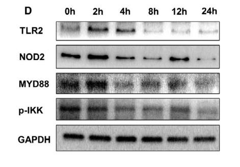
Application: WB Species: mice Sample: intestinal tissues

Application: WB Species: Human Sample: peripheral blood T cells

Application: WB Species: Mouse Sample:
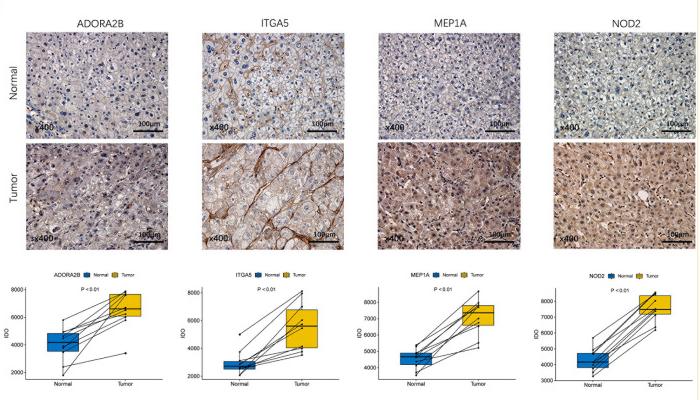
Application: IHC Species: Human Sample: HCC Tissues and Adjacent Non-Tumorous Tissues
Restrictive clause
Affinity Biosciences tests all products strictly. Citations are provided as a resource for additional applications that have not been validated by Affinity Biosciences. Please choose the appropriate format for each application and consult Materials and Methods sections for additional details about the use of any product in these publications.
For Research Use Only.
Not for use in diagnostic or therapeutic procedures. Not for resale. Not for distribution without written consent. Affinity Biosciences will not be held responsible for patent infringement or other violations that may occur with the use of our products. Affinity Biosciences, Affinity Biosciences Logo and all other trademarks are the property of Affinity Biosciences LTD.