| Product: | P2X7 Antibody |
| Catalog: | AF4626 |
| Description: | Rabbit polyclonal antibody to P2X7 |
| Application: | WB |
| Cited expt.: | WB |
| Reactivity: | Human, Mouse, Rat |
| Prediction: | Pig, Bovine, Horse, Sheep, Dog |
| Mol.Wt.: | 78kDa(Observed); 69kD(Calculated). |
| Uniprot: | Q99572 |
| RRID: | AB_2844573 |
Control Products

Related Downloads
Protocols
Product Info
*The optimal dilutions should be determined by the end user. For optimal experimental results, antibody reuse is not recommended.
*Tips:
WB: For western blot detection of denatured protein samples. IHC: For immunohistochemical detection of paraffin sections (IHC-p) or frozen sections (IHC-f) of tissue samples. IF/ICC: For immunofluorescence detection of cell samples. ELISA(peptide): For ELISA detection of antigenic peptide.
Cite Format: Affinity Biosciences Cat# AF4626, RRID:AB_2844573.
Fold/Unfold
ATP receptor; P2rx7; P2RX7_HUMAN; P2X purinoceptor 7; P2X7; P2Z receptor; Purinergic receptor; purinergic receptor P2X, ligand gated ion channel, 7;
Immunogens
A synthesized peptide derived from human P2X7, corresponding to a region within the internal amino acids.
- Q99572 P2RX7_HUMAN:
- Protein BLAST With
- NCBI/
- ExPASy/
- Uniprot
MPACCSCSDVFQYETNKVTRIQSMNYGTIKWFFHVIIFSYVCFALVSDKLYQRKEPVISSVHTKVKGIAEVKEEIVENGVKKLVHSVFDTADYTFPLQGNSFFVMTNFLKTEGQEQRLCPEYPTRRTLCSSDRGCKKGWMDPQSKGIQTGRCVVYEGNQKTCEVSAWCPIEAVEEAPRPALLNSAENFTVLIKNNIDFPGHNYTTRNILPGLNITCTFHKTQNPQCPIFRLGDIFRETGDNFSDVAIQGGIMGIEIYWDCNLDRWFHHCRPKYSFRRLDDKTTNVSLYPGYNFRYAKYYKENNVEKRTLIKVFGIRFDILVFGTGGKFDIIQLVVYIGSTLSYFGLAAVFIDFLIDTYSSNCCRSHIYPWCKCCQPCVVNEYYYRKKCESIVEPKPTLKYVSFVDESHIRMVNQQLLGRSLQDVKGQEVPRPAMDFTDLSRLPLALHDTPPIPGQPEEIQLLRKEATPRSRDSPVWCQCGSCLPSQLPESHRCLEELCCRKKPGACITTSELFRKLVLSRHVLQFLLLYQEPLLALDVDSTNSRLRHCAYRCYATWRFGSQDMADFAILPSCCRWRIRKEFPKSEGQYSGFKSPY
Predictions
Score>80(red) has high confidence and is suggested to be used for WB detection. *The prediction model is mainly based on the alignment of immunogen sequences, the results are for reference only, not as the basis of quality assurance.
High(score>80) Medium(80>score>50) Low(score<50) No confidence
Research Backgrounds
Receptor for ATP that acts as a ligand-gated ion channel. Responsible for ATP-dependent lysis of macrophages through the formation of membrane pores permeable to large molecules. Could function in both fast synaptic transmission and the ATP-mediated lysis of antigen-presenting cells. In the absence of its natural ligand, ATP, functions as a scavenger receptor in the recognition and engulfment of apoptotic cells.
Phosphorylation results in its inactivation.
ADP-ribosylation at Arg-125 is necessary and sufficient to activate P2RX7 and gate the channel.
Palmitoylation of several cysteines in the C-terminal cytoplasmic tail is required for efficient localization to cell surface.
Cell membrane>Multi-pass membrane protein.
Widely expressed with highest levels in brain and immune tissues.
Belongs to the P2X receptor family.
Research Fields
· Environmental Information Processing > Signal transduction > Calcium signaling pathway. (View pathway)
· Environmental Information Processing > Signaling molecules and interaction > Neuroactive ligand-receptor interaction.
· Organismal Systems > Immune system > NOD-like receptor signaling pathway. (View pathway)
References
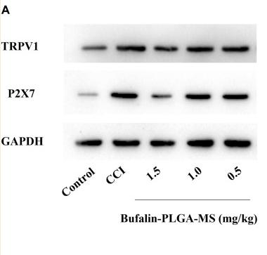
Application: WB Species: Rat Sample:
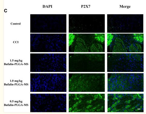
Application: IF/ICC Species: Rat Sample:

Application: IF/ICC Species: Rat Sample: brain cortex

Application: WB Species: Rat Sample: brain cortex
Restrictive clause
Affinity Biosciences tests all products strictly. Citations are provided as a resource for additional applications that have not been validated by Affinity Biosciences. Please choose the appropriate format for each application and consult Materials and Methods sections for additional details about the use of any product in these publications.
For Research Use Only.
Not for use in diagnostic or therapeutic procedures. Not for resale. Not for distribution without written consent. Affinity Biosciences will not be held responsible for patent infringement or other violations that may occur with the use of our products. Affinity Biosciences, Affinity Biosciences Logo and all other trademarks are the property of Affinity Biosciences LTD.