Product: Phospho-RIP3 (Ser232) Antibody
Catalog: AF7443
Description: Rabbit polyclonal antibody to Phospho-RIP3 (Ser232)
Application: WB
Reactivity: Mouse, Rat
Mol.Wt.: 57kDa.; 53kD(Calculated).
Uniprot: Q9QZL0
RRID: AB_2843883

View similar products>>

   Size Price Inventory
 100ul $350 In stock
 200ul $450 In stock

Lead Time: Same day delivery

For pricing and ordering contact:
Local distributors

Product Info

Source:
Rabbit
Application:
WB 1:500-1:2000
*The optimal dilutions should be determined by the end user.
*Tips:

WB: For western blot detection of denatured protein samples. IHC: For immunohistochemical detection of paraffin sections (IHC-p) or frozen sections (IHC-f) of tissue samples. IF/ICC: For immunofluorescence detection of cell samples. ELISA(peptide): For ELISA detection of antigenic peptide.

Reactivity:
Mouse,Rat
Clonality:
Polyclonal
Specificity:
Phospho-RIP3 (Ser232) Antibody detects endogenous levels of RIP3 only when phosphorylated at Ser232.
RRID:
AB_2843883
Cite Format: Affinity Biosciences Cat# AF7443, RRID:AB_2843883.
Conjugate:
Unconjugated.
Purification:
The antibody is from purified rabbit serum by affinity purification via sequential chromatography on phospho-peptide and non-phospho-peptide affinity columns.
Storage:
Rabbit IgG in phosphate buffered saline , pH 7.4, 150mM NaCl, 0.02% sodium azide and 50% glycerol. Store at -20 °C. Stable for 12 months from date of receipt.
Alias:

Fold/Unfold

Receptor interacting protein 3; Receptor interacting serine threonine kinase 3; Receptor interacting serine/threonine protein kinase 3; Receptor-interacting protein 3; Receptor-interacting serine/threonine-protein kinase 3; RIP 3; RIP like protein kinase 3; RIP-3; RIP-like protein kinase 3; RIPK 3; RIPK3; RIPK3_HUMAN;

Immunogens

Immunogen:

A synthesized peptide derived from mouse RIP3 around the phosphorylation site of Ser232.

Uniprot:
Expression:
Q9QZL0 RIPK3_MOUSE:

Expressed in embryo and in adult spleen, liver, testis, heart, brain and lung.

Sequence:
MSSVKLWPTGASAVPLVSREELKKLEFVGKGGFGVVFRAHHRTWNHDVAVKIVNSKKISWEVKAMVNLRNENVLLLLGVTEDLQWDFVSGQALVTRFMENGSLAGLLQPECPRPWPLLCRLLQEVVLGMCYLHSLNPPLLHRDLKPSNILLDPELHAKLADFGLSTFQGGSQSGSGSGSGSRDSGGTLAYLDPELLFDVNLKASKASDVYSFGILVWAVLAGREAELVDKTSLIRETVCDRQSRPPLTELPPGSPETPGLEKLKELMIHCWGSQSENRPSFQDCEPKTNEVYNLVKDKVDAAVSEVKHYLSQHRSSGRNLSAREPSQRGTEMDCPRETMVSKMLDRLHLEEPSGPVPGKCPERQAQDTSVGPATPARTSSDPVAGTPQIPHTLPFRGTTPGPVFTETPGPHPQRNQGDGRHGTPWYPWTPPNPMTGPPALVFNNCSEVQIGNYNSLVAPPRTTASSSAKYDQAQFGRGRGWQPFHK

PTMs - Q9QZL0 As Substrate

Site PTM Type Enzyme
S2 Phosphorylation
K5 Ubiquitination
S165 Phosphorylation
S184 Phosphorylation
T231 Phosphorylation
S232 Phosphorylation Q9QZL0 (Ripk3)
T257 Phosphorylation
K264 Ubiquitination
S304 Phosphorylation
S326 Phosphorylation
T338 Phosphorylation
S353 Phosphorylation
S369 Phosphorylation
T374 Phosphorylation
S380 Phosphorylation
T392 Phosphorylation
T399 Phosphorylation P63085 (Mapk1)
R477 Methylation
R479 Methylation

PTMs - Q9QZL0 As Enzyme

Substrate Site Source
Q6PHZ2 (Camk2d) T287 Uniprot
Q9D2Y4 (Mlkl) S345 Uniprot
Q9D2Y4 (Mlkl) S347 Uniprot
Q9D2Y4 (Mlkl) T349 Uniprot
Q9QZL0 (Ripk3) S232 Uniprot

Research Backgrounds

Function:

Serine/threonine-protein kinase that activates necroptosis and apoptosis, two parallel forms of cell death. Necroptosis, a programmed cell death process in response to death-inducing TNF-alpha family members, is triggered by RIPK3 following activation by ZBP1. Activated RIPK3 forms a necrosis-inducing complex and mediates phosphorylation of MLKL, promoting MLKL localization to the plasma membrane and execution of programmed necrosis characterized by calcium influx and plasma membrane damage. In addition to TNF-induced necroptosis, necroptosis can also take place in the nucleus in response to orthomyxoviruses infection: following ZBP1 activation, which senses double-stranded Z-RNA structures, nuclear RIPK3 catalyzes phosphorylation and activation of MLKL, promoting disruption of the nuclear envelope and leakage of cellular DNA into the cytosol. Also regulates apoptosis: apoptosis depends on RIPK1, FADD and CASP8, and is independent of MLKL and RIPK3 kinase activity. Phosphorylates RIPK1: RIPK1 and RIPK3 undergo reciprocal auto- and trans-phosphorylation (By similarity). In some cell types, also able to restrict viral replication by promoting cell death-independent responses. In response to flavivirus infection in neurons, promotes a cell death-independent pathway that restricts viral replication: together with ZBP1, promotes a death-independent transcriptional program that modifies the cellular metabolism via up-regulation expression of the enzyme ACOD1/IRG1 and production of the metabolite itaconate. Itaconate inhibits the activity of succinate dehydrogenase, generating a metabolic state in neurons that suppresses replication of viral genomes. RIPK3 binds to and enhances the activity of three metabolic enzymes: GLUL, GLUD1, and PYGL (By similarity). These metabolic enzymes may eventually stimulate the tricarboxylic acid cycle and oxidative phosphorylation, which could result in enhanced ROS production (By similarity).

PTMs:

RIPK1 and RIPK3 undergo reciprocal auto- and trans-phosphorylation (By similarity). Autophosphorylated following interaction with ZBP1. Phosphorylation of Ser-204 plays a role in the necroptotic function of RIPK3 (By similarity). Autophosphorylates at Thr-231 and Ser-232 following activation by ZBP1: phosphorylation at these sites is a hallmark of necroptosis and is required for binding MLKL. Phosphorylation at Thr-187 is important for its kinase activity, interaction with PELI1 and for its ability to mediate TNF-induced necroptosis (By similarity).

Polyubiquitinated with 'Lys-48' and 'Lys-63'-linked chains by BIRC2/c-IAP1 and BIRC3/c-IAP2, leading to activation of NF-kappa-B. Ubiquitinated by STUB1 leading to its subsequent proteasome-dependent degradation.

Subcellular Location:

Cytoplasm>Cytosol. Nucleus.
Note: Mainly cytoplasmic (PubMed:32200799, PubMed:32296175). Present in the nucleus in response to influenza A virus (IAV) infection (PubMed:32200799).

Extracellular region or secreted Cytosol Plasma membrane Cytoskeleton Lysosome Endosome Peroxisome ER Golgi apparatus Nucleus Mitochondrion Manual annotation Automatic computational assertionSubcellular location
Tissue Specificity:

Expressed in embryo and in adult spleen, liver, testis, heart, brain and lung.

Subunit Structure:

Interacts (via RIP homotypic interaction motif) with RIPK1 (via RIP homotypic interaction motif); this interaction induces RIPK1 phosphorylation and formation of a RIPK1-RIPK3 necrosis-inducing complex. Interacts with MLKL; the interaction is direct and triggers necroptosis. Interacts with ZBP1 (via RIP homotypic interaction motif); interaction with ZBP1 activates RIPK3, triggering necroptosis. Upon TNF-induced necrosis, the RIPK1-RIPK3 dimer further interacts with PGAM5 and MLKL; the formation of this complex leads to PGAM5 phosphorylation and increase in PGAM5 phosphatase activity (By similarity). Binds TRAF2 and is recruited to the TNFR-1 signaling complex (By similarity). Interacts with PYGL, GLUL and GLUD1; these interactions result in activation of these metabolic enzymes (By similarity). Interacts with BIRC2/c-IAP1, BIRC3/c-IAP2 and XIAP/BIRC4 (By similarity). Interacts with ARHGEF2 (By similarity). Interacts with PELI1 (via atypical FHA domain); the phosphorylated form at Thr-187 binds preferentially to PELI1. Interacts with BUB1B, TRAF2 and STUB1 (By similarity). Interacts with CASP6 (By similarity).

(Microbial infection) Interacts (via RIP homotypic interaction motif) with murid herpesvirus protein RIR1; this interaction disrupts RIP3-RIP1 interactions characteristic of TNF-alpha induced necroptosis, thereby suppressing this death pathway.

Family&Domains:

The RIP homotypic interaction motif (RHIM) mediates interaction with the RHIM motif of RIPK1. Both motifs form a hetero-amyloid serpentine fold, stabilized by hydrophobic packing and featuring an unusual Cys-Ser ladder of alternating Ser (from RIPK1) and Cys (from RIPK3).

Belongs to the protein kinase superfamily. TKL Ser/Thr protein kinase family.

References

1). RIPK1-RIPK3 mediates myocardial fibrosis in type 2 diabetes mellitus by impairing autophagic flux of cardiac fibroblasts. Cell Death & Disease, 2022 (PubMed: 35165268) [IF=9.0]

Application: WB    Species: Rat    Sample: apoptotic and necrotic cells

Fig. 1 RIPK1/RIPK3 silencing inhibited HGF-induced necroptosis. Representative images and percentages of RIPK1/ser166-p-RIPK1 (A–C) and RIPK3/ser232-p-RIPK3 (D–F). Percentages of apoptotic and necrotic cells (G–L). n = 3 per group. Means ± SD. *P < 0.05, **P < 0.01, ***P < 0.001. NGF normal glucose and fat group, HGF high glucose and high fat group.

2). Polystyrene nanoplastics and cadmium co-exposure aggravated cardiomyocyte damage in mice by regulating PANoptosis pathway. Environmental pollution (Barking, Essex : 1987), 2024 (PubMed: 38462200) [IF=8.9]

3). Polysaccharide from Strongylocentrotus nudus eggs regulates intestinal epithelial autophagy through CD36/PI3K-Akt pathway to ameliorate inflammatory bowel disease. International Journal of Biological Macromolecules, 2023 (PubMed: 37327932) [IF=8.2]

4). Cyclic helix B peptide promotes random‐pattern skin flap survival via TFE3‐mediated enhancement of autophagy and reduction of ROS levels. British Journal of Pharmacology, 2022 (PubMed: 34622942) [IF=7.3]

Application: WB    Species: Mouse    Sample: skin tissues

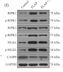
FIGURE 2 CHBP inhibits necroptosis in random-pattern skin flaps. (a,b) Representative immunohistochemical staining for RIPK3 (brown) in mouse skin tissues from the control, FLAP and FLAP + CHBP groups and quantification of integrated absorbance of RIPK3 signal (n = 6 mice per group). The tissues were counterstained with haematoxylin (blue; scale bar = 100 μm). (c,d) Representative images of immunofluorescence staining of mouse skin samples (DAPI staining of the nuclei) and quantification graph for RIPK1 (green)-positive cells (n = 6 mice per group). Scale bar: 10 μm. Skin samples were harvested from mice in the control, FLAP and FLAP + CHBP groups. (e–g) CHBP treatment attenuated the activation of RIPK1, RIPK3 and MLKL induced by ischaemia–reperfusion injury. The expression of caspase 8 (CASP8) was significantly increased, which has an inhibitory effect on necroptosis. Densitometric quantification is shown (n = 6 mice per group). Data shown are means ± SEM. *P 

5). Autophagy-related LC3 accumulation interacted directly with LIR containing RIPK1 and RIPK3, stimulating necroptosis in hypoxic cardiomyocytes. Frontiers in Cell and Developmental Biology, 2021 (PubMed: 34368130) [IF=5.5]

Application: WB    Species: mouse    Sample: myocardium

FIGURE 1 | Necroptosis mediates cardiac dysfunction caused by hypoxia. (B,C) Representative bands of western blotting and statistical analysis, which were performed to detect RIPK1, RIPK3, p-RIPK3, MLKL, and p-MLKL levels after hypoxia treatment in the myocardium, Means ± SEM, n = 5.*p < 0.05, **p < 0.01, and ***p < 0.001 versus the control group.

6). Tanshinone I exerts cardiovascular protective effects in vivo and in vitro through inhibiting necroptosis via Akt/Nrf2 signaling pathway. Chinese Medicine, 2021 (PubMed: 34183021) [IF=4.9]

Application: WB    Species: Rat    Sample: H9c2 cells

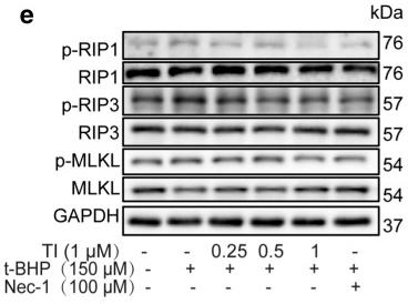
Fig. 2 TI ameliorated t‑BHP induced cell necroptosis via RIP1/RIP3/MLKL pathway. a H9c2 cells were cultured with Nec-1 for 12 h. b Cells were exposed to t-BHP (150 μM) for 10 h after treated with Nec-1 for 2 h. c Cells were exposed to t-BHP (150 μM) for 10 h after treated with TI (1 μM) or Nec-1 (100 μM) for 2 h. MTT was employed to detect cell viability. d Cells were treated with t-BHP (150 μM) for 10 h when pretreated with TI (1 μM) or Nec-1 (100 μM) for 2 h, the LDH level was monitored by LDH kit. e–h Pretreated with TI (0.25, 0.5, and 1 μM) or Nec-1 (100 μM) for 2 h respectively, then exposed to t-BHP (150 μM) for 4 h, the protein expression was determined by western blotting. n = 3. *p < 0.05, **p < 0.01, ***p < 0.001 vs. t-BHP group

7). Necrostatin-1 Against Sevoflurane-Induced Cognitive Dysfunction Involves Activation of BDNF/TrkB Pathway and Inhibition of Necroptosis in Aged Rats. NEUROCHEMICAL RESEARCH, 2022 (PubMed: 35040026) [IF=4.4]

8). Empagliflozin activates JAK2/STAT3 signaling and protects cardiomyocytes from hypoxia/reoxygenation injury under high glucose conditions. Journal of Thrombosis and Thrombolysis, 2022 (PubMed: 36396837) [IF=4.0]

9). Sugarcane leaf polysaccharide exerts a therapeutic effect on cardiovascular diseases through necroptosis. Heliyon, 2023 (PubMed: 38027563) [IF=4.0]

Application: WB    Species: Rat    Sample: H9c2 cells

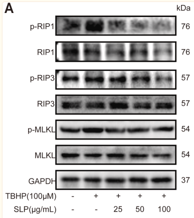
Fig. 5 SLP alleviated TBHP-stimulated cell necroptosis through the RIP1/RIP3/MLKL signaling pathway in H9c2 cells. (A) H9c2 cells were incubated with SLP (25, 50, and 100 μg/mL) and then treated with TBHP (100 μM). The protein expression of p-RIP1, RIP1, p-RIP3, RIP3, p-MLKL, and MLKL was determined using western blotting. (B–D) Statistical analysis of the above protein expressions. n = 3. ##p < 0.01, ###p < 0.001, vs. the control group. *p < 0.05, **p < 0.01, ***p < 0.001, vs. TBHP group.

10). RIP1/3-dependent programmed necrosis induces intestinal injury in septic rats: RIP1/3-dependent programmed necrosis induces intestinal injury. Acta Biochimica et Biophysica Sinica, 2024 (PubMed: 38151997) [IF=3.7]

Application: WB    Species: Rat    Sample:

Figure 2 Nec-1 relieves intestinal injury in septic rats CLP was performed to establish the rat model of sepsis, and the septic rats were intraperitoneally injected with 0.2 mL normal saline with Nec-1 after the procedures. The rats were sacrificed, and the ileum and serum were collected. (A) Western blot analysis was performed to assess the protein levels of RIP1, p-RIP3, and p-MLKL in the ileum of rats. (B) HE staining was performed to assess the pathological changes in the ileum of all rats. (C, D) ELISA was performed to detect IL-6 and TNF-α levels in the serum of rats. Data are presented as the mean±SD.

Load more

Restrictive clause


Affinity Biosciences tests all products strictly. Citations are provided as a resource for additional applications that have not been validated by Affinity Biosciences. Please choose the appropriate format for each application and consult Materials and Methods sections for additional details about the use of any product in these publications.

For Research Use Only.
Not for use in diagnostic or therapeutic procedures. Not for resale. Not for distribution without written consent. Affinity Biosciences will not be held responsible for patent infringement or other violations that may occur with the use of our products. Affinity Biosciences, Affinity Biosciences Logo and all other trademarks are the property of Affinity Biosciences LTD.