Product: Phospho-PI3 Kinase Class III (Ser244/Ser249) Antibody
Catalog: AF7421
Description: Rabbit polyclonal antibody to Phospho-PI3 Kinase Class III (Ser244/Ser249)
Application: WB IHC
Reactivity: Human, Mouse, Rat
Prediction: Pig, Bovine, Horse, Sheep, Rabbit, Chicken, Xenopus
Mol.Wt.: 100kDa; 102kD(Calculated).
Uniprot: Q8NEB9
RRID: AB_2843861

View similar products>>

   Size Price Inventory
 100ul $350 In stock
 200ul $450 In stock

Lead Time: Same day delivery

For pricing and ordering contact:
Local distributors

Product Info

Source:
Rabbit
Application:
WB 1:500-1:2000, IHC 1:50-1:200
*The optimal dilutions should be determined by the end user.
*Tips:

WB: For western blot detection of denatured protein samples. IHC: For immunohistochemical detection of paraffin sections (IHC-p) or frozen sections (IHC-f) of tissue samples. IF/ICC: For immunofluorescence detection of cell samples. ELISA(peptide): For ELISA detection of antigenic peptide.

Reactivity:
Human,Mouse,Rat
Prediction:
Pig(92%), Bovine(100%), Horse(100%), Sheep(100%), Rabbit(100%), Chicken(91%), Xenopus(82%)
Clonality:
Polyclonal
Specificity:
Phospho-PI3 Kinase Class III (Ser244/Ser249) Antibody detects endogenous levels of PI3 Kinase Class III only when phosphorylated at Ser244/249.
RRID:
AB_2843861
Cite Format: Affinity Biosciences Cat# AF7421, RRID:AB_2843861.
Conjugate:
Unconjugated.
Purification:
The antibody is from purified rabbit serum by affinity purification via sequential chromatography on phospho-peptide and non-phospho-peptide affinity columns.
Storage:
Rabbit IgG in phosphate buffered saline , pH 7.4, 150mM NaCl, 0.02% sodium azide and 50% glycerol. Store at -20 °C. Stable for 12 months from date of receipt.
Alias:

Fold/Unfold

hVps34; MGC61518; Phosphatidylinositol 3 kinase catalytic subunit type 3; Phosphatidylinositol 3 kinase class 3; Phosphatidylinositol 3 kinase p100 subunit; Phosphatidylinositol 3-kinase catalytic subunit type 3; Phosphatidylinositol 3-kinase p100 subunit; Phosphoinositide 3 kinase class 3; Phosphoinositide-3-kinase class 3; PI3 kinase type 3; PI3-kinase type 3; PI3K type 3; Pik3c3; PK3C3_HUMAN; PtdIns 3 kinase type 3; PtdIns-3-kinase type 3; Vps 34; Vps34;

Immunogens

Immunogen:
Uniprot:
Gene(ID):
Expression:
Q8NEB9 PK3C3_HUMAN:

Ubiquitously expressed, with a highest expression in skeletal muscle.

Sequence:
MGEAEKFHYIYSCDLDINVQLKIGSLEGKREQKSYKAVLEDPMLKFSGLYQETCSDLYVTCQVFAEGKPLALPVRTSYKAFSTRWNWNEWLKLPVKYPDLPRNAQVALTIWDVYGPGKAVPVGGTTVSLFGKYGMFRQGMHDLKVWPNVEADGSEPTKTPGRTSSTLSEDQMSRLAKLTKAHRQGHMVKVDWLDRLTFREIEMINESEKRSSNFMYLMVEFRCVKCDDKEYGIVYYEKDGDESSPILTSFELVKVPDPQMSMENLVESKHHKLARSLRSGPSDHDLKPNAATRDQLNIIVSYPPTKQLTYEEQDLVWKFRYYLTNQEKALTKFLKCVNWDLPQEAKQALELLGKWKPMDVEDSLELLSSHYTNPTVRRYAVARLRQADDEDLLMYLLQLVQALKYENFDDIKNGLEPTKKDSQSSVSENVSNSGINSAEIDSSQIITSPLPSVSSPPPASKTKEVPDGENLEQDLCTFLISRACKNSTLANYLYWYVIVECEDQDTQQRDPKTHEMYLNVMRRFSQALLKGDKSVRVMRSLLAAQQTFVDRLVHLMKAVQRESGNRKKKNERLQALLGDNEKMNLSDVELIPLPLEPQVKIRGIIPETATLFKSALMPAQLFFKTEDGGKYPVIFKHGDDLRQDQLILQIISLMDKLLRKENLDLKLTPYKVLATSTKHGFMQFIQSVPVAEVLDTEGSIQNFFRKYAPSENGPNGISAEVMDTYVKSCAGYCVITYILGVGDRHLDNLLLTKTGKLFHIDFGYILGRDPKPLPPPMKLNKEMVEGMGGTQSEQYQEFRKQCYTAFLHLRRYSNLILNLFSLMVDANIPDIALEPDKTVKKVQDKFRLDLSDEEAVHYMQSLIDESVHALFAAVVEQIHKFAQYWRK

Predictions

Predictions:

Score>80(red) has high confidence and is suggested to be used for WB detection. *The prediction model is mainly based on the alignment of immunogen sequences, the results are for reference only, not as the basis of quality assurance.

Species
Results
Score
Horse
100
Bovine
100
Sheep
100
Rabbit
100
Pig
92
Chicken
91
Xenopus
82
Zebrafish
73
Dog
0
Model Confidence:
High(score>80) Medium(80>score>50) Low(score<50) No confidence

PTMs - Q8NEB9 As Substrate

Site PTM Type Enzyme
S25 Phosphorylation
K29 Acetylation
K29 Ubiquitination
K36 Methylation
K36 Ubiquitination
K96 Ubiquitination
K132 Ubiquitination
K144 Ubiquitination
K158 Ubiquitination
T159 Phosphorylation P06493 (CDK1) , Q00535 (CDK5)
T163 Phosphorylation
S164 Phosphorylation
S165 Phosphorylation
S168 Phosphorylation
K209 Ubiquitination
K229 Ubiquitination
Y231 Phosphorylation
K238 Ubiquitination
S244 Phosphorylation
S249 Phosphorylation
S261 Phosphorylation
S268 Phosphorylation
K269 Ubiquitination
S276 Phosphorylation
S282 Phosphorylation
K287 Methylation
K287 Ubiquitination
T305 Phosphorylation
Y310 Phosphorylation
Y321 Phosphorylation
Y322 Phosphorylation
T324 Phosphorylation
K328 Ubiquitination
K346 Ubiquitination
Y379 Phosphorylation
Y395 Phosphorylation
K412 Ubiquitination
S448 Phosphorylation
S454 Phosphorylation
S455 Phosphorylation
K463 Ubiquitination
T488 Phosphorylation
K582 Ubiquitination
K600 Ubiquitination
K613 Methylation
T625 Phosphorylation
K630 Ubiquitination
Y631 Phosphorylation
K666 Ubiquitination
T668 Phosphorylation Q00535 (CDK5)
T677 Phosphorylation
K706 Ubiquitination
K771 Acetylation
K781 Acetylation
K781 Ubiquitination
Y803 Phosphorylation
K840 Sumoylation
K845 Sumoylation
Y884 Phosphorylation

Research Backgrounds

Function:

Catalytic subunit of the PI3K complex that mediates formation of phosphatidylinositol 3-phosphate; different complex forms are believed to play a role in multiple membrane trafficking pathways: PI3KC3-C1 is involved in initiation of autophagosomes and PI3KC3-C2 in maturation of autophagosomes and endocytosis. Involved in regulation of degradative endocytic trafficking and required for the abcission step in cytokinesis, probably in the context of PI3KC3-C2. Involved in the transport of lysosomal enzyme precursors to lysosomes. Required for transport from early to late endosomes (By similarity).

Subcellular Location:

Midbody. Late endosome. Cytoplasmic vesicle>Autophagosome.
Note: As component of the PI3K complex I localized to pre-autophagosome structures. As component of the PI3K complex II localized predominantly to endosomes (Probable). Localizes also to discrete punctae along the ciliary axoneme and to the base of the ciliary axoneme (By similarity).

Extracellular region or secreted Cytosol Plasma membrane Cytoskeleton Lysosome Endosome Peroxisome ER Golgi apparatus Nucleus Mitochondrion Manual annotation Automatic computational assertionSubcellular location
Tissue Specificity:

Ubiquitously expressed, with a highest expression in skeletal muscle.

Subunit Structure:

Component of the PI3K (PI3KC3/PI3K-III/class III phosphatidylinositol 3-kinase) complex the core of which is composed of the catalytic subunit PIK3C3, the regulatory subunit PIK3R4 and BECN1 associating with additional regulatory/auxilliary subunits to form alternative complex forms. Alternative complex forms containing a forth regulatory subunit in a mutually exclusive manner are: the PI3K complex I (PI3KC3-C1) containing ATG14, and the PI3K complex II (PI3KC3-C2) containing UVRAG. PI3KC3-C1 displays a V-shaped architecture with PIK3R4 serving as a bridge between PIK3C3 and the ATG14:BECN1 subcomplex. Both, PI3KC3-C1 and PI3KC3-C2, can associate with further regulatory subunits such as RUBCN, SH3GLB1/Bif-1 and AMBRA1. PI3KC3-C1 probably associates with PIK3CB (By similarity). Interacts with RAB7A in the presence of PIK3R4. Interacts with AMBRA1 (By similarity). Interacts with BECN1P1/BECN2. Interacts with SLAMF1. May be a component of a complex composed of RAB5A (in GDP-bound form), DYN2 and PIK3C3 (By similarity). Interacts with NCKAP1L.

Family&Domains:

Belongs to the PI3/PI4-kinase family.

Research Fields

· Cellular Processes > Transport and catabolism > Autophagy - other.   (View pathway)

· Cellular Processes > Transport and catabolism > Autophagy - animal.   (View pathway)

· Cellular Processes > Transport and catabolism > Phagosome.   (View pathway)

· Environmental Information Processing > Signal transduction > Phosphatidylinositol signaling system.

· Environmental Information Processing > Signal transduction > Apelin signaling pathway.   (View pathway)

· Human Diseases > Infectious diseases: Bacterial > Tuberculosis.

· Metabolism > Carbohydrate metabolism > Inositol phosphate metabolism.

· Metabolism > Global and overview maps > Metabolic pathways.

References

1). Inhibition of reinstatement of alcohol-induced conditioned place preference in mice by Lonicera japonica polysaccharide. Food & Function, 2022 (PubMed: 35899807) [IF=6.1]

2). Preclinical efficacy of TZG in myofascial pain syndrome by impairing PI3K-RAC2 signaling-mediated neutrophil extracellular traps. iScience, 2023 (PubMed: 37860777) [IF=5.8]

Application: WB    Species: Rat    Sample:

Figure 5 TZG significantly regulated the expression of NETs formation-related indicators, and the corresponding proteins in PI3K-RAC2 signaling (A–E). Serum levels of dsDNA, IL-8, TNF-α, CitH3, and MPO detected by ELISA analyses, respectively. Samples in (A)∼(E) were obtained from the blood supernatant of rats in different groups. (F–I) The ratios of p-PI3K/PI3K and p-P47/P47, and the expression levels of RAC2 and PAD4 proteins detected by western blot analyses, respectively. Samples in (F)∼(I) were obtained from MTrPs tissues of rats in different groups. The number of rats in each group was 8. Data are expressed as the mean ± S.D. “∗,” “∗∗,” and “∗∗∗,” p < 0.05, p < 0.01, and p < 0.001, respectively, comparison with the normal control group. “#,” “##,” and “###,” p < 0.05, p < 0.01, and p < 0.001, respectively, comparison with the MPS model group.

3). Flavonoids from Smilax china L. Rhizome improve chronic pelvic inflammatory disease by promoting macrophage reprogramming via the NLRP3 inflammasome-autophagy pathway. Journal of Functional Foods, 2023 [IF=5.6]

Application: WB    Species: Rat    Sample:

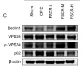
Fig. 3. Effect of FSCR on the NLRP3 inflammasome-related autophagy signaling pathway in uterine tissue of CPID rats. Detection of mRNA expression in NLRP3 inflammatory bodies by RT-PCR: NLRP3, CASP1, ASC, IL-1β, and IL-18 (A). Western blot detection of proteins in the NLRP3 inflammatory body: NLRP3, cleaved-CASP1, and cleaved-IL-1β (B). Western blot detection of proteins in the autophagy pathway: Beclin1, p-VPS34, and p62 (C). The expression of NLRP3 inflammatory bodies and autophagic vesicles in uterine tissues was observed by IF: NLRP3 was labeled with red fluorescence, VPS34 was labeled with green fluorescence, and nuclei were labeled with DAPI with blue fluorescence (D). Western blot detection of proteins in the lysosomal pathway: LAMP2, Ubiquitin and LC3 Ⅱ. (E). Results are presented as the mean ± S.E.M.

Restrictive clause


Affinity Biosciences tests all products strictly. Citations are provided as a resource for additional applications that have not been validated by Affinity Biosciences. Please choose the appropriate format for each application and consult Materials and Methods sections for additional details about the use of any product in these publications.

For Research Use Only.
Not for use in diagnostic or therapeutic procedures. Not for resale. Not for distribution without written consent. Affinity Biosciences will not be held responsible for patent infringement or other violations that may occur with the use of our products. Affinity Biosciences, Affinity Biosciences Logo and all other trademarks are the property of Affinity Biosciences LTD.