Product: Phospho-Beclin 1 (Ser90/Ser93/Ser96) Antibody
Catalog: AF7386
Description: Rabbit polyclonal antibody to Phospho-Beclin 1 (Ser90/Ser93/Ser96)
Application: WB IHC IF/ICC
Reactivity: Human, Mouse, Rat
Prediction: Pig, Bovine, Horse, Sheep, Rabbit, Dog, Chicken, Xenopus
Mol.Wt.: 60kDa; 52kD(Calculated).
Uniprot: Q14457
RRID: AB_2843826

View similar products>>

   Size Price Inventory
 100ul $350 In stock
 200ul $450 In stock

Lead Time: Same day delivery

For pricing and ordering contact:
Local distributors

Product Info

Source:
Rabbit
Application:
WB 1:500-1:2000, IHC 1:50-1:200, IF/ICC 1:100-1:500
*The optimal dilutions should be determined by the end user.
*Tips:

WB: For western blot detection of denatured protein samples. IHC: For immunohistochemical detection of paraffin sections (IHC-p) or frozen sections (IHC-f) of tissue samples. IF/ICC: For immunofluorescence detection of cell samples. ELISA(peptide): For ELISA detection of antigenic peptide.

Reactivity:
Human,Mouse,Rat
Prediction:
Pig(100%), Bovine(100%), Horse(100%), Sheep(100%), Rabbit(100%), Dog(100%), Chicken(100%), Xenopus(90%)
Clonality:
Polyclonal
Specificity:
Phospho-Beclin 1 (Ser90/Ser93/Ser96) Antibody detects endogenous levels of Beclin 1 only when phosphorylated at Ser90/93/96.
RRID:
AB_2843826
Cite Format: Affinity Biosciences Cat# AF7386, RRID:AB_2843826.
Conjugate:
Unconjugated.
Purification:
The antibody is from purified rabbit serum by affinity purification via sequential chromatography on phospho-peptide and non-phospho-peptide affinity columns.
Storage:
Rabbit IgG in phosphate buffered saline , pH 7.4, 150mM NaCl, 0.02% sodium azide and 50% glycerol. Store at -20 °C. Stable for 12 months from date of receipt.
Alias:

Fold/Unfold

APG6; ATG 6; ATG6; ATG6 autophagy related 6 homolog; Bcl-2-interacting protein beclin; Beclin 1 (coiled coil moesin like BCL2 interacting protein); Beclin 1 autophagy related; Beclin-1; Beclin1; BECN 1; Becn1; BECN1_HUMAN; Coiled coil myosin like BCL2 interacting protein; Coiled-coil myosin-like BCL2-interacting protein; GT197; Protein GT197; VPS 30; VPS30;

Immunogens

Immunogen:
Uniprot:
Gene(ID):
Expression:
Q14457 BECN1_HUMAN:

Ubiquitous.

Sequence:
MEGSKTSNNSTMQVSFVCQRCSQPLKLDTSFKILDRVTIQELTAPLLTTAQAKPGETQEEETNSGEEPFIETPRQDGVSRRFIPPARMMSTESANSFTLIGEASDGGTMENLSRRLKVTGDLFDIMSGQTDVDHPLCEECTDTLLDQLDTQLNVTENECQNYKRCLEILEQMNEDDSEQLQMELKELALEEERLIQELEDVEKNRKIVAENLEKVQAEAERLDQEEAQYQREYSEFKRQQLELDDELKSVENQMRYAQTQLDKLKKTNVFNATFHIWHSGQFGTINNFRLGRLPSVPVEWNEINAAWGQTVLLLHALANKMGLKFQRYRLVPYGNHSYLESLTDKSKELPLYCSGGLRFFWDNKFDHAMVAFLDCVQQFKEEVEKGETRFCLPYRMDVEKGKIEDTGGSGGSYSIKTQFNSEEQWTKALKFMLTNLKWGLAWVSSQFYNK

Predictions

Predictions:

Score>80(red) has high confidence and is suggested to be used for WB detection. *The prediction model is mainly based on the alignment of immunogen sequences, the results are for reference only, not as the basis of quality assurance.

Species
Results
Score
Pig
100
Horse
100
Bovine
100
Sheep
100
Dog
100
Chicken
100
Rabbit
100
Xenopus
90
Zebrafish
0
Model Confidence:
High(score>80) Medium(80>score>50) Low(score<50) No confidence

PTMs - Q14457 As Substrate

Site PTM Type Enzyme
Ubiquitination
M1 Acetylation
S15 Phosphorylation
K26 Ubiquitination
S30 Phosphorylation
K32 Ubiquitination
T38 Phosphorylation
K53 Ubiquitination
T57 Phosphorylation
T62 Phosphorylation
S64 Phosphorylation
S90 Phosphorylation P49137 (MAPKAPK2) , Q16644 (MAPKAPK3)
T91 Phosphorylation
S93 Phosphorylation
S96 Phosphorylation
T108 Phosphorylation Q13043 (STK4)
K117 Ubiquitination
T119 Phosphorylation P53355 (DAPK1)
T141 Phosphorylation
K203 Ubiquitination
K206 Acetylation
K214 Ubiquitination
Y229 Phosphorylation P00533 (EGFR)
Y233 Phosphorylation P00533 (EGFR)
S234 Phosphorylation
K237 Ubiquitination
K248 Ubiquitination
K263 Ubiquitination
S279 Phosphorylation
S295 Phosphorylation P31749 (AKT1)
K324 Ubiquitination
S337 Phosphorylation
Y352 Phosphorylation P00533 (EGFR)
T388 Phosphorylation
K402 Ubiquitination
T406 Phosphorylation P78368 (CSNK1G2)
S409 Phosphorylation P78368 (CSNK1G2)
K427 Ubiquitination
K430 Acetylation
K437 Acetylation
K437 Ubiquitination

Research Backgrounds

Function:

Plays a central role in autophagy. Acts as core subunit of the PI3K complex that mediates formation of phosphatidylinositol 3-phosphate; different complex forms are believed to play a role in multiple membrane trafficking pathways: PI3KC3-C1 is involved in initiation of autophagosomes and PI3KC3-C2 in maturation of autophagosomes and endocytosis. Involved in regulation of degradative endocytic trafficking and required for the abcission step in cytokinesis, probably in the context of PI3KC3-C2. Essential for the formation of PI3KC3-C2 but not PI3KC3-C1 PI3K complex forms. Involved in endocytosis. Protects against infection by a neurovirulent strain of Sindbis virus. May play a role in antiviral host defense.

Beclin-1-C 35 kDa localized to mitochondria can promote apoptosis; it induces the mitochondrial translocation of BAX and the release of proapoptotic factors.

PTMs:

Phosphorylation at Thr-119 by DAPK1 reduces its interaction with BCL2 and BCL2L1 and promotes induction of autophagy. In response to autophagic stimuli, phosphorylated at serine residues by AMPK in an ATG14-dependent manner, and this phosphorylation is critical for maximally efficient autophagy.

Polyubiquitinated by NEDD4, both with 'Lys-11'- and 'Lys-63'-linkages. 'Lys-11'-linked polyubiquitination leads to degradation and is enhanced when the stabilizing interaction partner VPS34 is depleted. Deubiquitinated by USP10 and USP13, leading to stabilize the PIK3C3/VPS34-containing complexes. Polyubiquitinated at Lys-402 with 'Lys-48'-linkages. 'Lys-48'-linked polyubiquitination of Lys-402 leads to degradation. Deubiquitinated by ATXN3, leading to stabilization.

Proteolytically processed by caspases including CASP8 and CASP3; the C-terminal fragments lack autophagy-inducing capacity and are proposed to induce apoptosis. Thus the cleavage is proposed to be an determinant to switch from autophagy to apoptosis pathways affecting cellular homeostasis including viral infections and survival of tumor cells.

Subcellular Location:

Cytoplasm. Golgi apparatus>trans-Golgi network membrane>Peripheral membrane protein. Endosome membrane>Peripheral membrane protein. Endoplasmic reticulum membrane>Peripheral membrane protein. Mitochondrion membrane>Peripheral membrane protein. Endosome. Cytoplasmic vesicle>Autophagosome.
Note: Interaction with ATG14 promotes translocation to autophagosomes. Expressed in dendrites and cell bodies of cerebellar Purkinje cells (By similarity).

Mitochondrion. Nucleus. Cytoplasm.

Mitochondrion.

Extracellular region or secreted Cytosol Plasma membrane Cytoskeleton Lysosome Endosome Peroxisome ER Golgi apparatus Nucleus Mitochondrion Manual annotation Automatic computational assertionSubcellular location
Tissue Specificity:

Ubiquitous.

Subunit Structure:

A homodimeric form is proposed to exist; this metastable form readily transits to ATG14- or UVRAG-containing complexes with BECN1:UVRAG being more stable than BECN1:ATG14 (By similarity). Component of the PI3K (PI3KC3/PI3K-III/class III phosphatidylinositol 3-kinase) complex the core of which is composed of the catalytic subunit PIK3C3, the regulatory subunit PIK3R4 and BECN1 associating with additional regulatory/auxilliary subunits to form alternative complex forms. Alternative complex forms containing a forth regulatory subunit in a mutually exclusive manner are PI3K complex I (PI3KC3-C1) containing ATG14, and PI3K complex II (PI3KC3-C2) containing UVRAG. PI3KC3-C1 displays a V-shaped architecture with PIK3R4 serving as a bridge between PIK3C3 and the ATG14:BECN1 subcomplex. Both, PI3KC3-C1 and PI3KC3-C2, can associate with further regulatory subunits, such as RUBCN, SH3GLB1/Bif-1 and AMBRA1. PI3KC3-C1 probably associates with PIK3CB (By similarity). Interacts with AMBRA1, GOPC, GRID2 (By similarity). Interacts with BCL2 and BCL2L1 isoform Bcl-X(L); the interaction inhibits BECN1 function in promoting autophagy by interfering with the formation of the PI3K complex. Interacts with cytosolic HMGB1; inhibits the interaction of BECN1 and BCL2 leading to promotion of autophagy. Interacts with USP10, USP13, VMP1, DAPK1, RAB39A. Interacts with the poly-Gln domain of ATXN3; the interaction causes deubiquitination at Lys-402 and stabilizes BECN1. Interacts with SLAMF1. Interacts with TRIM5; the interaction causes activation of BECN1 by causing its dissociation from its inhibitors BCL2 and TAB2. Interacts with active ULK1 (phosphorylated on 'Ser-317') and MEFV simultaneously. Interacts with WDR81 and WDR91; negatively regulates the PI3 kinase/PI3K activity associated with endosomal membranes. Interacts with LAPTM4B; competes with EGFR for LAPTM4B binding; regulates EGFR activity. Interacts with TRIM50. Interacts with TRIM16.

(Microbial infection) Interacts with human cytomegalovirus/HHV-5 protein TRS1.

(Microbial infection) Interacts with murine gammaherpesvirus 68 M11.

(Microbial infection) Interacts with herpes simplex virus 1 (HHV-1) protein ICP34.5; this interaction antagonizes the host autophagy response.

Family&Domains:

The coiled coil domain can form antiparallel homodimers and mediates dimerization with the coiled coil domains of ATG14 or UVRAG involved in the formation of PI3K complexes.

The C-terminal evolutionary conserved domain (ECD) contains poly-Gln-binding domains such as the ATXN3 poly-Gln motif, consistent with structural docking models revealing two highly scored poly-Gln-binding pockets in the ECD (PubMed:28445460). As some binding is observed with BECN1 lacking the ECD, other domains of BECN1 may also interact with ATXN3 (PubMed:28445460).

Belongs to the beclin family.

Research Fields

· Cellular Processes > Transport and catabolism > Autophagy - other.   (View pathway)

· Cellular Processes > Transport and catabolism > Autophagy - animal.   (View pathway)

· Cellular Processes > Cell growth and death > Apoptosis - multiple species.   (View pathway)

· Environmental Information Processing > Signal transduction > Apelin signaling pathway.   (View pathway)

References

1). Bunyavirus SFTSV exploits autophagic flux for viral assembly and egress. Autophagy (PubMed: 34747299) [IF=13.3]

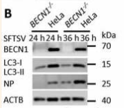
Application: WB    Species: Human    Sample: HeLa cells and WT cells

Figure 3. Autophagy is essential for the replication of SFTSV. (A-E) BECN1, atg5, atg7, RB1CC1, ATG16L1 knockout MEF or HeLa cells and WT cells were infected with SFTSV at an MOI of 1 for 24 h and 36 h. Cell lysates were evaluated via WB. (F-G) BECN1, atg5, atg7, RB1CC1, ATG16L1 knockout MEF or HeLa cells and WT cells were infected with SFTSV at an MOI of 1 for 24 h. Nuclear DNA, endogenous LC3, and SFTSV NP were stained as blue, green, and violet respectively. Single plane type of images was present. Scale bar: 20 μm. (H) BECN1, atg5, atg7, RB1CC1, ATG16L1 knockout MEF or HeLa cells and WT cells were infected with SFTSV at an MOI of 1 for 2 h. Then cells were washed once by PBS for three times and internalized SFTSV were detected via RT-qPCR. (I) BECN1, atg5, atg7, RB1CC1, ATG16L1 knockout MEF or HeLa cells and WT cells were infected with SFTSV at an MOI of 1 for 24 h. Endpoint 10-fold dilutions of an SFTSV stock were titrated. Values presented in the graph are calculated and expressed as the log10 of TCID50 units per ml of supernatant. Error bars, mean ± SD of three experiments. Student’s t test;

Application: IF/ICC    Species: Human    Sample: HeLa cells and WT cells

Figure 3. Autophagy is essential for the replication of SFTSV. (A-E) BECN1, atg5, atg7, RB1CC1, ATG16L1 knockout MEF or HeLa cells and WT cells were infected with SFTSV at an MOI of 1 for 24 h and 36 h. Cell lysates were evaluated via WB. (F-G) BECN1, atg5, atg7, RB1CC1, ATG16L1 knockout MEF or HeLa cells and WT cells were infected with SFTSV at an MOI of 1 for 24 h. Nuclear DNA, endogenous LC3, and SFTSV NP were stained as blue, green, and violet respectively. Single plane type of images was present. Scale bar: 20 μm. (H) BECN1, atg5, atg7, RB1CC1, ATG16L1 knockout MEF or HeLa cells and WT cells were infected with SFTSV at an MOI of 1 for 2 h. Then cells were washed once by PBS for three times and internalized SFTSV were detected via RT-qPCR. (I) BECN1, atg5, atg7, RB1CC1, ATG16L1 knockout MEF or HeLa cells and WT cells were infected with SFTSV at an MOI of 1 for 24 h. Endpoint 10-fold dilutions of an SFTSV stock were titrated. Values presented in the graph are calculated and expressed as the log10 of TCID50 units per ml of supernatant. Error bars, mean ± SD of three experiments. Student’s t test;

2). Inhibition of reinstatement of alcohol-induced conditioned place preference in mice by Lonicera japonica polysaccharide. Food & Function (PubMed: 35899807) [IF=6.1]

3). Blocking transmembrane219 protein signaling inhibits autophagy and restores normal cell death. PLoS One (PubMed: 31220095) [IF=3.7]

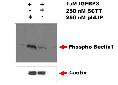
Application: WB    Species: human    Sample: MCF7 cells

Fig 6. |TM219 short cytoplasmic protein tail peptide disrupts the interaction between calmodulin and TM219 protein. C-MCF7 cells pretreated with either SCTTpHLIP or pHLIP peptide alone were treated with 1μM IGFBP3 for 1hour in DMEM serum free medium. Cell lysates were subjected to immunoblot using anti-human phosphor-beclin1 antibody. Treatment with SCTT-pHLIP reduced drastically phosphorylation of Beclin1 protein.

Restrictive clause


Affinity Biosciences tests all products strictly. Citations are provided as a resource for additional applications that have not been validated by Affinity Biosciences. Please choose the appropriate format for each application and consult Materials and Methods sections for additional details about the use of any product in these publications.

For Research Use Only.
Not for use in diagnostic or therapeutic procedures. Not for resale. Not for distribution without written consent. Affinity Biosciences will not be held responsible for patent infringement or other violations that may occur with the use of our products. Affinity Biosciences, Affinity Biosciences Logo and all other trademarks are the property of Affinity Biosciences LTD.