Phospho-ULK1 (Ser555)[Ser556] Antibody - #AF7148
![Phospho-ULK1 (Ser555)[Ser556] Antibody - AF7148 at 1/100 staining human kidney tissue sections by IHC-P.](http://img.affbiotech.cn/images/201806/thumb_img/13870_thumb_P_1528817062328.jpg "AF7148 at 1/100 staining human kidney tissue sections by IHC-P. The tissue was formaldehyde fixed and a heat mediated antigen retrieval step in citrate buffer was performed. The tissue was then blocked and incubated with the antibody for 1.5 hours at 22°C. An HRP conjugated goat anti-rabbit antibody was used as the secondary antibody.")
![Phospho-ULK1 (Ser555)[Ser556] Antibody - AF7148 at 1/100 staining rat heart tissue sections by IHC-P.](http://img.affbiotech.cn/images/201806/thumb_img/13870_thumb_P_1528817062213.jpg "AF7148 at 1/100 staining rat heart tissue sections by IHC-P. The tissue was formaldehyde fixed and a heat mediated antigen retrieval step in citrate buffer was performed. The tissue was then blocked and incubated with the antibody for 1.5 hours at 22°C. An HRP conjugated goat anti-rabbit antibody was used as the secondary antibody.")
![Phospho-ULK1 (Ser555)[Ser556] Antibody - AF7148 at 1/100 staining mouse heart tissue sections by IHC-P.](http://img.affbiotech.cn/images/201806/thumb_img/13870_thumb_P_1528817062686.jpg "AF7148 at 1/100 staining mouse heart tissue sections by IHC-P. The tissue was formaldehyde fixed and a heat mediated antigen retrieval step in citrate buffer was performed. The tissue was then blocked and incubated with the antibody for 1.5 hours at 22°C. An HRP conjugated goat anti-rabbit antibody was used as the secondary antibody.")
![Phospho-ULK1 (Ser555)[Ser556] Antibody - peptide-ELISA analysis of AF7148.](http://img.affbiotech.cn/images/pelisa/809/af7148-peptide-elisa.png "peptide-ELISA analysis of AF7148. showing specificity to antigen peptide. Peptides concentration: 1ug/ml.<br>
P-peptide: phospho-peptide. N-peptide: non-phospho-peptide.")
| Product: | Phospho-ULK1 (Ser555)[Ser556] Antibody |
| Catalog: | AF7148 |
| Description: | Rabbit polyclonal antibody to Phospho-ULK1 (Ser555)[Ser556] |
| Application: | WB IHC |
| Cited expt.: | WB, IHC |
| Reactivity: | Human, Mouse, Rat |
| Prediction: | Pig, Bovine, Horse, Sheep, Rabbit, Dog, Chicken, Xenopus |
| Mol.Wt.: | 150kD(Observed); 113kD(Calculated). |
| Uniprot: | O75385 |
| RRID: | AB_2843588 |
Related Downloads
Protocols
Product Info
*The optimal dilutions should be determined by the end user. For optimal experimental results, antibody reuse is not recommended.
*Tips:
WB: For western blot detection of denatured protein samples. IHC: For immunohistochemical detection of paraffin sections (IHC-p) or frozen sections (IHC-f) of tissue samples. IF/ICC: For immunofluorescence detection of cell samples. ELISA(peptide): For ELISA detection of antigenic peptide.
Cite Format: Affinity Biosciences Cat# AF7148, RRID:AB_2843588.
Fold/Unfold
ATG 1; ATG1; ATG1 autophagy related 1 homolog; ATG1A; Autophagy related protein 1 homolog; Autophagy-related protein 1 homolog; FLJ38455; FLJ46475; hATG1; KIAA0722; Serine/threonine protein kinase ULK1; Serine/threonine protein kinase Unc51.1; Serine/threonine-protein kinase ULK1; ULK 1; ULK1; ULK1_HUMAN; Unc 51 (C. elegans) like kinase 1; UNC 51; Unc 51 like kinase 1; Unc-51 like kinase 1 (C. elegans); Unc-51-like kinase 1; UNC51; UNC51, C. elegans, homolog of; Unc51.1;
Immunogens
A synthesized peptide derived from human ULK1 around the phosphorylation site of Ser556.
Ubiquitously expressed. Detected in the following adult tissues: skeletal muscle, heart, pancreas, brain, placenta, liver, kidney, and lung.
- O75385 ULK1_HUMAN:
- Protein BLAST With
- NCBI/
- ExPASy/
- Uniprot
MEPGRGGTETVGKFEFSRKDLIGHGAFAVVFKGRHREKHDLEVAVKCINKKNLAKSQTLLGKEIKILKELKHENIVALYDFQEMANSVYLVMEYCNGGDLADYLHAMRTLSEDTIRLFLQQIAGAMRLLHSKGIIHRDLKPQNILLSNPAGRRANPNSIRVKIADFGFARYLQSNMMAATLCGSPMYMAPEVIMSQHYDGKADLWSIGTIVYQCLTGKAPFQASSPQDLRLFYEKNKTLVPTIPRETSAPLRQLLLALLQRNHKDRMDFDEFFHHPFLDASPSVRKSPPVPVPSYPSSGSGSSSSSSSTSHLASPPSLGEMQQLQKTLASPADTAGFLHSSRDSGGSKDSSCDTDDFVMVPAQFPGDLVAEAPSAKPPPDSLMCSGSSLVASAGLESHGRTPSPSPPCSSSPSPSGRAGPFSSSRCGASVPIPVPTQVQNYQRIERNLQSPTQFQTPRSSAIRRSGSTSPLGFARASPSPPAHAEHGGVLARKMSLGGGRPYTPSPQVGTIPERPGWSGTPSPQGAEMRGGRSPRPGSSAPEHSPRTSGLGCRLHSAPNLSDLHVVRPKLPKPPTDPLGAVFSPPQASPPQPSHGLQSCRNLRGSPKLPDFLQRNPLPPILGSPTKAVPSFDFPKTPSSQNLLALLARQGVVMTPPRNRTLPDLSEVGPFHGQPLGPGLRPGEDPKGPFGRSFSTSRLTDLLLKAAFGTQAPDPGSTESLQEKPMEIAPSAGFGGSLHPGARAGGTSSPSPVVFTVGSPPSGSTPPQGPRTRMFSAGPTGSASSSARHLVPGPCSEAPAPELPAPGHGCSFADPITANLEGAVTFEAPDLPEETLMEQEHTEILRGLRFTLLFVQHVLEIAALKGSASEAAGGPEYQLQESVVADQISLLSREWGFAEQLVLYLKVAELLSSGLQSAIDQIRAGKLCLSSTVKQVVRRLNELYKASVVSCQGLSLRLQRFFLDKQRLLDRIHSITAERLIFSHAVQMVQSAALDEMFQHREGCVPRYHKALLLLEGLQHMLSDQADIENVTKCKLCIERRLSALLTGICA
Predictions
Score>80(red) has high confidence and is suggested to be used for WB detection. *The prediction model is mainly based on the alignment of immunogen sequences, the results are for reference only, not as the basis of quality assurance.
High(score>80) Medium(80>score>50) Low(score<50) No confidence
Research Backgrounds
Serine/threonine-protein kinase involved in autophagy in response to starvation. Acts upstream of phosphatidylinositol 3-kinase PIK3C3 to regulate the formation of autophagophores, the precursors of autophagosomes. Part of regulatory feedback loops in autophagy: acts both as a downstream effector and negative regulator of mammalian target of rapamycin complex 1 (mTORC1) via interaction with RPTOR. Activated via phosphorylation by AMPK and also acts as a regulator of AMPK by mediating phosphorylation of AMPK subunits PRKAA1, PRKAB2 and PRKAG1, leading to negatively regulate AMPK activity. May phosphorylate ATG13/KIAA0652 and RPTOR; however such data need additional evidences. Plays a role early in neuronal differentiation and is required for granule cell axon formation. May also phosphorylate SESN2 and SQSTM1 to regulate autophagy.
Autophosphorylated. Phosphorylated under nutrient-rich conditions; dephosphorylated during starvation or following treatment with rapamycin. Under nutrient sufficiency, phosphorylated by MTOR/mTOR, disrupting the interaction with AMPK and preventing activation of ULK1 (By similarity). In response to nutrient limitation, phosphorylated and activated by AMPK, leading to activate autophagy.
Acetylated by KAT5/TIP60 under autophagy induction, promoting protein kinase activity.
Cytoplasm>Cytosol. Preautophagosomal structure.
Note: Under starvation conditions, is localized to puncate structures primarily representing the isolation membrane that sequesters a portion of the cytoplasm resulting in the formation of an autophagosome.
Ubiquitously expressed. Detected in the following adult tissues: skeletal muscle, heart, pancreas, brain, placenta, liver, kidney, and lung.
Belongs to the protein kinase superfamily. Ser/Thr protein kinase family. APG1/unc-51/ULK1 subfamily.
Research Fields
· Cellular Processes > Transport and catabolism > Autophagy - animal. (View pathway)
· Environmental Information Processing > Signal transduction > mTOR signaling pathway. (View pathway)
· Environmental Information Processing > Signal transduction > AMPK signaling pathway. (View pathway)
· Organismal Systems > Aging > Longevity regulating pathway. (View pathway)
References
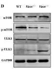
Application: WB Species: Mice Sample: liver
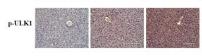
Application: IHC Species: Mice Sample: liver

Application: WB Species: Mouse Sample: stomach tissue
Restrictive clause
Affinity Biosciences tests all products strictly. Citations are provided as a resource for additional applications that have not been validated by Affinity Biosciences. Please choose the appropriate format for each application and consult Materials and Methods sections for additional details about the use of any product in these publications.
For Research Use Only.
Not for use in diagnostic or therapeutic procedures. Not for resale. Not for distribution without written consent. Affinity Biosciences will not be held responsible for patent infringement or other violations that may occur with the use of our products. Affinity Biosciences, Affinity Biosciences Logo and all other trademarks are the property of Affinity Biosciences LTD.