

Control Products

Related Downloads
Protocols
Product Info
*The optimal dilutions should be determined by the end user. For optimal experimental results, antibody reuse is not recommended.
*Tips:
WB: For western blot detection of denatured protein samples. IHC: For immunohistochemical detection of paraffin sections (IHC-p) or frozen sections (IHC-f) of tissue samples. IF/ICC: For immunofluorescence detection of cell samples. ELISA(peptide): For ELISA detection of antigenic peptide.
Cite Format: Affinity Biosciences Cat# DF9919, RRID:AB_2843113.
Fold/Unfold
AMCFII; C-X-C motif chemokine 5; C-X-C motif chemokine ligand 5; chemokine (C-X-C motif) ligand 5; Cxcl5; CXCL5_HUMAN; ENA 78; ENA-78 (8-78); ENA-78(1-78); ENA-78(9-78); ENA78; Epithelial derived neutrophil activating protein 78; Epithelial-derived neutrophil-activating protein 78; Lipopolysaccharide-induced CXC chemokine; Neutrophil activating peptide ENA 78; Neutrophil-activating peptide ENA-78; neutrophil-activating protein 78; SCYB5; Small inducible cytokine B5; small inducible cytokine subfamily B (Cys-X-Cys), member 5 (epithelial-derived neutrophil-activating peptide 78); small inducible cytokine subfamily B, member 5; Small-inducible cytokine B5;
Immunogens
A synthesized peptide derived from human CXCL5, corresponding to a region within the internal amino acids.
- P42830 CXCL5_HUMAN:
- Protein BLAST With
- NCBI/
- ExPASy/
- Uniprot
MSLLSSRAARVPGPSSSLCALLVLLLLLTQPGPIASAGPAAAVLRELRCVCLQTTQGVHPKMISNLQVFAIGPQCSKVEVVASLKNGKEICLDPEAPFLKKVIQKILDGGNKEN
Predictions
Score>80(red) has high confidence and is suggested to be used for WB detection. *The prediction model is mainly based on the alignment of immunogen sequences, the results are for reference only, not as the basis of quality assurance.
High(score>80) Medium(80>score>50) Low(score<50) No confidence
Research Backgrounds
Involved in neutrophil activation. In vitro, ENA-78(8-78) and ENA-78(9-78) show a threefold higher chemotactic activity for neutrophil granulocytes.
N-terminal processed forms ENA-78(8-78) and ENA-78(9-78) are produced by proteolytic cleavage after secretion from peripheral blood monocytes.
Secreted.
Belongs to the intercrine alpha (chemokine CxC) family.
Research Fields
· Environmental Information Processing > Signaling molecules and interaction > Cytokine-cytokine receptor interaction. (View pathway)
· Environmental Information Processing > Signal transduction > TNF signaling pathway. (View pathway)
· Human Diseases > Infectious diseases: Bacterial > Pertussis.
· Human Diseases > Immune diseases > Rheumatoid arthritis.
· Organismal Systems > Immune system > Chemokine signaling pathway. (View pathway)
· Organismal Systems > Immune system > IL-17 signaling pathway. (View pathway)
References

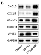
Application: WB Species: human Sample: BxPC-3 and PATU 8988t cells

Application: IF/ICC Species: Mouse Sample:

Application: IHC Species: Human Sample: placental villous tissue

Application: IHC Species: Human Sample: placental villous tissue

Application: WB Species: Mouse Sample:

Application: IHC Species: Mouse Sample:

Application: WB Species: human Sample:

Application: WB Species: Mouse Sample: lung cancer cells

Application: IHC Species: Mouse Sample: lung cancer cells

Application: WB Species: human Sample: A549 cells
Application: WB Species: Human Sample:
Restrictive clause
Affinity Biosciences tests all products strictly. Citations are provided as a resource for additional applications that have not been validated by Affinity Biosciences. Please choose the appropriate format for each application and consult Materials and Methods sections for additional details about the use of any product in these publications.
For Research Use Only.
Not for use in diagnostic or therapeutic procedures. Not for resale. Not for distribution without written consent. Affinity Biosciences will not be held responsible for patent infringement or other violations that may occur with the use of our products. Affinity Biosciences, Affinity Biosciences Logo and all other trademarks are the property of Affinity Biosciences LTD.